A novel perspective on dissemination: somatic metastasis of germ cell tumors from the central nervous system
- PMID: 40533624
- PMCID: PMC12176715
- DOI: 10.1007/s12672-025-02467-6
A novel perspective on dissemination: somatic metastasis of germ cell tumors from the central nervous system
Abstract
Purpose: Primary germ cell tumors (GCTs) of the central nervous system are rare malignancies, with somatic metastasis being even less common. This study aims to propose a novel classification framework for dissemination patterns in CNS GCTs.
Methods: A group of 4 patients with somatic metastasis were identified among 284 germ cell tumors. The characteristics of patients were identified through clinical history, imaging, and histopathology. Hematoxylin-eosin (H&E) and Immunohistochemistry (IHC) confirmed the pathological features, while radiology was used to determine the temporal and spatial characteristics of metastasis. Univariate analysis was conducted to assess the factors contributing to abdominal spread. Fisher's exact test was used for categorical data analysis. The dissemination process was systematically analyzed.
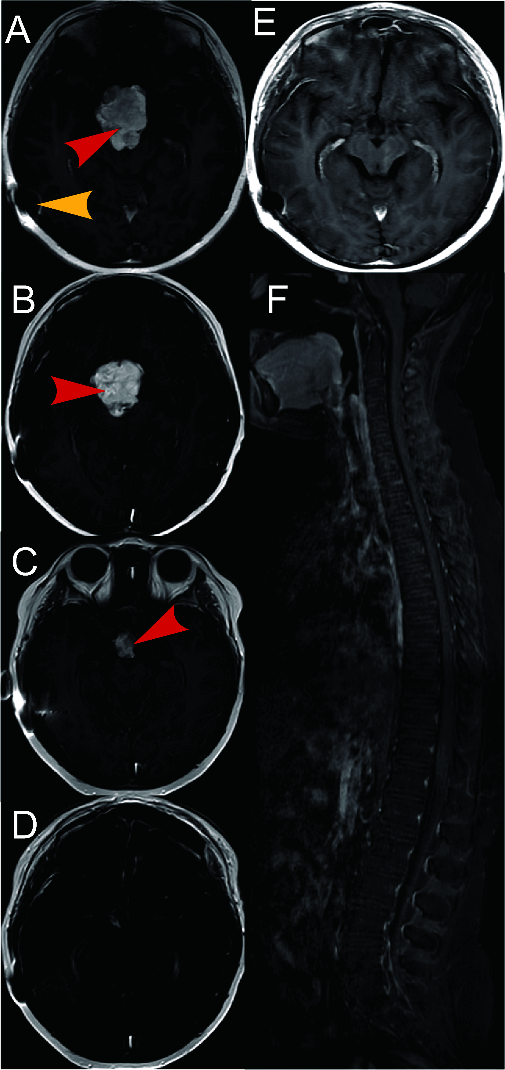
Results: This disease is extremely rare, and the progression of metastatic germ cell tumors is often complex. All four patients underwent surgery, radiotherapy, and chemotherapy. In one case, the tumor spread to the anterior superior mediastinum via hematogenous metastasis; in two cases, it spread to the abdominal cavity through a ventriculoperitoneal shunt; and in one case, intraspinal spread occurred via cerebrospinal fluid. Over a follow-up period of 9 to 67 months, only one patient died from the disease. H&E and IHC staining confirmed pathological features. Analyzing dissemination introduced concepts like dynamics, components, mediums, ranges, portals, and patterns, creating a framework for understanding disease spread.
Conclusion: Our findings offer valuable insights for guiding treatment strategies and improving patient outcomes. CNS GCTs can be managed effectively with accurate diagnosis and appropriate treatment. The introduction of new concepts and classifications of dissemination can inform and guide treatment strategies.
Keywords: Dissemination; Germ cell tumor; Somatic metastasis; Treatment.
© 2025. The Author(s).
Conflict of interest statement
Declarations. Ethics approval and consent to participants: All procedures performed in studies were in accordance with the ethical standards of the institutional and/or national research committee and comparable ethical standards. All experimental protocols were approved by the Ethics Committee of Sanbo Brain Hospital and conducted in accordance with relevant regulations. Informed consent was obtained from all participating subjects. For subjects under 18 years of age or deceased, consent was provided by their parent(s) and/or legal guardian(s). Competing interests: The authors declare no competing interests.
Figures


Similar articles
-
Molecular feature-based classification of retroperitoneal liposarcoma: a prospective cohort study.Elife. 2025 May 23;14:RP100887. doi: 10.7554/eLife.100887. Elife. 2025. PMID: 40407808 Free PMC article.
-
Surveillance for Violent Deaths - National Violent Death Reporting System, 50 States, the District of Columbia, and Puerto Rico, 2022.MMWR Surveill Summ. 2025 Jun 12;74(5):1-42. doi: 10.15585/mmwr.ss7405a1. MMWR Surveill Summ. 2025. PMID: 40493548 Free PMC article.
-
Assessing the comparative effects of interventions in COPD: a tutorial on network meta-analysis for clinicians.Respir Res. 2024 Dec 21;25(1):438. doi: 10.1186/s12931-024-03056-x. Respir Res. 2024. PMID: 39709425 Free PMC article. Review.
-
Transcriptome analysis and artificial intelligence for predicting lymph node metastasis of esophageal squamous cell carcinoma.J Thorac Dis. 2025 May 30;17(5):3283-3296. doi: 10.21037/jtd-2025-662. Epub 2025 May 28. J Thorac Dis. 2025. PMID: 40529736 Free PMC article.
-
Sociodemographic and Socioeconomic Determinants for the Usage of Digital Patient Portals in Hospitals: Systematic Review and Meta-Analysis on the Digital Divide.J Med Internet Res. 2025 Jun 3;27:e68091. doi: 10.2196/68091. J Med Internet Res. 2025. PMID: 40460427 Free PMC article. Review.
References
-
- Ostrom Q, Price M, Ryan K, Edelson J, Neff C, Cioffi G, Waite K, Kruchko C, Barnholtz-Sloan J. CBTRUS statistical report: pediatric brain tumor foundation childhood and adolescent primary brain and other central nervous system tumors diagnosed in the United States in 2014–2018. Neuro Oncol. 2022;24:1–38. 10.1093/neuonc/noac161. - PMC - PubMed
-
- Bowzyk Al-Naeeb A, Murray M, Horan G, Harris F, Kortmann RD, Nicholson J, Ajithkumar T. Current management of intracranial germ cell tumours. Clin Oncol (R Coll Radiol). 2018;30:204–14. 10.1016/j.clon.2018.01.009. - PubMed
LinkOut - more resources
Full Text Sources
