. 2025 Jun 18;24(1):181.
doi: 10.1186/s12943-025-02382-y.

Biologically targeted dual adaptive and innate nano-Immunotherapy for clear cell renal cell carcinoma treatment

Affiliations

Biologically targeted dual adaptive and innate nano-Immunotherapy for clear cell renal cell carcinoma treatment

Kin Man Au et al. Mol Cancer. .

Abstract

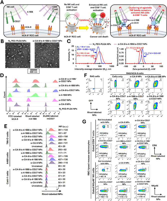
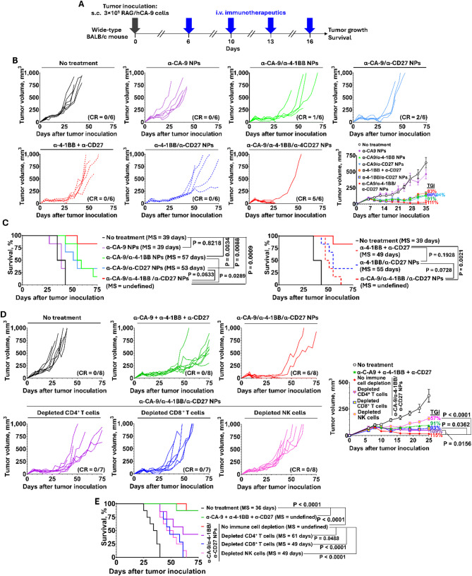
Background: Immunotherapy treatments have significantly improved metastatic renal cell carcinoma (RCC) treatment outcomes. Despite recent advancements, the rates of durable response to immunotherapy remain low, and the toxicity profiles of treatment continue to be high. To address these challenges, we report the development of a human carbonic anhydrase-IX (hCA-9)-targeted multifunctional immunotherapy nanoparticles (MINPs) aimed at improving treatment efficacy and reducing toxicity. We hypothesized that these MINPs will facilitate the recognition and elimination of hCA-9-expressing tumor cells by both adaptive immune cells (cytotoxic CD8+ T cells) and innate immune cells (natural killer (NK) cells).

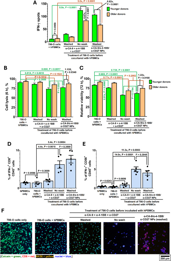
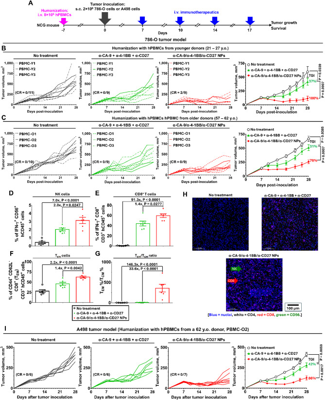
Methods: Non-targeted and hCA-9-targeted MINPs were prepared by conjugating anti-CA-9, anti-4-1BB, and anti-CD27 antibodies to poly(ethylene glycol)-block-poly(lactic-co-glycolic acid) diblock copolymer NPs. The abilities of different MINPs in activating CD8+ T cells, NK cells, and human peripheral blood mononuclear cells (hPBMCs) were assessed. In vivo efficacy and mechanistic studies were conducted to evaluate the anticancer activities of different MINPs in immunocompetent hCA-9-transfected mouse RCC tumor models and human ccRCC xenograft models using humanized mice. We also investigated the impact of aging on anticancer efficacy of hCA-9-targeted MINPs in humanized mice. The immune-related side effects associated with the systemic administration of hCA-9-targeted MINPs were characterized.

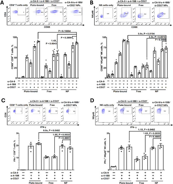
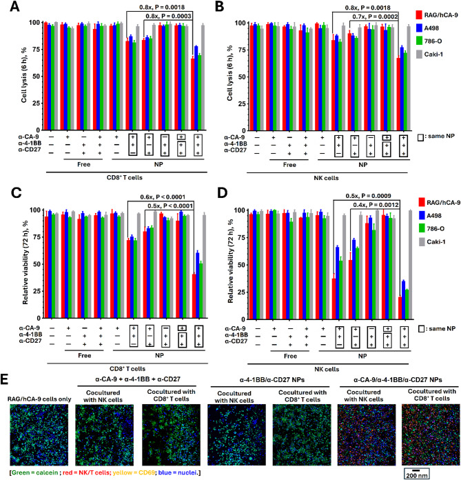
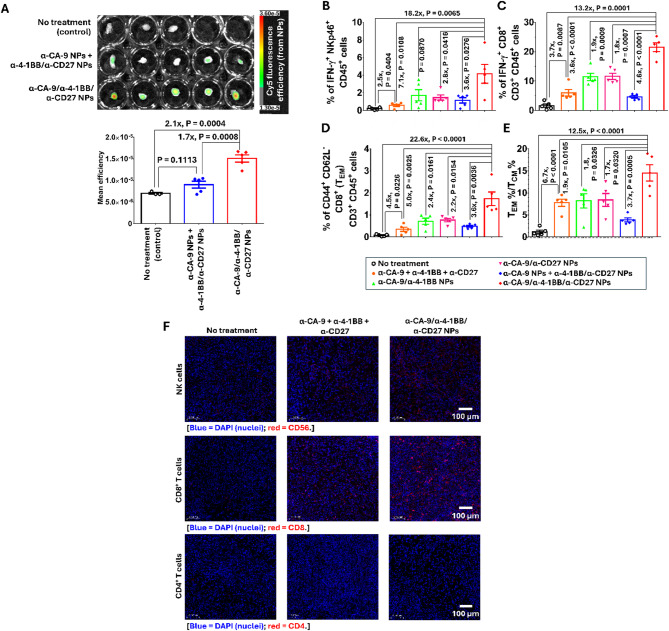
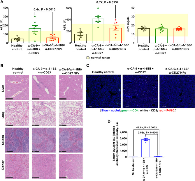
Results: Human CA-9-targeted multifunctionalized immunotherapy NPs (MINPs) functionalized with anti-CA-9, anti-4-1BB, and anti-CD27 antibodies outperformed hCA-9-targeted bifunctionalized immunotherapy NPs (BINPs), non-targeted BINPs, and the combination of free antibodies in activating mouse CD8+ T cells and NK cells to kill hCA-9-expressing RCC cells in vitro. In vivo correlative study confirmed that tumor targeting and effective spatiotemporal coactivation of the 4-1BB and CD27 pathways in CD8+ T cells and NK cells are essential for robust antitumor activity. Furthermore, hCA-9-targeted MINPs, but not the combination of free antibodies, inhibited the growth of human ccRCC in hPBMC-humanized mouse models. The anticancer activity of MINPs in mice humanized with hPBMCs from older donors was slightly weaker than in those humanized with younger donors. More importantly, the MINP formulation effectively prevented the hepatotoxicity associated with the systemic administration of immune checkpoint agonistic antibodies.

Conclusion: This study demonstrates that MINPs are a versatile platform capable of facilitating immune cell engagement and the eradication of targeted ccRCC without causing systemic immune-related side effects.


Conflict of interest statement

Declarations. Ethics approval and consent to participate: All animal experiments were approved by the University of Texas Southwestern Medical Center Institutional Animal Care (protocol 2023-103631) following the Guideline for the Care and Use of Laboratory Animals (NIH publication no. 86 − 23, revised 1985). Consent for publication: All authors have consented to submit this article for publication. Competing interests: The authors declare no competing interests.

Figures

Fig. 1
Fig. 1
Design and characterization of hCA-9-targeted MINPs. (A) Schematic illustration of the design of hCA-9-targeted MINPs and their proposed mechanism of action against hCA-9-expressing RCC cells. (B) TEM images of azide-functionalized PEG-PLGA NPs (N3 PEG-PLGA NPs) and α-CA-9/α-4-1BB/α-CD27 NPs (chimeric MINPs, functionalized with anti-human α-CA-9 antibody and anti-mouse 4-1BB and CD27 agonistic antibodies). This subfigure was created using BioRender. (C) Intensity-average diameter distribution curves and zeta potential distribution curves recorded for N3 PEG-PLGA NPs and α-CA-9/α-4-1BB/α-CD27 NPs, as determined through DLS and aqueous electrolysis methods. (D) Representative fluorescence histograms recorded for various MINPs stained with fluorescently labeled hCA-9, mouse 4-1BB (m4-1BB), and mouse CD27 (mCD27). (E) Representative fluorescence histograms of 786-0, A498, and Caki-1 cells after incubation with different targeted rhodamine-labeled MINPs (n = 3). (F) Representative FACS density plots of wild-type RAG cells and hCA-9-transfected RAG/hCA-9 cells after incubation with targeted and non-targeted rhodamine-labeled MINPs. The hCA-9 gene was transfected with the green fluorescent protein (GFP) gene into the parental RAG cells. Consequently, the RAG/hCA-9 cells became fluorescent in the GFP channel. (G) FACS density plots of CD8+ T cell-gated mouse splenocytes and NKp46+ NK cell-gated mouse splenocytes after incubation with different rhodamine-labeled MINPs (n = 3)
Fig. 2
Fig. 2
The MINP platform mimics plate-bound agonistic antibodies to crosslink and activate 4-1BB and/or CD27 receptors on mouse CD8+ T cells and NK cells in vitro. (A) Frequencies of CD69+ activated mouse CD8+ T cells after incubation for 6 h with different formulations of α-CA-9, α-4-1BB, and/or α-CD27 (n = 4). (B) Frequencies of CD69+ activated mouse NKp46+ NK cells after incubation for 6 h with different formulations of α-CA-9, α-4-1BB, and/or α-CD27 (n = 4). (C) Frequencies of IFN-γ+ activated mouse CD8+ T cells after incubation for 72 h with different formulations of α-CA-9, α-4-1BB, and/or α-CD27 (n = 4). (D) Frequencies of IFN-γ+ activated mouse NK cells after incubation for 72 h with different formulations of α-CA-9, α-4-1BB, and/or α-CD27 (n = 4). Anti-human CA-9, anti-mouse 4-1BB, and CD27 agonistic antibodies were used in these studies. Data are presented as mean ± SEM. All p-values were analyzed using two-way ANOVA with Tukey’s HSD multiple comparisons post-hoc test
Fig. 3
Fig. 3
Human CA-9-targeted chimeric MINPs mediate mouse CD8+ T cells and NK cells to kill hCA-9-expressing RCC cells in vitro. (A) In vitro lysis of free antibody- and chimeric MINP-pretreated calcein-loaded RCC cells after coculture with CD8+ T cells for 6 h. The effector cell-to-target (E: T) ratio was 5:1. (B) In vitro lysis of free antibody- and chimeric MINP-pretreated calcein-loaded RCC cells after coculturing for 6 h with mouse NK cells. The E: T ratio was 5:1. (C) Relative viability of free antibody- and chimeric MINP-pretreated RCC cells after coculture for 72 h with mouse CD8+ T cells, as determined through an MTS assay (E: T ratio = 5:1). (D) Relative viability of free antibody- and chimeric MINP-pretreated RCC cells after coculture for 72 h with mouse CD8+ T cells, as determined through an MTS assay (E: T ratio = 5:1). The chimeric MINPs were functionalized with anti-human CA-9, anti-mouse 4-1BB, and/or CD27 agonistic antibodies in these in vitro studies. (E) Representative CLSM images of free antibody- and chimeric MINP-pretreated calcein-loaded RAG/hCA-9 cells after coculture for 6 h with CD8+ T cells or NK cells. Calcein-free RAG/hCA-9 cells and calcein-loaded RAG/hCA-9 cells were used as negative and positive controls, respectively, for the calcein channel. The E: T ratio was 5:1. The merge channel images are the same as those shown in Fig. S12. Data are presented as mean ± SEM. All p-values were analyzed using two-way ANOVA with Tukey’s HSD multiple comparisons post-hoc test
Fig. 4
Fig. 4
Fully humanized MINPs effectively activate hPBMCs to eradicate 786-O cells in vitro. (A) IFN-γ positive spots developed after hPBMCs were incubated with untreated, antibody-pretreated, antibody-cocultured, or MINP-pretreated 786-O cells for 24 h, as determined through an IFN-γ/IL-2 ELISpot assay (n = 4 per hPBMC sample). (B) In vitro lysis of antibody- and MINP-pretreated, calcein-loaded 786-O cells occurred after coculture for 6 h with hPBMCs (E: T ratio = 20:1) (n = 6). (C) In vitro viabilities of free antibody- and MINP-pretreated 786-O cells were assessed after coculture for 72 h with human PBMCs (E: T ratio = 20:1) (n = 6). (D) Frequencies of IFN-γ+ CD8+ T cells in hPBMCs (PBMC-O2) measured after cocultured with different untreated, antibody-pretreated or MINP-pretreated 786-O cells for 24 h (n = 5). (E) Frequencies of IFN-γ+ CD56+ NK cells in hPBMCs (PBMC-O2) measured after cocultured with different untreated, antibody-pretreated or MINP-pretreated 786-O cells for 24 h (n = 5). The fully humanized MINPs were functionalized with anti-human CA-9, anti-human 4-1BB, and CD27 agonistic antibodies in these in vitro studies. (F) Representative CLSM images of calcein-loaded 786-O cells after cocultured with hPBMCs (PBMC-O2). The 786-O cells in the experimental group were pretreated with a combination of free antibodies or fully humanized MINPs before cocultured with hPBMCs. The PHA-stimulated hPBMCs contain 23–42% of CD8+ T cells and 0.5–15% of NK cells (Fig. S13, and Tables S1 and S2). Data are presented as mean ± SEM. All p-values were analyzed using two-way ANOVA with Tukey’s HSD multiple comparisons post-hoc test. The merged channel CLSM images shown in Fig. 4F are identical to those shown in Fig. S17
Fig. 5
Fig. 5
Human CA-9-targeted chimeric MINPs are more effective than the combination of free antibodies for inhibiting RAG/hCA-9 tumor growth in immunocompetent mice. (A) In vivo correlative study in a RAG/hCA-9 xenograft tumor model in BALB/c mice. Mice received treatments with either free antibodies or different hCA-9-targeted or non-targeted MINPs starting on day 7 post-inoculation. (n = 6 per group; TGI– tumor growth inhibition). (B) Tumor growth curves of RAG/hCA-9 xenograft tumor-bearing mice after different targeted and non-targeted treatments. (C) Survival curves of RAG/hCA-9 xenograft tumor-bearing mice following different targeted and non-targeted treatments. (D) In vivo anticancer efficacy study in immune cell-depleted BALB/c mice with RAG/hCA-9 xenograft tumors. Mice were treated with a combination of free antibodies or hCA-9-targeted MINPs. CD8+ T cells, CD4+ T cells, and NK cells were depleted using α-CD8, α-CD4, and α-Asialo-GM1 on days 5, 9, 12, 16, and 19 post-inoculation (n = 7 or 8 per group). (E) Survival curves of immune cell-depleted mice after various treatments. Chimeric MINPs were functionalized with anti-human CA-9, anti-mouse 4-1BB, and/or CD27 agonistic antibodies in these in vivo studies. All p-values, except for the survival data, were analyzed using two-way ANOVA with Tukey’s HSD multiple comparisons post-hoc test. The p-values for the survival data were analyzed using the log-rank (Mantel–Cox) test
Fig. 6
Fig. 6
Human CA-9-targeted chimeric MINPs inhibit RAG/hCA-9 tumor growth by activating tumor-infiltrating CD8+ T cells and NK cells in immunocompetent mice. (A) Ex vivo fluorescence images of RAG/hCA-9 tumors preserved 48 h after i.v. administration of Cy5-labeled α-CA-9 NPs plus Cy5-labeled α-4-1BB/α-CD27 NPs, or Cy5-labeled α-CA-9/α-4-1BB/α-CD27 NPs (n = 5, except for the non-treatment control group where n = 3). (B) Frequencies of active IFN-γ+ tumor-infiltrating NK cells. (C) Frequencies of active IFN-γ+ tumor-infiltrating CD8+ T cells. (D) frequencies of tumor-infiltrating TEM cells. (E) The TEM-to-TCM ratio. The phenotypes of tumor-infiltrating lymphocytes were determined by the FACS method 4 days after 2 targeted or non-targeted MINP treatments (n = 5). (F) Representative immunohistochemistry images of RAG/hCA-9 tumors after treatment with free combinational antibodies or hCA-9-targeted MINPs. The chimeric MINPs were functionalized using anti-human CA-9, anti-mouse 4-1BB, and/or CD27 agonistic antibodies in these in vivo studies. Data are presented as mean ± SEM. All p-values were analyzed using two-way ANOVA with Tukey’s HSD multiple comparisons post-hoc test
Fig. 7
Fig. 7
Fully humanized MINPs effectively inhibit 786-O and A498 tumor growth in hPBMC-humanized mice. (A) Mouse humanization, tumor inoculation, and treatment schedule. (B) 786-O tumor growth curves recorded for mice humanized with hPBMCs from younger donors after treatment with free combinational antibodies or MINPs (n = 3 for treatment groups per donor and n = 3 or 4 for non-treatment groups per donor. There were three hPBMC donors for each control and experimental group. Two mice in the non-treatment group and 1 mouse in each treatment group died before the study endpoint). (C) 786-O tumor growth curves recorded for mice humanized with hPBMCs from older donors after treatment with free combinational antibodies or MINPs (n = 3 for treatment groups per donor and n = 3 or 4 for non-treatment groups per donor; 3 hPBMC donors for each control and experimental group). In vivo studies in (B) and (C) were conducted separately. (DG) Analysis of 786-O tumor-infiltrating lymphocytes in mice humanized with PBMCs from older donors. (D) Frequencies of active IFN-γ+ tumor-infiltrating NK cells. (E) Frequencies of active IFN-γ+ tumor-infiltrating CD8+ T cells. (F) Frequencies of tumor-infiltrating TEM cells and (G) the TEM/TCM ratio, as determined through the FACS method on a dissociated 786-O tumor preserved 4 days after 2 treatments (n = 2 per donor. Three donors per control and treatment group). (H) Representative immunohistochemistry images of a 786-O tumor after treatment with free combinational antibodies or MINPs in mice humanized with hPBMCs from an older donor. (I) A498 tumor growth curves recorded for humanized mice (hPBMCs from a 62-year-old donor) after treatment with free combinational antibodies or MINPs (n = 6, except n = 7 for mice treated with MINPs). The fully humanized MINPs were functionalized using anti-human CA-9, anti-human 4-1BB, and CD27 agonistic antibodies in these in vivo studies. Data are presented as mean ± SEM. All p-values were analyzed using two-way ANOVA with Tukey’s HSD multiple comparisons post-hoc test
Fig. 8
Fig. 8
The MINP platform reduces hepatotoxicity induced by agonistic α-4-1BB and α-CD25 antibodies in healthy immunocompetent mice. (A) Serum ALT, AST, and BUN levels recorded for healthy BALB/c mice 48 h after 4 i.v. administrations of free α-CA-9, α-4-1BB, and α-CD27, or α-CA-9/α-4-1BB/α-CD27 NPs (chimeric MINPs) (n = 7, except for mice treated with MINPs, where n = 8). (B) Representative H&E-stained images of the liver, lung, spleen, and kidney preserved from mice at the study endpoint. (C) Representative immunohistochemistry images of liver sections preserved at the study endpoint after different treatments. (D) Serum DyLight 650 fluorescent intensity 6 h after i.v. administration of DyLight 650 labeled α-CA-9, α-4-1BB, and α-CD27 or α-CA-9/α-4-1BB/α-CD27 NPs (functionalized with DyLight 650-labeled antibodies) (n = 5, except for the non-treatment control group, where n = 3). The chimeric MINPs were functionalized using anti-human CA-9, anti-mouse 4-1BB, and CD27 agonistic antibodies in these in vivo studies. Data are presented as mean ± SEM. All p-values were analyzed using two-way ANOVA with Tukey’s HSD multiple comparisons post-hoc test

Similar articles

References

    1. Siegel RL, Giaquinto AN, Jemal A. Cancer statistics, 2024. Cancer J Clin. 2024;74:12–49. - PubMed
    1. Tan HJ, Norton EC, Ye Z, Hafez KS, Gore JL, Miller DC. Long-term survival following partial vs radical nephrectomy among older patients with early-stage kidney cancer. JAMA. 2012;307:1629–35. - PMC - PubMed
    1. Quinlan M, Wei G, Davis N, Poyet C, Perera M, Bolton D, Lawrentschuk N. Renal cell carcinoma Follow-Up - Is it time to abandon ultrasound? Curr Urol. 2019;13:19–24. - PMC - PubMed
    1. Fahey CC, Shevach JW, Flippot R, Albiges L, Haas NB, Beckermann KE. Triplet strategies in metastatic clear cell renal cell carcinoma: A worthy option in the First-Line setting? Am Soc Clin Oncol Educ Book. 2023;43:e389650. - PMC - PubMed
    1. Makhov P, Joshi S, Ghatalia P, Kutikov A, Uzzo RG, Kolenko VM. Resistance to systemic therapies in clear cell renal cell carcinoma: mechanisms and management strategies. Mol Cancer Ther. 2018;17:1355–64. - PMC - PubMed

MeSH terms