Review
. 2025 Jun 18;22(1):60.
doi: 10.1186/s12987-025-00671-8.

Brain fluid physiology in ischaemic stroke; more than just oedema

Affiliations
Review

Brain fluid physiology in ischaemic stroke; more than just oedema

Kirsten G Coupland et al. Fluids Barriers CNS. .

Abstract

Background: Cerebrospinal fluid and interstitial fluid dynamics are critical for maintaining homeostasis in the central nervous system. These fluids facilitate waste clearance, micronutrient distribution, and provide a tightly regulated ionic environment. Ischaemic stroke, a leading cause of morbidity and mortality, disrupts this delicate system, compounding the physiological challenges posed by the condition. Despite recent advances in our understanding of the importance of cerebrospinal fluid (CSF) and interstitial fluid (ISF) movement and exchange, the role of this system in stroke pathophysiology remains underexplored.

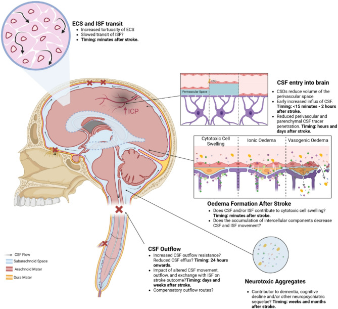
Main body: Emerging evidence indicates that ischaemic stroke acutely alters CSF and ISF movement and exchange, with effects observed at both local and brain-wide levels. In the hyper-acute phase, there is an influx of CSF into perivascular spaces, potentially contributing to early cell swelling. Over time, impaired clearance mechanisms exacerbate ionic and vasogenic oedema, elevating intracranial pressure and further compromising perfusion in the ischaemic penumbra. Mechanistic studies suggest that disruptions in arterial pulsatility, extracellular space microstructure, and aquaporin 4 localisation may underlie these changes. Experimental models have revealed decreased CSF and ISF exchange, movement and outflow in the hours to days following stroke, with implications for waste clearance and secondary injury processes. The interplay between these dynamics and cortical spreading depolarisations, stroke severity, and cerebrovascular physiology adds complexity to understanding the condition's progression.

Conclusion: The disruption of CSF and ISF movement and exchange may represent a significant, yet underappreciated contributor to post-stroke pathology. Addressing these alterations could offer novel therapeutic avenues to mitigate secondary damage, improve central nervous system (CNS) homeostasis, and enhance recovery outcomes. Future research must focus on elucidating the precise mechanisms of CSF and ISF movement and exchange disturbance and exploring targeted interventions to restore normal fluid dynamics in the CNS post-stroke.

Keywords: Cerebral oedema; Cerebrospinal fluid; Cerebrospinal fluid efflux; Glymphatics; Intracranial pressure; Stroke; Stroke pathophysiology.


Conflict of interest statement

Declarations. Ethics approval and consent to participate: Not applicable. Consent for publication: Not applicable. Competing interests: The authors declare no competing interests.

Figures

Fig. 1
Fig. 1
An overview of our current, broad understanding of the production and net movement of CSF. CSF is predominantly produced in the choroid plexuses of the four ventricles and from there migrates into either the periventricular space or the subarachnoid space of the brain and spinal cord via the aqueduct of Sylvius and the foramens of Luschka and Magendie. CSF then permeates the brain via Virchow-Robin spaces around penetrating arteries and by crossing the pia mater to enter the parenchyma where it mixes with ISF. Evidence indicates that outflow of the mixed CSF and ISF appears to occur via perivenous spaces (glymphatic hypothesis), but there is some evidence that it may occur through the arterial basement membrane (IPAD hypothesis). From the subarachnoid space CSF exits the cranial compartment via several routes including around cranial and spinal nerves and the meningeal lymphatics, particularly those at the cribriform plate (reviewed in 18) where it then returns to the peripheral circulation. Created in BioRender. Spratt, N. (2025) https://BioRender.com/y18u546
Fig. 2
Fig. 2
A putative timeline of the events that impact on CSF and ISF movement and exchange in the context of ischaemic stroke. Cortical spreading depolarisation events start within minutes of loss of blood supply and appear to correspond with vasoconstriction and the influx of CSF into the perivascular space [58]. This potentially occurs alongside increased tortuosity of the extracellular space due to cytotoxic cell swelling [–152] which may slow ISF transit and restrict CSF and ISF exchange in this space. There is an overall decrease in movement of CSF in the perivascular space and penetration of CSF tracers into the parenchyma within hours of a stroke event [32, 55, 59] and there is also evidence of reduced CSF outflow at similar time points [32, 87, 88]. The reduced efflux of CSF from the cranial compartment corresponds temporally with oedema-independent ICP elevation. Created in BioRender [–96]. Spratt, N. (2025) https://BioRender.com/y16b797
Fig. 3
Fig. 3
Schematic of the arteries that supply the choroid plexuses of the ventricles. The choroid plexus of the lateral ventricles is supplied by the anterior choroidal artery and posterior choroidal arteries. The posterior choroidal artery also supplies the choroid plexus of the third ventricle. The fourth ventricle choroid plexus is supplied by inferior cerebellar artery. Created in BioRender. Spratt, N. (2025) https://BioRender.com/j95t466
Fig. 4
Fig. 4
A schematic summarising the ways that ischaemic stroke impacts on CSF and ISF movement and efflux from the cranial compartment. Ischaemic stroke triggers cortical spreading depolarisations (CSDs) which are associated with an initial increase, and then decrease, in the perivascular space that appears to initially increase, and then restrict, CSF movement into this space. There is also evidence that there is increased tortuosity of the extracellular space (ECS) which hinders the movement of ISF-bourne solutes. This overall reduction in the movement of CSF and ISF might result in reduced clearance of waste products which may contribute to the increased prevalence of cognitive decline noted in stroke patients. CSF and ISF are likely contributors to early cell swelling and cytotoxic/ionic oedema, which may exacerbate vasogenic oedema. Furthermore, there is evidence that CSF outflow is reduced at the cribriform plate and potentially other sites which may lead to elevated intracranial pressure (ICP) and reduced perfusion of the at-risk penumbra. Created in BioRender. Spratt, N. (2025) https://BioRender.com/y16b797

References

    1. Cserr HF. Physiology of the choroid plexus. Physiol Rev. 1971;51(2):273–311. - PubMed
    1. Damkier HH, Brown PD, Praetorius J. Cerebrospinal fluid secretion by the choroid plexus. Physiol Rev. 2013;93(4):1847–92. - PubMed
    1. Jensen DB, Toft-Bertelsen TL, Barbuskaite D, Stubbe J, Nietzsche S, Capion T et al. The Na+,K+,2Cl cotransporter, not Aquaporin 1, sustains cerebrospinal fluid secretion while controlling brain K+ homeostasis. Adv Sci. 2024;2409120. - PMC - PubMed
    1. Rosenberg GA, Kyner WT, Estrada E. Bulk flow of brain interstitial fluid under normal and hyperosmolar conditions. Am J Physiology-Renal Physiol. 1980;238(1):F42–9. - PubMed
    1. McComb JG, McComb JG, McComb JG. Recent research into the nature of cerebrospinal fluid formation and absorption. J Neurosurg. 1983;59(3):369–83. - PubMed

MeSH terms

LinkOut - more resources