

Using radiomics model for predicting extraprostatic extension with PSMA PET/CT studies: a comparative study with the Mehralivand grading system
- PMID: 40533824
- PMCID: PMC12177976
- DOI: 10.1186/s40644-025-00894-w
Using radiomics model for predicting extraprostatic extension with PSMA PET/CT studies: a comparative study with the Mehralivand grading system
Abstract
Purpose: This study aimed to evaluate the effectiveness of using a radiomics model to predict extraprostatic extension (EPE) in prostate cancer from PSMA PET/CT, and to directly compare its performance with the Mehralivand Grading System, an MRI-based method for EPE assessment.
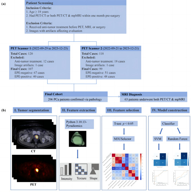
Methods: A total of 206 patients who underwent radical prostatectomy were included in this study. Radiomics features were extracted from PSMA PET/CT images to construct predictive models using Support Vector Machine (SVM) and Random Forest algorithms. In addition, among the 63 patients who underwent both PSMA PET/CT and multiparametric MRI (mpMRI), the performance of the radiomics model was compared with that of the Mehralivand Grading System. Key performance metrics, including the area under the curve (AUC), sensitivity, specificity, positive predictive value (PPV), and negative predictive value (NPV), were reported.
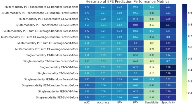
Results: Among the 63 patients who underwent both PSMA PET/CT and multiparametric MRI (mpMRI), the radiomics model achieved an AUC of 76.8% (95% CI: 64.4-86.5%), sensitivity of 72.0%, specificity of 81.5%, PPV of 72.0%, and NPV of 81.6%. In comparison, the Mehralivand Grading System yielded AUCs of 66.8%, 63.5%, and 60.2% from three independent readers. DeLong's test showed that the radiomics model significantly outperformed all three readers in terms of AUC (p = 0.013, 0.003, and 0.001, respectively).
Conclusion: The radiomics model derived from PSMA PET/CT can better capture features associated with EPE and shows promise for aiding preoperative assessment in prostate cancer. However, further validation in larger, independent cohorts is necessary to confirm its stability and clinical utility.
Keywords: Extraprostatic extension (EPE); PSMA PET/CT; Radiomics.
© 2025. The Author(s).
Conflict of interest statement
Declarations. Ethics approval and consent to participate: The study was approved by the Ethics Committee of Fudan University Shanghai Cancer Center (Ethical approval number: 1612167-18). Consent to participate: A specifc informed consent was waived because of the observational and retrospective study design. Competing interests: The authors declare no competing interests.
Figures
Similar articles
-
Comparing Magnetic Resonance Imaging and Prostate-Specific Membrane Antigen-Positron Emission Tomography for Prediction of Extraprostatic Extension of Prostate Cancer and Surgical Guidance: A Prospective Nonrandomized Clinical Trial.J Urol. 2024 Aug;212(2):290-298. doi: 10.1097/JU.0000000000004032. Epub 2024 May 24. J Urol. 2024. PMID: 38785259 Free PMC article. Clinical Trial.
-
Can Negative Prostate-specific Membrane Antigen Positron Emission Tomography/Computed Tomography Avoid the Need for Pelvic Lymph Node Dissection in Newly Diagnosed Prostate Cancer Patients? A Systematic Review and Meta-analysis with Backup Histology as Reference Standard.Eur Urol Oncol. 2022 Feb;5(1):1-17. doi: 10.1016/j.euo.2021.08.001. Epub 2021 Sep 17. Eur Urol Oncol. 2022. PMID: 34538770
-
Machine learning-based construction and validation of an radiomics model for predicting ISUP grading in prostate cancer: a multicenter radiomics study based on [68Ga]Ga-PSMA PET/CT.Eur J Nucl Med Mol Imaging. 2025 Jun 24. doi: 10.1007/s00259-025-07412-x. Online ahead of print. Eur J Nucl Med Mol Imaging. 2025. PMID: 40553115
-
Diagnostic Performance of 68Ga-PSMA-11 PET/CT Versus Multiparametric MRI for Detection of Intraprostatic Radiorecurrent Prostate Cancer.J Nucl Med. 2024 Mar 1;65(3):379-385. doi: 10.2967/jnumed.123.266527. J Nucl Med. 2024. PMID: 38212074
-
The diagnostic accuracy of radiolabeled PSMA-ligand PET for tumour staging in newly diagnosed prostate cancer patients compared to histopathology: a systematic review and meta-analysis.Eur J Nucl Med Mol Imaging. 2023 Dec;51(1):281-294. doi: 10.1007/s00259-023-06392-0. Epub 2023 Aug 19. Eur J Nucl Med Mol Imaging. 2023. PMID: 37597010
References
-
- Parker CC, Clarke NW, Cook AD, Petersen PM, Catton CN, Cross WR et al. Randomised trial of no, Short-term, or Long-term androgen deprivation therapy with postoperative radiotherapy after radical prostatectomy: results from the Three-way comparison of RADICALS-HD (NCT00541047). Eur Urol. 2024. - PMC - PubMed
-
- Sood A, Jeong W, Palma-Zamora I, Abdollah F, Butaney M, Corsi N, et al. Description of surgical technique and oncologic and functional outcomes of the precision prostatectomy procedure (IDEAL stage 1-2b Study). Eur Urol. 2022;81(4):396–406. - PubMed
-
- Bianchi L, Chessa F, Angiolini A, Cercenelli L, Lodi S, Bortolani B, et al. The use of augmented reality to guide the intraoperative frozen section during Robot-assisted radical prostatectomy. Eur Urol. 2021;80(4):480–8. - PubMed
-
- Moschovas MC, Bhat S, Sandri M, Rogers T, Onol F, Mazzone E, et al. Comparing the approach to radical prostatectomy using the multiport Da Vinci Xi and Da Vinci SP robots: A propensity score analysis of perioperative outcomes. Eur Urol. 2021;79(3):393–404. - PubMed
Publication types
MeSH terms
Grants and funding
LinkOut - more resources
Full Text Sources
Medical
Miscellaneous
