

Spatial-temporal radiogenomics in predicting neoadjuvant chemotherapy efficacy for breast cancer: a comprehensive review
- PMID: 40533825
- PMCID: PMC12178003
- DOI: 10.1186/s12967-025-06641-w
Spatial-temporal radiogenomics in predicting neoadjuvant chemotherapy efficacy for breast cancer: a comprehensive review
Abstract
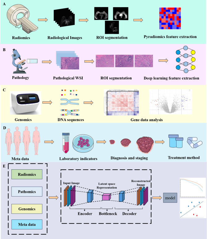
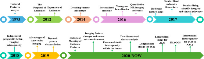
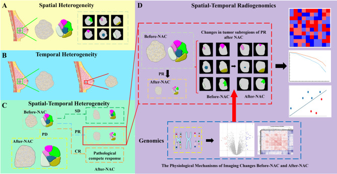
Radiomics is undergoing a paradigm shift from single-omics to multi-omics, from single-temporal to multi-temporal analysis, and from global to subregional analysis. These transformations have shown great potential in addressing key challenges related to imaging changes before and after neoadjuvant chemotherapy (NAC) in breast cancer. Furthermore, radiomics has achieved remarkable progress in tasks such as exploring tumor heterogeneity and uncovering underlying biological mechanisms. Integrating imaging data with gene data offers novel perspectives for understanding imaging changes driven by specific genetic alterations. However, current radiomics studies on neoadjuvant chemotherapy for breast cancer have not yet achieved a close integration of imaging changes with underlying biological mechanisms. They are largely limited to simple associations between models and genomic data, without in-depth interpretation of the biological significance inherent in imaging features, which is essential to directly link these features with the dynamic progression of the disease. This review seeks to explore the spatial-temporal heterogeneity of imaging alterations observed during NAC for breast cancer, while assessing their biological implications using established analytical approaches. It highlights the distinct advantages of spatial-temporal radiomics in predictive model development and examines potential correlations between imaging dynamics and gene expression profiles before and after NAC. Additionally, we critically examines previous radiogenomics studies, providing theoretical insights into their limitations. Finally, the review proposes future directions and innovative approaches for applying spatial-temporal radiogenomics in NAC for breast cancer, serving as a valuable reference and roadmap for researchers and clinical practitioners in this field.
Keywords: Breast cancer; Magnetic resonance imaging; Neoadjuvant chemotherapy; Radiogenomics; Spatial-temporal heterogeneity.
© 2025. The Author(s).
Conflict of interest statement
Declarations. Ethics approval and consent to participate: Not applicable. Consent for publication: Not applicable. Competing interests: The authors have declared that no competing interest exists.
Figures
Similar articles
-
Integrative Radiogenomics Using MRI Radiomics and Microarray Gene Expression Analysis to Predict Pathological Complete Response in Patients with Breast Cancer Undergoing Neoadjuvant Chemotherapy.Cureus. 2025 Jun 18;17(6):e86287. doi: 10.7759/cureus.86287. eCollection 2025 Jun. Cureus. 2025. PMID: 40688842 Free PMC article.
-
Assessing the comparative effects of interventions in COPD: a tutorial on network meta-analysis for clinicians.Respir Res. 2024 Dec 21;25(1):438. doi: 10.1186/s12931-024-03056-x. Respir Res. 2024. PMID: 39709425 Free PMC article. Review.
-
Prehabilitation during neoadjuvant chemotherapy results in an enhanced immune response in esophageal adenocarcinoma tumors: A randomized controlled trial.J Sport Health Sci. 2025 Jun 3:101063. doi: 10.1016/j.jshs.2025.101063. Online ahead of print. J Sport Health Sci. 2025. PMID: 40499717
-
Exploring engagement patterns within a mobile health intervention for women at risk of gestational diabetes.Womens Health (Lond). 2025 Jan-Dec;21:17455057251327510. doi: 10.1177/17455057251327510. Epub 2025 Jun 5. Womens Health (Lond). 2025. PMID: 40470610 Free PMC article. Clinical Trial.
-
The accuracy of breast MRI radiomic methodologies in predicting pathological complete response to neoadjuvant chemotherapy: A systematic review and network meta-analysis.Eur J Radiol. 2022 Dec;157:110561. doi: 10.1016/j.ejrad.2022.110561. Epub 2022 Oct 17. Eur J Radiol. 2022. PMID: 36308849
References
-
- Bray F, Laversanne M, Sung H, Ferlay J, Siegel RL, Soerjomataram I, et al. Global cancer statistics 2022: GLOBOCAN estimates of incidence and mortality worldwide for 36 cancers in 185 countries. CA Cancer J Clin. 2024;74(3):229–63. 10.3322/caac.21834. - PubMed
-
- Aristokli N, Polycarpou I, Themistocleous SC, Sophocleous D, Mamais I. Comparison of the diagnostic performance of magnetic resonance imaging (MRI), ultrasound and mammography for detection of breast cancer based on tumor type, breast density and patient’s history: A review. Radiography (Lond). 2022;28(3):848–56. 10.1016/j.radi.2022.01.006. - PubMed
-
- Filho OM, Viale G, Stein S, Trippa L, Yardley DA, Mayer IA, et al. Impact of HER2 heterogeneity on treatment response of Early-Stage HER2-Positive breast cancer: phase II neoadjuvant clinical trial of T-DM1 combined with Pertuzumab. Cancer Discov. 2021;11(10):2474–87. 10.1158/2159-8290.Cd-20-1557. - PMC - PubMed
Publication types
MeSH terms
Grants and funding
LinkOut - more resources
Full Text Sources
Medical
