. 2025 Jun 18;162(1):108.
doi: 10.1186/s41065-025-00478-5.

circ-NOLC1 inhibits the development of cervical cancer by regulating miR-330-5p-PALM signaling axis

Affiliations

circ-NOLC1 inhibits the development of cervical cancer by regulating miR-330-5p-PALM signaling axis

Yali Duo et al. Hereditas. .

Abstract

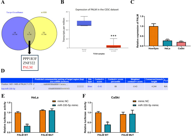
Background: Recent studies have increasingly demonstrated that circular RNAs (circRNAs) play significant roles in the occurrence and progression of cervical cancer (CC). In CC, circRNAs act as ceRNAs by sponging miRNAs to regulate genes associated with proliferation, migration, and apoptosis, exhibiting both promoting and inhibiting effects on tumor progression. The aim of this study was to clarify the role of hsa_circ_0019686 (named circ-NOLC1) in CC.

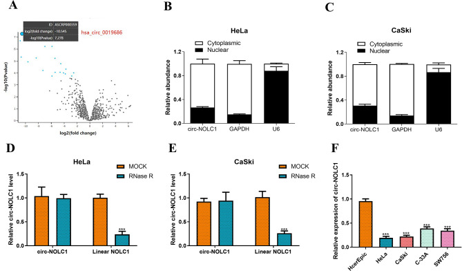
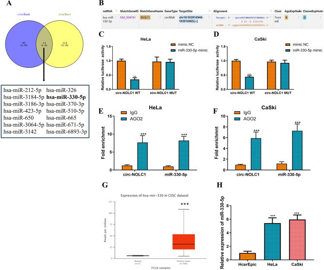
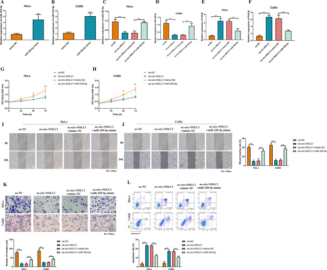
Methods: By conducting an online GEO2R analysis of the expression profile GSE113696 in the GEO database, circ-NOLC1 was selected. The expression levels of circ-NOLC1 in CC cell lines were measured using real-time quantitative PCR (RT-qPCR). The role of circ-NOLC1 in CC was validated through both in vitro and in vivo gain-of-function assays. Bioinformatic analysis, combined with luciferase reporter and RNA Immunoprecipitation (RIP) assays, confirmed that circ-NOLC1 acts as a sponge for miR-330-5p and regulates the expression of paralemmin-1 (PALM). The role of the circ-NOLC1-miR-330-5p-PALM signaling axis in CC was elucidated through the rescue experiments. Relative gene expression levels were measured using RT-qPCR, while relative protein levels were assessed through immunohistochemistry (IHC). CCK-8, wound healing, Transwell, and flow cytometry assays were employed to evaluate CC cell proliferation, migration, and invasion, respectively.

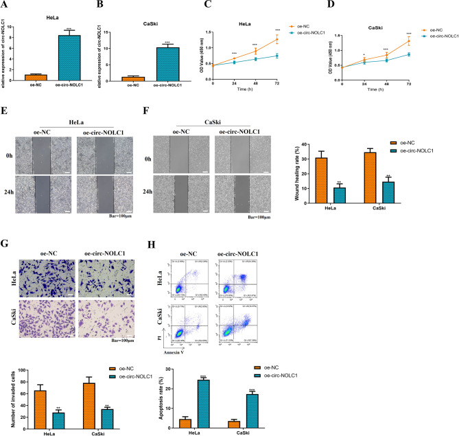
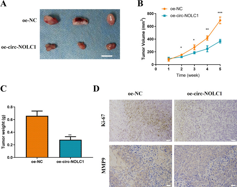
Results: The expression levels of circ-NOLC1 were dramatically downregulated in CC cells (P < 0.001). Up-regulation of circ-NOLC1 significantly inhibited cell proliferation (P < 0.001), migration (P < 0.01) and invasion (P < 0.01), while promoting cell apoptosis (P < 0.001). In vivo studies showed that up-regulation of circ-NOLC1 suppressed tumor growth (tumor volume: P < 0.001; tumor weight: P < 0.01). Additionally, miR-330-5p was found to be up-regulated in CC (P < 0.001), whereas PALM was downregulated in CC (P < 0.001). The up-regulation of circ-NOLC1 inhibited the expression of miR-330-5p (P < 0.001) and enhanced the expression of PALM (P < 0.001). Rescue experiments further demonstrated that the up-regulation of circ-NOLC1 inhibited CC cell proliferation (P < 0.001), migration (P < 0.001), invasion (P < 0.001), while promoting apoptosis (P < 0.001) through the regulation of the miR-330-5p-PALM pathway.

Conclusion: The circ-NOLC1 inhibits CC development through regulating the miR-330-5p-PALM signaling axis. This finding reveals a novel mechanism and identifies potential therapeutic targets, emphasizing the necessity for further regulatory studies and clinical validation.

Keywords: Cervical cancer; Circular RNAs; PALM; circ-NOLC1; miR-330-5p.


Conflict of interest statement

Declarations. Ethical approval: The present study was approved by The IACUC of the Harrison International Peace Hospital (approval no. 2020-2-004-1). All experiments involving animals were admitted and performed according to the requirements of The Medical Ethics Committee of the Harrison International Peace Hospital. Consent to participate: Not applicable. Consent for publication: Not applicable. Competing interests: The authors declare no competing interests.

Figures

Fig. 1
Fig. 1
circ-NOLC1 was downregulated in CC cells. (A) The Gene Expression Omnibus dataset (accession no. GSE113696) analysis of differential circRNAs in CC cell lines (HeLa, CaSki, C-33 A, and SW756) compared to a normal cervical epithelial cell line (HcerEpic). Volcano map of differentially expressed genes (hsa_circ_0019686 is the most significantly downregulated gene in this dataset, adj.P.Val = 0.000246; logFC=-29.599789). RT-qPCR was used to detect the circ-NOLC1 expression level in subcellular components of (B) HeLa and (C) CaSki cells, including the nucleus (marker is U6) and cytoplasm (marker is GAPDH). The levels of circ-NOLC1 and linear NOLC1 in (D) HeLa and (E) CaSki cells treated with RNase R were detected by RT-qPCR. (F) RT-qPCR was applied to measure circ-NOLC1 expression in CC (HeLa, CaSki, C-33 A and SW756) and normal cervical epithelial (HcerEpic) cell lines. ***P < 0.001
Fig. 2
Fig. 2
Overexpression of circ-NOLC1 inhibits the proliferation, migration, and invasion of CC cells while promoting their apoptosis. RT-qPCR was employed to assess the transfection efficiency of the circ-NOLC1 overexpression plasmid (based on the pCD5-ciR vector) in (A) HeLa and (B) CaSki cells. Cell Counting Kit-8 assay was conducted to evaluate the viability of (C) HeLa and (D) CaSki cells. A scratch wound healing assay was performed to estimate cell migration in (E) HeLa and (F) CaSki cells (scale bar, 100 μm). (G) Transwell assay was utilized to determine the cell invasion of HeLa and CaSki cells (scale bar, 100 μm). (H) Flow cytometry was used to detect the cell apoptosis rate of HeLa and CaSki cells. **P < 0.01, ***P < 0.001
Fig. 3
Fig. 3
Overexpression of circ-NOLC1 inhibits the growth of CC in vivo. (A) Representative images from the tumorigenicity assay conducted in nude mice (n = 3 per group; scale bar, 1 cm). (B) Relative tumor growth curves. (C) Tumor weight plot. (D) The expression levels of Ki-67 and MMP9 proteins in the tumors was detected by immunohistochemistry (scale bar, 50 μm). **P < 0.01, ***P < 0.001
Fig. 4
Fig. 4
circ-NOLC1 sponges miR-330-5p. (A) A Venn diagram illustrating the analysis of the circBank and starBase databases to predict circ-NOLC1 target miRNAs. (B) Binding sites between circ-NOLC1 and miR-330-5p were predicted using the starBase database. A dual luciferase reporter gene assay was conducted to elucidate the targeting relationship between circ-NOLC1 and miR-330-5p in (C) HeLa and (D) CaSki cells. RIP assay was performed to validate the interaction between circ-NOLC1 and miR-330-5p in (E) HeLa and (F) CaSki cells. (G) The expression of miR-330-5p in CESC was predicted using the TCGA database (P = 1.62447832963153E-12). (H) RT-qPCR was employed to determine miR-330-5p expression in cervical cancer (HeLa and CaSki) and normal cervical epithelial (HcerEpic) cell lines. **P < 0.01, ***P < 0.001
Fig. 5
Fig. 5
miR-330-5p interacts with PALM. (A) A Venn diagram illustrating the analysis of the TargetScanHuman 7.2 and miRDB databases to predict the target mRNAs of miR-330-5p. (B) The expression of PALM in CESC was predicted using the TCGA database (P = 2.14740003556813E-10). (C) RT-qPCR was employed to measure PALM expression in cervical cancer (HeLa and CaSki) and normal cervical epithelial (HcerEpic) cell lines. (D) Binding sites between miR-330-5p and PALM were predicted using the TargetScanHuman database. A dual luciferase reporter gene assay was conducted to elucidate the targeting relationship between miR-330-5p and PALM in (E) HeLa and (F) CaSki cells. **P < 0.01, ***P < 0.001
Fig. 6
Fig. 6
circ-NOLC1 inhibits the proliferation, migration, and invasion of CC cells while promoting apoptosis by regulating the miR-330-5p/PALM signaling axis. (A-F) RT-qPCR was utilized to measure miR-330-5p expression in HeLa and CaSki cells. Cell Counting Kit-8 assay was applied to evaluate the viability of (G) HeLa and (H) CaSki cells. Scratch wound healing assay was applied to estimate the migration of (I) HeLa and (J) CaSki cells (scale bar, 100 μm). (K) Transwell assay was used to determine the invasion of HeLa and CaSki cells (scale bar, 100 μm). (L) Flow cytometry was used to detect the cell apoptosis rate of HeLa and CaSki cells. **P < 0.01, ***P < 0.001

Similar articles

References

    1. Hill EK. Updates in Cervical Cancer Treatment. Clin Obstet Gynecol. 2020;63(1):3–11. - PubMed
    1. Buskwofie A, David-West G, Clare CA. A review of cervical cancer: incidence and disparities. J Natl Med Assoc. 2020;112(2):229–32. - PubMed
    1. Siegel RL, Miller KD, Jemal A. Cancer statistics, 2020. CA Cancer J Clin. 2020;70(1):7–30. - PubMed
    1. Fakhr E, Modic Z, Cid-Arregui A. Recent developments in immunotherapy of cancers caused by human papillomaviruses. Immunology. 2021;163(1):33–45. - PMC - PubMed
    1. Simms KT, Hanley SJB, Smith MA, Keane A, Canfell K. Impact of HPV vaccine hesitancy on cervical cancer in japan: a modelling study. Lancet Public Health. 2020;5(4):e223–34. - PubMed