Review
. 2025 Jun 11;6(8):1233-1251.
doi: 10.1039/d5cb00050e. eCollection 2025 Jul 30.

Emerging gut microbial glycoside hydrolase inhibitors

Affiliations
Review

Emerging gut microbial glycoside hydrolase inhibitors

Mark E Kowalewski et al. RSC Chem Biol. .

Abstract

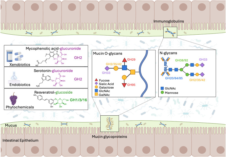
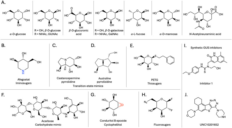
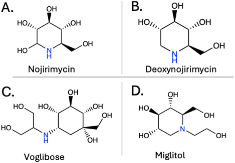
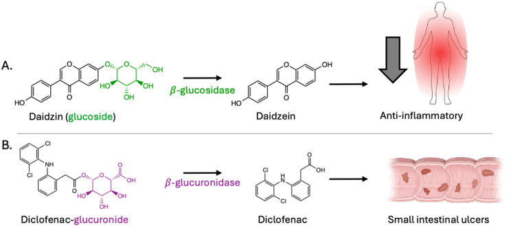
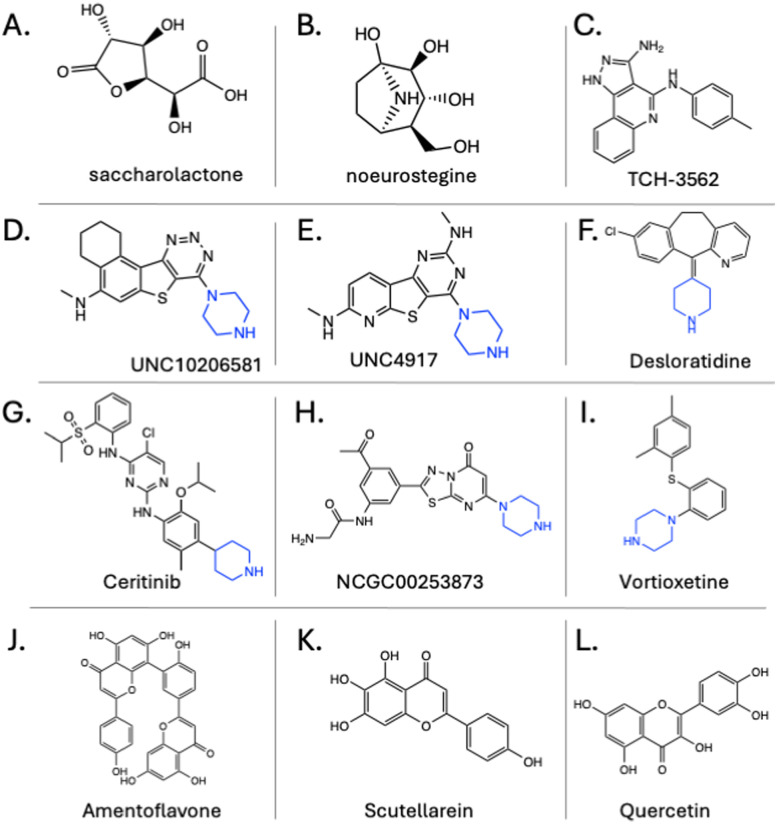
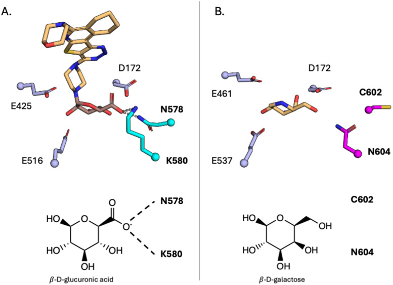
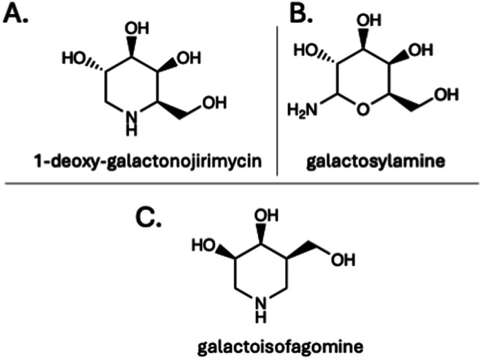
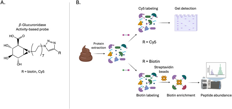
The human gut microbiota has been linked to numerous diseases through their metabolism of molecules in the gastrointestinal tract. Post-translational glycosylation is applied to many secreted proteins, including mucins and immunoglobulins, and glycosides are present in diet and generated by host metabolism systems. Thus, glycosides are key targets for degradation by gut microbial glycoside hydrolases (GHs). Indeed, diverse xenobiotic compounds, including therapeutics and dietary phytochemicals, along with endobiotics like neurotransmitters and hormones, are conjugated to monosaccharides making them substrates for GH enzymes. A range of GH inhibitors have been developed to study lysosomal storage diseases, treat viral infections, and to address type II diabetes. Recently, GH inhibitors have offered promising avenues for investigating gut microbial GHs and their influence on host health and disease. In this review we describe the growing classes of GH inhibitors and their applications in studying gut microbial GHs that target host-derived glycans and dietary and drug-xenobiotic molecules. We also review the use of GH-targeting activity-based probes to pinpoint specific proteins expressed by the gut microbiota that influence molecular and phenotypic outcomes. As we deepen our understanding of gut microbial GH function, we will further elucidate the roles played by the microbiota in host physiology and disease toward potential therapeutic interventions that target non-host factors in acute and chronic disorders.


Conflict of interest statement

MRR is a founder of Symberix, Inc., and has received research funding from Lilly and Merck.

Figures

Fig. 1
Fig. 1. Substrates for glycoside hydrolases (GH) in the gastrointestinal (GI) tract. The range of carbohydrate substrates present in the GI tract including host-derived glycans (mucins & immunoglobulins), xenobiotic compounds (therapeutics and phytochemicals), and host-generated endobiotic compounds. The GH family responsible for removing the glycans are indicated, showing the diverse enzymes needed to degrade carbohydrate substrates.
Fig. 2
Fig. 2. (A) Chemical structures of monosaccharides, which are the targets for the glycoside hydrolases described in this review. Glycoside hydrolase inhibitor structural classes with representative chemical structures for each class: (B) afegostat, an iminosugar β-glucosidase inhibitor; (C) castanospermine, a pyrrolidine β-glucosidase inhibitor; (D) australine, a pyrrolizidine α-glucosidase inhibitor; (E) PETG, a thiosugar β-galactosidase inhibitor; (F) acarbose, a carbohydrate mimic α-glucosidase inhibitor (G) conduritol B epoxide, a cyclophellitol β-glucosidase inhibitor; (H) 6-azido-2,6-dideoxy-2-fluoro-β-d-galactosyl fluoride a fluorosugar β-galactosidase inhibitor and the synthetic β-glucuronidase inhibitors (I) inhibitor 1 and (J) UNC10201652.
Fig. 3
Fig. 3. Binding mode for (A) acarbose in complex with an α-glucosidase (3PHA), (B) PETG in complex with a β-galactosidase (6CVM), (C) deoxynojirimycin bound to an α-glucosidase (2JKE), (D) InhR1 in complex with a β-glucuronidase (5CZK), (E) UNC10206581 in complex with a β-glucuronidase (8UGT), (F) cyclophellitol in complex with a β-glucuronidase (6NZG), (G) fluorosugar in complex with an α-mannosidase (1QX1), (H) castanospermine bound to a β-glucosidase (2PWG). Inhibitors are coloured light orange and active site residues are coloured light blue.
Fig. 4
Fig. 4. Chemical structures for representative α-glucosidase inhibitors (A) nojirimycin, (B) deoxynojirimycin, (C) voglibose, and (D) miglitol.
Fig. 5
Fig. 5. (A) Daidzin activation pathway by gut microbial β-glucosidases, producing daidzein which has anti-inflammatory properties. (B) Diclofenac reactivation pathway by gut microbial β-glucuronidases resulting in intestinal ulcers.
Fig. 6
Fig. 6. Chemical structures for representative β-glucuronidase inhibitors (A) saccharolactone, (B) noeurostegine, (C) TCH-3562, (D) UNC10206581, (E) UNC4917, (F) desloratidine, (G) ceritinib, (H) NCGC00253873, (I) vortioxetine, (J) amentoflavone, (K) scutellarein, and (L) quercetin. Reactive piperazine and piperadine moieties are highlighted in blue.
Fig. 7
Fig. 7. Active site differences for GH2 β-glucuronidases (GUS) and β-galactosidases (GAL). (A) Active site of a GH2 GUS (PDB 8GEN) bound to UNC10201652-glucuronide, with residues N578 and K580 highlighted in cyan. These residues recognize the carboxyl on glucuronic acid and differentiate enzymes within the GH2 family. (B) Active site of a GH2 GAL bound to deoxygalactonojirimycin (PDB 6TSH) with C602 and N604 highlighted in magenta. These residues differentiate the active sites of these proteins and can therefore be exploited to develop selective inhibitors.
Fig. 8
Fig. 8. Chemical structures for representative β-galactosidase inhibitors (A) 1-deoxy-galactonojirimycin, (B) galactosylamine, and (C) galactoisofagomine.
Fig. 9
Fig. 9. Chemical structures for representative α-fucosidase inhibitors (A) 1-deoxy-fuconojirimycin, and (B) compound 2.
Fig. 10
Fig. 10. Chemical structures for representative α-sialidase inhibitors (A) DANA, (B) zanamivir, (C) oseltamivir, and (D) peramivir.
Fig. 11
Fig. 11. Chemical structures for representative β-hexosaminidase inhibitors (A) PUGNAc, and (B) NAGT.
Fig. 12
Fig. 12. Chemical structures for representative α-mannosidase inhibitors (A) 1-deoxymannojirimycin, (B) swainsonine, (C) mannoimidazole, and (D) kifunensine.
Fig. 13
Fig. 13. (A) Chemical structure of a β-glucuronidase activity-based probe. (B) Activity-based protein profiling pipeline, illustrating the ability to label specific GHs in complex biological samples. Probes contain a reactive warhead, mimicking the native substrate, linked to either a fluorophore for in-gel fluorescence detection or biotin for enrichment using streptavidin beads. These probes allow researchers to selectively identify and profile active GHs, enabling the study of GHs in complex biological samples.

Similar articles

References

    1. Grice E. A. Segre J. A. Annu. Rev. Genomics Hum. Genet. 2012;13:151–170. doi: 10.1146/annurev-genom-090711-163814. - DOI - PMC - PubMed
    1. Bäckhed F. Ley R. E. Sonnenburg J. L. Peterson D. A. Gordon J. I. Science. 2005;307:1915–1920. doi: 10.1126/science.1104816. - DOI - PubMed
    1. Ley R. E. Turnbaugh P. J. Klein S. Gordon J. I. Nature. 2006;444:1022–1023. doi: 10.1038/4441022a. - DOI - PubMed
    1. Caruso R. Lo B. C. Núñez G. Nat. Rev. Immunol. 2020;20:411–426. doi: 10.1038/s41577-019-0268-7. - DOI - PubMed
    1. Graboski A. L. Kowalewski M. E. Simpson J. B. Cao X. Ha M. Zhang J. Walton W. G. Flaherty D. P. Redinbo M. R. Cell Chem. Biol. 2023;30:1402–1413.e7. doi: 10.1016/j.chembiol.2023.07.015. - DOI - PMC - PubMed

LinkOut - more resources