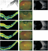
Efficacy of combining posterior scleral contraction and intravitreal C3F8 injection in high myopia with macular hole retinal detachment
- PMID: 40534814
- PMCID: PMC12120467
- DOI: 10.18240/ijo.2025.06.14
Efficacy of combining posterior scleral contraction and intravitreal C3F8 injection in high myopia with macular hole retinal detachment
Abstract
Aim: To evaluate the efficacy and safety of combining posterior scleral contraction (PSC) with intravitreal perfluoropropane (C3F8) injection in high myopia with macular hole retinal detachment (MHRD).
Methods: A total of 22 participants (22 eyes) with high myopia [axial length (AL)≥26.5 mm] and MHRD who underwent PSC combined with intravitreal C3F8 injection, with at least 6mo of follow-up were retrospectively recruited. Outcome measures included best-corrected visual acuity (BCVA), AL, optical coherence tomography (OCT) findings, and adverse events. Retinal recovery was categorized as type I (macular hole bridging with retinal reattachment) or type II (reattachment without hole bridging).
Results: The mean age of participants was 62.1±8.8y and mean follow-up duration was 9.18±4.21mo. Complete retinal reattachment was observed in 11 eyes (50%) at postoperative day 1, 19 eyes (86.3%) at week 1, and all 22 eyes at month 1. Ten eyes (45.5%) achieved type I recovery and 12 eyes (54.5%) achieved type II. Mean BCVA improved from 1.68±0.84 logMAR before surgery to 1.21±0.65 logMAR after surgery (P<0.001), and AL was significantly reduced compared to baseline (29.07±2.05 vs 30.8±2.2 mm; P<0.001). No serious complications were reported.
Conclusion: PSC combined with intravitreal C3F8 injection is a safe and effective treatment for MHRD in highly myopic eyes, especially for retinal detachment limited within the vascular arcade.
Keywords: C3F8; macular hole; myopia; posterior scleral contraction; retinal detachment.
International Journal of Ophthalmology Press.
Conflict of interest statement
Conflicts of Interest: Chen S, None; Ye J, None; Pan QT, None; Huang F, None; Zheng LY, None; Ye HF, None; Su YF, None; Li Y, None; Zhu SQ, None.
Figures


Similar articles
-
Drainage from intentional extramacular holes after internal limiting membrane insertion to treat macular hole retinal detachment in highly myopic eyes.Int Ophthalmol. 2025 Jan 29;45(1):43. doi: 10.1007/s10792-025-03406-8. Int Ophthalmol. 2025. PMID: 39880997
-
Internal Limiting Membrane Flap and Insertion Techniques Improve Prognosis in Macular Hole-Associated Retinal Detachment.Ophthalmol Retina. 2025 Jul 11:S2468-6530(25)00316-1. doi: 10.1016/j.oret.2025.07.006. Online ahead of print. Ophthalmol Retina. 2025. PMID: 40653047
-
Efficacy of a simple intravitreal perfluoropropane injection in treating unclosed idiopathic macular holes following vitrectomy.BMC Ophthalmol. 2025 Feb 5;25(1):61. doi: 10.1186/s12886-024-03839-2. BMC Ophthalmol. 2025. PMID: 39910548 Free PMC article.
-
Macular epiretinal membrane in high myopia: timing and prognosis of pars plana vitrectomy surgery.Int J Ophthalmol. 2025 Sep 18;18(9):1689-1696. doi: 10.18240/ijo.2025.09.10. eCollection 2025. Int J Ophthalmol. 2025. PMID: 40881444
-
Prenatal administration of progestogens for preventing spontaneous preterm birth in women with a multiple pregnancy.Cochrane Database Syst Rev. 2019 Nov 20;2019(11):CD012024. doi: 10.1002/14651858.CD012024.pub3. Cochrane Database Syst Rev. 2019. PMID: 31745984 Free PMC article.
References
-
- Lim LS, Tsai A, Wong D, et al. Prognostic factor analysis of vitrectomy for retinal detachment associated with myopic macular holes. Ophthalmology. 2014;121(1):305–310. - PubMed
-
- Wang ZQ, Ni ZZ, Zhang XL, et al. Vitrectomy for retinal detachment associated with macular hole: Prognostic factor analysis under different axial length conditions. Acta Ophthalmol. 2024;102(4):e557–e564. - PubMed
-
- Arias L, Caminal JM, Rubio MJ, et al. Autofluorescence and axial length as prognostic factors for outcomes of macular hole retinal detachment surgery in high myopia. Retina. 2015;35(3):423–428. - PubMed
LinkOut - more resources
Full Text Sources
