. 2025 Jun 4:16:1596598.
doi: 10.3389/fimmu.2025.1596598. eCollection 2025.

Temporal changes in the protein cargo of extracellular vesicles and resultant immune reprogramming after severe burn injury in humans and mice

Affiliations

Temporal changes in the protein cargo of extracellular vesicles and resultant immune reprogramming after severe burn injury in humans and mice

Micah L Willis et al. Front Immunol. .

Abstract

Introduction: Severe injury, including burn trauma, leads to profound immune dysfunction, yet the mechanisms driving these changes remain incompletely defined. This lack of understanding has hindered efforts to modulate the immune response effectively. Additionally, a clear biomarker profile to guide clinicians in identifying burn patients at high risk for poor clinical outcomes is lacking. Extracellular vesicles (EVs) have emerged as novel mediators of immune dysfunction in various pathologies. Prior studies in mouse models have demonstrated that plasma EVs increase following burn injury and contribute to immune dysfunction. Furthermore, EVs have potential as biomarkers for predicting extended hospital stays in burn patients. This study hypothesizes that human EVs, purified early and late after burn injury, will exhibit immune reprogramming effects similar to those observed in mice and that specific EV protein cargo may serve as biomarkers of immune and physiological responses to burn injury.

Methods: EVs were isolated from the plasma of burn-injury patients at early (<72h) and late (≥14 days) time points post-injury. Using unbiased immune transcriptome and bioinformatic causal network analyses, the immunomodulatory effects of these EVs were assessed in human THP-1 macrophages. Mass spectrometry-based quantitative proteomics and pathway analyses were conducted to characterize the protein cargo of EVs from both human and mouse models at different post-burn phases.

Results: Early post-burn human EVs induced significant immune reprogramming in macrophages, increasing pro-inflammatory signaling while suppressing anti-inflammatory pathways. In contrast, late post-burn EVs exhibited an immunosuppressive profile, with downregulation of pro-inflammatory pathways and upregulation of anti-inflammatory signaling. Proteomic analyses revealed that human and mouse EVs contained unique and overlapping protein cargo across different time points. At day 7 post-burn, mouse EVs were enriched in circulation/complement and neuronal proteins, whereas by day 14, reductions in membrane and metabolism-associated proteins were observed. Similarly, in human EVs at 14 days post-burn, increased levels of circulation/complement, immune, and transport proteins were detected.

Conclusions: EVs from burn-injury patients at distinct time points differentially modulate immune responses in macrophages, mirroring the temporal immune phenotypes observed in clinical settings. These findings suggest that EV-macrophage interactions play a crucial role in burn-induced immune dysfunction and highlight the potential of EV protein cargo as biomarkers for immune status and patient outcomes following burn injury.

Keywords: burn injury; exosomes; extracellular vesicles; macrophages; microvesicles; thermal injury.


Conflict of interest statement

The authors declare that the research was conducted in the absence of any commercial or financial relationships that could be construed as a potential conflict of interest. The author(s) declared that they were an editorial board member of Frontiers, at the time of submission. This had no impact on the peer review process and the final decision.

Figures
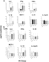
Figure 1
Figure 1
Human extracellular vesicles (EVs) isolated after burn injury regulate cytokine and chemokine release from PMA-conditioned THP-1 macrophages. EVs (3x10⁷ EVs) were isolated either “early”, 0–72 hrs, or “late” 14–20 days after injury or from healthy controls and applied to cultures for 48 hours (A) in the absence of or (B) presence of LPS to mimic burn-induced sepsis. Media cytokines and chemokines were measured by multiplex ELISA. Data shown as mean ± SEM; *p < 0.05, **p<0.01, ***p<0.0001.
Figure 2
Figure 2
Human extracellular vesicles (EVs) isolated early after burn injury reprogram THP-1 macrophages to a proinflammatory state. PMA-conditioned THP-1 cells were exposed to 3x10⁷ EVs/well from human burn patients (<72 hrs after injury) or healthy controls in the absence (A, B) or presence (C) of LPS. Gene expression was evaluated using nanoString barcoding spanning 561 mRNAs (nCounter Human Immunology CodeSet v2.0). Data are presented as the log2-transformed differential fold change in immune gene expression (A, C), with associated p-value significance (using Welch’s t test), after data normalization to housekeeping and internal control genes by nSolver v4.0. Differential fold change is shown as early EV versus healthy EVs (each group represents cell cultures stimulated with EVs from 6 patients). Differential gene expression and pathway Z-scores between early burn-induced EVs and healthy controls were analyzed via Ingenuity Pathway Analysis (B); only significantly altered (p<0.05) pathways are shown. There were not enough differentially-expressed genes to perform IPA for the +LPS cultures (N/A).
Figure 3
Figure 3
Human extracellular vesicles (EVs) isolated late after burn injury reprogram THP-1 macrophages to an anti-inflammatory state. PMA-conditioned THP-1 cells were exposed to 3x10⁷ EVs/well from human burn patients (14–20 days after injury) or healthy controls in the absence (A, B) or presence (C, D) of LPS. Gene expression was evaluated using nanoString and are presented as the log2-transformed differential fold change in immune gene expression (A, C), with associated p-value significance (using Welch’s t test), after data normalization to housekeeping and internal control genes by nSolver v4.0. Differential fold change shown early EV versus healthy EVs (each group represents cell cultures stimulated with EVs from 6 source patients, with n=6 different EV preparations from 2 experiments). Differential gene expression and pathway Z-scores between early burn-induced EVs and healthy controls were analyzed via Ingenuity Pathway Analysis (B, D); only significantly altered (p<0.05) pathways are shown.
Figure 4
Figure 4
Human extracellular vesicles (EVs) isolated early and late after burn injury induce differential reprogramming in THP-1 macrophages. Gene changes induced in THP-1 cells by EVs from human burn patients isolated late (14–20 days) compared to human burn patients isolated early (0–3 days), in the absence (A, B) or presence (C, D) of LPS. Differential fold change is shown as late versus early EVs (each group represents cell cultures stimulated with EVs from 6 source patients, with n=6 different EV preparations from 2 experiments). Differential gene expression and pathway Z-scores between early burn-induced EVs and healthy controls were analyzed via Ingenuity Pathway Analysis (B, D); only significantly altered (p<0.05) pathways are shown.
Figure 5
Figure 5
Burn injury in mice alters protein cargo of plasma EVs 7 and 14 days after injury. Adult mice underwent a 20% total body surface area (TBSA) burn and plasma EVs were collected 7 or 14 days after injury and protein content measured by LC-MS/MS. (A) Differentially expressed protein peptides in burn versus sham EVs (LFQ ratio, p-value Student’s t-test, N=3 per group). (B) Characteristics of proteins that are decreased in burn EVs relative to controls. (C) Characteristics of proteins that are increased in burn EVs relative to sham controls.
Figure 6
Figure 6
EV protein cargo-induced pathways altered over time after burn injury in mice. Adult mice underwent a 20% total body surface area (TBSA) burn and plasma EVs were collected at different time points after injury and protein content measured by LC-MS/MS (p-value Student’s t-test, N=3 per group). Differential protein expression and pathway Z-scores between burn at different time points past injury; (A) 3 days following injury (data reanalyzed from Maile et al., 2021 (6)), (B) 7 days following injury and (C) 14 days following injury and sham were analyzed via Ingenuity Pathway Analysis; only significantly altered (p < 0.05) pathways are shown.
Figure 7
Figure 7
Comparison of significantly expressed proteins in mouse plasma EVs over time after burn injury. Adult mice underwent a 20% total body surface area (TBSA) burn and plasma EVs were collected at early (3 days), intermediate (7 days) and late (14 days) time points after injury. Protein content was measured by LC-MS/MS. A Venn diagram in (A) summarizes the number and identity of proteins between the different time points after injury (p-value Student’s t-test, N=3 per group). (B) Heatmap showing Z-scores of protein expression between burn and sham, red shows positive Z-score > 1, and green shows negative Z-score < -1. (C) Heatmap showing Z-scores of pathways from EV proteome between burn and sham, red shows positive Z-score > 1, and green shows negative Z-score < -1. (D) Heatmap showing Z-scores of disease function from EV proteome between burn and sham, red shows positive Z-score > 1, and green shows negative Z-score < -1. White denotes no significant fold change detected.
Figure 8
Figure 8
Burn injury alters protein cargo of human plasma EVs 14 days after injury. Plasma EVs from human burn patients with severe burn injury isolated 14–20 or 3 days after admission. Protein content was measured by LC-MS/MS. (A) Differentially expressed protein peptides in late after burn (14–20 days) vs 3 days following admission (LFQ ratio, p-value Student’s t-test, N=3 per group). (B) Characteristics of proteins that are increased in burn EVs 14–20 days relative to 3 days. (C) Characteristics of proteins that are decreased in burn EVs relative to controls. Ingenuity Pathway Analysis quantified pathway Z-scores between burn at different time points past injury; (D) 3 days following injury compared to healthy, (E) 14 days following injury compared to 3 days following injury; only significantly altered (p < 0.5, n=3 per group) pathways are shown.

Update of

Similar articles

References

    1. Jeschke MG, van Baar ME, Choudhry MA, Chung KK, Gibran NS, Logsetty S. Burn injury. Nat Rev Dis Primers. (2020) 6:11. doi: 10.1038/s41572-020-0145-5 - DOI - PMC - PubMed
    1. van Duin D, Strassle PD, DiBiase LM, Lachiewicz AM, Rutala WA, Eitas T, et al. Timeline of health care-associated infections and pathogens after burn injuries. Am J Infect Control. (2016) 44(12):1511–6. doi: 10.1016/j.ajic.2016.07.027 - DOI - PMC - PubMed
    1. Wasserman D, Ioannovich JD, Hinzmann RD, Deichsel G, Steinmann GG. Interferon-gamma in the prevention of severe burn-related infections: a European phase III multicenter trial. The Severe Burns Study Group. Crit Care Med. (1998) 26:434–9. doi: 10.1097/00003246-199803000-00010 - DOI - PubMed
    1. Seim RF, Willis ML, Wallet SM, Maile R, Coleman LG, Jr. Extracellular vesicles as regulators of immune function in traumatic injuries and sepsis. Shock. (2023) 59:180–9. doi: 10.1097/SHK.0000000000002023 - DOI - PMC - PubMed
    1. Abels ER, Breakefield XO. Introduction to extracellular vesicles: biogenesis, RNA cargo selection, content, release, and uptake. Cell Mol Neurobiol. (2016) 36:301–12. doi: 10.1007/s10571-016-0366-z - DOI - PMC - PubMed

LinkOut - more resources