Review
. 2025 Jun 4:16:1597462.
doi: 10.3389/fimmu.2025.1597462. eCollection 2025.

Mechanism of cell death and its application in the repair of inflammatory bowel disease by mesenchymal stem cells

Affiliations
Review

Mechanism of cell death and its application in the repair of inflammatory bowel disease by mesenchymal stem cells

Francis Atim Akanyibah et al. Front Immunol. .

Abstract

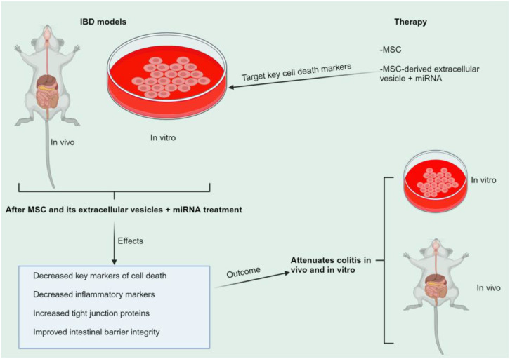
The onset and progression of inflammatory bowel disease (IBD), which encompasses ulcerative colitis and Crohn's disease, are influenced by the immune system, environmental factors, genetics, and intestinal flora. Cell death is a biological phenomenon that occurs in all living organisms; nevertheless, excessive cell death has been linked to IBD, including increased immune and intestinal epithelial cell death and intestinal barrier abnormalities. Anti-tumor necrosis factor medication, which has made significant progress in treating IBD cell death, may fail in some individuals or lose effectiveness over time, necessitating the search for a safe and effective treatment. One of the novel and emerging areas in regenerative and nanomedicine used to regulate cell death is mesenchymal stem cells (MSCs) and their mediators (extracellular vesicles). MSCs and their mediators have been found to attenuate cell death in several illnesses, including IBD. This review explores cell death mechanisms and their implications in IBD, focusing on the potential ameliorative effects of MSCs and their mediators on cell death.

Keywords: IBD; MSc; cell death; exosome; extracellular vesicles.


Conflict of interest statement

The authors declare that the research was conducted in the absence of any commercial or financial relationships that could be construed as a potential conflict of interest.

Figures

Figure 1
Figure 1
Apoptosis pathway. Intrinsic stimuli like hypoxia, DNA damage, and ER stress all contribute to this pathway. Extrinsic stimuli like TNF alpha and FASL contribute to the extrinsic pathway. Ultimately, both routes activate the effector caspases, resulting in apoptosis. APAF1, apoptotic protease activating factor-1; BAX, bcl-2 associated x protein; BCL2, b-cell lymphoma-2; BH3, bcl2 homology domain 3; CIAPs, cellular inhibitor of apoptosis proteins; FADD, fas-associated death domain protein; FASL/R, Fas ligand/receptor; PRC: procaspase; SMAC, second mitochondrial-derived activator of caspase; tBid, truncated bid; TNFα/R, tumor necrosis alpha/receptor; TRADD, TNF receptor-associated death domain protein; TRAF2, TNF receptor-associated factor 2; XIAP, x-linked inhibitor of apoptosis protein.
Figure 2
Figure 2
Mechanism of pyroptosis. PAMP and DAMP commence the canonical route, whereas bacteria and LPS synthesis initiate the non-canonical route. These two routes eventually cleave GSDMD, resulting in the GSDMD channel that leaks IL-1β and IL-18, causing pyroptosis. ASC, apoptosis-associated speck-like protein containing card; ATP, adenosine triphosphate; DAMP: damage-associated molecular patterns; GSDMD, gasdermin d; IL, interleukin; NLRP3: nod-like receptor pyrin domain-containing protein 3; PRR, pattern-recognition receptor; PAMP, pathogen-associated molecular patterns.
Figure 3
Figure 3
Necroptosis mechanism. The canonical and non-canonical paths lead to RIP3/MLKL activation, triggering the oligomerization of MLKL, resulting in the rupture of the plasma membrane, the release of intracellular chemicals, and cell death and inflammation promotion. FADD, fas-associated death domain protein; IFN, Interferon;IFNARI, ifn-α receptor type I; IRF9, interferon regulatory factor 9; ISGF3, ifn-stimulated gene factor 3; JAK1, janus kinase 1; LPS, lipopolysaccharides; MLKL, mixed lineage kinase domain-like; mt, mitochondrial; RIP/RIPK, receptor-interacting protein kinase; STAT, signal transducer and activator of transcription; TLR3/4, toll-like receptor-3/4; TNFα, tumor necrosis factor (TNF) alpha; TRAIL/R, TNF-related apoptosis-inducing ligand/receptor; TRIF, TIR domain-containing adapter-inducing interferon-β; ZBP1 (DAI), Z-DNA binding protein 1; VDNA, DNA viruses.
Figure 4
Figure 4
Autophagy pathway. Nutrient starvation and energy depletion cause several cascades that alter the phagophore’s initiation and elongation to the autophagosome. During this process, ubiquitin-like conjugation cascade systems such as the LC3s and ATGs facilitate autophagosome formation. AMPK, amp-activated protein kinase; ATG, autophagy-related gene; FIP200, focal adhesion kinase family interacting protein of 200 kDa; HOPS, homotypic fusion and protein sorting; LAMP-2, lysosome-associated membrane protein 2; LC3, microtubule-associated protein 1 light chain 3; mTOR: mammalian target of rapamycin; Nrf2, nuclear factor erythroid 2-related factor 2; PI3P, phosphatidylinositol 3-phosphate; SNARE, soluble N-ethylmaleimide-sensitive factor attachment protein receptor; ULK1, unc-51-like kinase 1; VPS 34, vacuolar protein sorting 34.
Figure 5
Figure 5
Mechanism of ferroptosis. TFRI/TF, FAT/FATP/PUFAs, and other pathways influence ferroptosis formation. Lip-1, DFO, Fer-1, the GCH1/BH4 and GSH/GPX4 pathways, and FSP1 suppress ferroptosis. ACSL4, long-chain acyl-coenzyme A synthase 4; ALOX12/5, arachidonate 12/5-lipoxygenase; ATF4, activating transcription factor 4; BH2, dihydrobiopterin; BH4, tetrahydrobiopterin; CHACI: chac glutathione specific γ−glutamylcyclotransferase 1; DFO, deferoxamine; DHFR, dihydrofolate reductase; DMT1, divalent metal ion transporter 1; ER, endoplasmic reticulum; Fer-1, ferrostatin-1; FPN, ferroportin; FSP1, ferroptosis suppressor protein 1; GCH1, cyclohydrolase-1; GPX4, glutathione peroxidase-4; GSH, glutathione; GTP, guanosine 5′-triphosphate; HO-1, heme oxygenase-1; iPLA2β, independent phospholipase A2β; LIP, labile iron pool; Lip-1, liproxstatin-1; MVA, Mevalonate; NADP+,nicotinamide adenine dinucleotide phosphate; NADPH, nicotinamide adenine dinucleotide phosphate; NCOA4, nuclear receptor coactivator 4; Nrf2, nuclear factor erythroid 2-related factor 2; PUFAs, polyunsaturated fatty acids; ROS, reactive oxygen species; SATI, spermine N1-acetyltransferase 1; SLC, solute carrier family; STEAP3, six-transmembrane epithelial antigen of prostate family member 3; TCA, tricarboxylic acid; TFR1, transferrin receptor 1; TS, transsulfuration.
Figure 6
Figure 6
Summary of MSC + EV regulation on key cell death markers of IBD. The figure summarizes how MSC + EV regulates specific cell death markers to prevent or activate subsequent downstream signaling, as shown in Figures 1 , 2 , 3 , 4 , 5 . (A) Ferroptosis, (B) Pyroptosis, (C) Autophagy (D) Apoptosis. ACSL4, long-chain acyl-coenzyme A synthase 4; ASC, apoptosis-associated speck-like protein containing card; ATG, autophagy-related gene; BAX, bcl-2 associated x protein; BH3, bcl2 homology domain 3; Casp, caspase; CIAPs, cellular inhibitor of apoptosis proteins; DAMP, damage-associated molecular patterns; DMT1, divalent metal ion transporter 1; E, effector; FADD, fas-associated death domain protein; GPX4, glutathione peroxidase-4; GSDMD, gasdermin d; GSH, glutathione; IL, interleukin; LC3, microtubule-associated protein 1 light chain 3; NLRP3, nod-like receptor pyrin domain-containing protein 3; PAMP, pathogen-associated molecular patterns; PI3P, phosphatidylinositol 3-phosphate; PRC, procaspase; PRR, pattern-recognition receptor; PUFA, polyunsaturated fatty acid; TRADD, TNF receptor-associated death domain protein; TRAF2, TNF receptor-associated factor 2; VPS, vacuolar protein sorting.
Figure 7
Figure 7
Summary of the role of MSCs in targeting cell death markers of IBD. MSCs and their mediators target cell death indicators in vitro and in vivo to reduce inflammatory markers, disrupt tight junction proteins, and improve intestinal barrier integrity, thereby alleviating colitis. IBD, inflammatory bowel disease; miRNA, microRNA; MSC, mesenchymal stem cell.

Similar articles

References

    1. Akrapovic Olic I, Vukovic J, Radic M, Sundov Z. Enthesitis in IBD patients. J Clin Med. (2024) 13:4540. doi: 10.3390/jcm13154540 - DOI - PMC - PubMed
    1. Akanyibah FA, Zhu Y, Wan A, Ocansey DKW, Xia Y, Fang AN, et al. Effects of DNA methylation and its application in inflammatory bowel disease (Review). Int J Mol Med. (2024) 53:55. doi: 10.3892/ijmm.2024.5379 - DOI - PMC - PubMed
    1. Nóbrega VG, Silva INN, Brito BS, Silva J, Silva M, Santana GO. THE ONSET OF CLINICAL MANIFESTATIONS IN INFLAMMATORY BOWEL DISEASE PATIENTS. Arq Gastroenterol. (2018) 55:290–5. doi: 10.1590/s0004-2803.201800000-73 - DOI - PubMed
    1. Ng SC, Shi HY, Hamidi N, Underwood FE, Tang W, Benchimol EI, et al. Worldwide incidence and prevalence of inflammatory bowel disease in the 21st century: a systematic review of population-based studies. Lancet (London England). (2017) 390:2769–78. doi: 10.1016/S0140-6736(17)32448-0 - DOI - PubMed
    1. Wang S, Dong Z, Wan X. Global, regional, and national burden of inflammatory bowel disease and its associated anemia, 1990 to 2019 and predictions to 2050: An analysis of the global burden of disease study 2019. Autoimmun Rev. (2024) 23:103498. doi: 10.1016/j.autrev.2023.103498 - DOI - PubMed

MeSH terms

LinkOut - more resources