. 2025 Jun 5;30(2):385.
doi: 10.3892/ol.2025.15131. eCollection 2025 Aug.

Pan-cancer analysis reveals the potential role of DHCR24 in bladder cancer via interactions with HRAS to facilitate cholesterol synthesis

Affiliations

Pan-cancer analysis reveals the potential role of DHCR24 in bladder cancer via interactions with HRAS to facilitate cholesterol synthesis

Zhibin Wang et al. Oncol Lett. .

Abstract

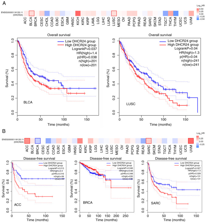
There is a strong association between cholesterol reprogramming and cancer development. However, 3β-hydroxysteroid Δ24-reductase (DHCR24), the final enzyme in the cholesterol biosynthesis pathway, has been relatively understudied in cancer progression. The present study aimed to perform a comprehensive pan-cancer analysis of DHCR24 to elucidate its role across different malignancies. The interacting proteins of DHCR24 were identified by molecular docking node dynamics simulation. Duolink proximity ligation, cell viability and filipin staining assays were used to assess the function of DHCR24 in cancer cells and its underlying oncogenic mechanisms. The findings revealed that DHCR24 exhibits high expression in seven cancer types (bladder cancer, breast invasive carcinoma, liver hepatocellular carcinoma, prostate adenocarcinoma, cervical squamous cell carcinoma and endocervical adenocarcinoma, uterine corpus endometrial carcinoma and stomach adenocarcinoma), and low expression in five others (glioblastoma multiforme, kidney chromophobe, kidney renal clear cell carcinoma, lung adenocarcinoma and lung squamous cell carcinoma), suggesting that DHCR24 serves distinct roles depending on the cancer type. Notably, it was demonstrated that DHCR24 expression consistently increases with tumor stage and serves as an independent prognostic factor in BLCA. Moreover, molecular docking and kinetic modeling identified HRAS as a key interacting protein of DHCR24. The Duolink assay further demonstrated that DHCR24 interacts with HRAS outside the nucleus in 5637 human BLCA cells. Filipin fluorescence staining and cell proliferation assays also revealed that this interaction promoted cholesterol synthesis, contributing to cancer cell proliferation in the 5637 cells. In conclusion, the results of the present study provide novel insights into the oncogenic role of DHCR24 in BLCA and demonstrates its interaction with HRAS for the first time to the best of our knowledge, highlighting a potential mechanism driving tumor progression.

Keywords: BLCA; DHCR24; HRAS; cholesterol synthesis; pan-cancer analysis.


Conflict of interest statement

The authors declare that they have no competing interests.

Figures

Figure 1. Upregulated mRNA expression of DHCR24 in pan–cancer. The results obtained from Tumor Immune Estimation Resource 2.0 were used for preliminary pan–cancer analysis of cancer data in The Cancer...
Figure 1.
Upregulated mRNA expression of DHCR24 in pan-cancer. The results obtained from Tumor Immune Estimation Resource 2.0 were used for preliminary pan-cancer analysis of cancer data in The Cancer Genome Atlas, and they revealed that DHCR24 expression was significantly increased in 12 tumors compared with that in normal tissue. The red and blue boxes represent tumor tissues and normal tissues, respectively. **P<0.01; ***P<0.001. DHCR24, 3β-hydroxysteroid Δ24-reductase; TPM, transcripts per million. BLCA, bladder cancer; BRCA, breast invasive carcinoma; CESC, cervical squamous cell carcinoma and endocervical adenocarcinoma; GBM, glioblastoma multiforme; KICH, kidney chromophobe; KIRC, kidney renal clear cell carcinoma; LIHC, liver hepatocellular carcinoma; LUAD, lung adenocarcinoma; LUSC, lung squamous cell carcinoma; PRAD, prostate adenocarcinoma; UCEC, uterine corpus endometrial carcinoma; STAD, stomach adenocarcinoma.
Figure 2. Expression of DHCR24 in different types of cancer. (A) Expression distribution of the DHCR24 gene in tumor and normal tissues. The differences between the two groups were compared. (B) Pan–c...
Figure 2.
Expression of DHCR24 in different types of cancer. (A) Expression distribution of the DHCR24 gene in tumor and normal tissues. The differences between the two groups were compared. (B) Pan-cancer differential expression of DHCR24 in paired tumor and adjacent normal tissues in the indicated tumor types from The Cancer Genome Atlas database. *P<0.05, **P<0.01; ***P<0.001. DHCR24, 3β-hydroxysteroid Δ24-reductase; BLCA, bladder cancer; BRCA, breast invasive carcinoma; CESC, cervical squamous cell carcinoma and endocervical adenocarcinoma; GBM, glioblastoma multiforme; KICH, kidney chromophobe; KIRC, kidney renal clear cell carcinoma; LIHC, liver hepatocellular carcinoma; LUAD, lung adenocarcinoma; LUSC, lung squamous cell carcinoma; PRAD, prostate adenocarcinoma; UCEC, uterine corpus endometrial carcinoma; STAD, stomach adenocarcinoma.
Figure 3. DHCR24 as a survival factor in some cancer types. The GEPIA2 tool was used to perform (A) overall survival and (B) disease–free survival analyses of different tumors in The Cancer Genome Atl...
Figure 3.
DHCR24 as a survival factor in some cancer types. The GEPIA2 tool was used to perform (A) overall survival and (B) disease-free survival analyses of different tumors in The Cancer Genome Atlas by DHCR24 gene expression. Survival maps and Kaplan-Meier curves with positive results are presented. DHCR24, 3β-hydroxysteroid Δ24-reductase; BLCA, bladder cancer; LUSC, lung squamous cell carcinoma; ACC, adrenocortical carcinoma; BRCA, breast cancer; SARC, sarcoma.
Figure 4. DHCR24 is a factor for clinical stage and the prognosis of cancer. (A) Based on The Cancer Genome Atlas data, the association between expression levels of the DHCR24 gene and the main pathol...
Figure 4.
DHCR24 is a factor for clinical stage and the prognosis of cancer. (A) Based on The Cancer Genome Atlas data, the association between expression levels of the DHCR24 gene and the main pathological stages (stages I–IV) of BLCA and LUSC were analyzed. Log2 (transcripts per million +1) was applied for log-scale. (B) Univariate and multifactorial proportional hazards model analyses. (C) Receiver operating characteristic curves demonstrated efficacy in predicting risk scores for the 1-, 3- and 5-year survival of patients, and the nomogram revealed the probability of predicting the 1-, 3- and 5-year survival of patients. DHCR24, 3β-hydroxysteroid Δ24-reductase; BLCA, bladder cancer; LUSC, lung squamous cell carcinoma; AUC, area under the curve; N, lymph node stage; M, metastasis stage; T, tumor stage.
Figure 5. Important role of DHCR24 in bladder cancer. (A) Circos plot shows the correlation between the expression of several genes and DHCR24 in The Cancer Genome Atlas dataset. (B) Expression heat m...
Figure 5.
Important role of DHCR24 in bladder cancer. (A) Circos plot shows the correlation between the expression of several genes and DHCR24 in The Cancer Genome Atlas dataset. (B) Expression heat map of differential genes in bladder cancer. Gene Ontology analysis of the differently expressed genes was performed, and (C) bubble, (D) transverse bar and (E) circle charts were generated. KEGG analysis of the differently expressed genes was performed and (F) bubble and (G) transverse bar charts were generated. (H) Results of the Gene Set Enrichment Analysis of the differential genes. DHCR24, 3β-hydroxysteroid Δ24-reductase; KEGG, Kyoto Encyclopedia of Genes and Genomes.
Figure 6. DHCR24 changes the tumor microenvironment of bladder cancer. (A) Differential analysis of immune cells in the DHCR24 low and high expression groups. Analysis of the correlation between DHCR2...
Figure 6.
DHCR24 changes the tumor microenvironment of bladder cancer. (A) Differential analysis of immune cells in the DHCR24 low and high expression groups. Analysis of the correlation between DHCR24 expression and (B) immune cells and (C) tumor immune checkpoints. (D) Tumor mutation compliance analysis. *P<0.05, **P<0.01. DHCR24, 3β-hydroxysteroid Δ24-reductase; NK, natural killer.
Figure 7. In the HPA database, DHCR24 is highly expressed in the immunohistochemical sections of bladder cancer. Immunohistochemical images of (A) cancerous bladder tissue from patients with bladder c...
Figure 7.
In the HPA database, DHCR24 is highly expressed in the immunohistochemical sections of bladder cancer. Immunohistochemical images of (A) cancerous bladder tissue from patients with bladder cancer and (B) normal human bladder tissue from the Human Protein Atlas database.
Figure 8. DHCR24 may interact with HRAS to play a role in cancer. (A) Bubble map and (B) histogram of the Kyoto Encyclopedia of Genes and Genomes analysis of the DHCR24–interacting proteome. (C) Prote...
Figure 8.
DHCR24 may interact with HRAS to play a role in cancer. (A) Bubble map and (B) histogram of the Kyoto Encyclopedia of Genes and Genomes analysis of the DHCR24-interacting proteome. (C) Protein-protein interaction network formed by DHCR24 and HRAS. (D) Correlation analysis of DHCR24 and HRAS expression in the GEPIA database. DHCR24, 3β-hydroxysteroid Δ24-reductase; tRNA, transfer RNA; TPM, transcripts per million.
Figure 9. DHCR24 interacts with HRAS in vivo . (A) Modeller multi–template modeling of the constructed DHCR24 structure. (B) Pulled graph analysis of DHCR24 with multiple template modeling. (C) Sequen...
Figure 9.
DHCR24 interacts with HRAS in vivo. (A) Modeller multi-template modeling of the constructed DHCR24 structure. (B) Pulled graph analysis of DHCR24 with multiple template modeling. (C) Sequence alignment between the receptor-binding domain region of PI3KCG and DHCR24. (D) ZDOCK results show that DHCR24 and HRAS are able to interact with each other. Random docking results: Blue for HRAS and green for DHCR24; hot spot docking results: Red for HRAS and orange for DHCR24; both comparison results. (E) Trunk plot of RMSD vs. time for DHCR24 in complex with HRAS. (F) Stable three-dimensional structure of DHCR24 complexed with HRAS after 70 nsec of kinetic simulation. (G) Stable three-dimensional structure of DHCR24 complexed with HRAS after 70 nsec of kinetic simulation. (H) 5637 cells were stained with Duolink using mouse-derived anti-HRAS with rabbit-derived anti-DHCR24 and their nuclei were stained with DAPI. Images were captured using fluorescence microscopy. DHCR24, 3β-hydroxysteroid Δ24-reductase; RMSD, root mean square deviation; PI3KCG, phosphatidylinositol-4,5-bisphosphate 3-kinase catalytic subunit γ.
Figure 10. Role of DHCR24–HRAS interaction in bladder carcinogenesis. (A) 5637 cells were stimulated with several drugs, and cell proliferation was observed after 72 h using light microscopy and cell ...
Figure 10.
Role of DHCR24-HRAS interaction in bladder carcinogenesis. (A) 5637 cells were stimulated with several drugs, and cell proliferation was observed after 72 h using light microscopy and cell counting. normal: 5637 cells cultured in normal medium without drug stimulation; U18666A: 5637 cells in normal culture with U18666A added; lonafarnib: 5637 cells in normal culture with lonafarnib added; EGF: 5637 cells in normal culture with the addition of EGF; EGF + U18666A: 5637 cells in normal culture with the addition of both EGF and U18666A; and EGF + Lonafarnib: 5637 cells in normal culture with the addition of both EGF and lonafarnib. (B) Cell proliferation measured by MTT assay (left) and number of adherent cells at 72 h (right). (C) Fluorescence staining of Filipin in 5637 cells after EGF stimulation. (D) Analysis of filipin fluorescence intensity of 5637 cells with (EGF group) and without (Normal group) EGF stimulation. (E) Fluorescence staining of Filipin in 5637 cells after the administration of U18666A and lonafarnib. (F) Filipin fluorescence intensity analysis. *P<0.05; **P<0.01; ***P<0.001 normal vs. negative control group; #P<0.05; ##P<0.01; ###P<0.001; ####P<0.001 EGF vs. experimental group. EGF, epidermal growth factor; ns, not significant.

Similar articles

References

    1. Haas MJ, Mooradian AD. Potential therapeutic agents that target ATP binding cassette A1 (ABCA1) gene expression. Drugs. 2022;82:1055–1075. doi: 10.1007/s40265-022-01743-x. - DOI - PubMed
    1. Wahida A, Buschhorn L, Fröhling S, Jost PJ, Schneeweiss A, Lichter P, Kurzrock R. The coming decade in precision oncology: Six riddles. Nat Rev Cancer. 2023;23:43–54. doi: 10.1038/s41568-022-00529-3. - DOI - PubMed
    1. Zhou C, Solomon B, Loong HH, Park K, Pérol M, Arriola E, Novello S, Han B, Zhou J, Ardizzoni A, et al. First-line selpercatinib or chemotherapy and pembrolizumab in RET Fusion-positive NSCLC. N Engl J Med. 2023;389:1839–1850. doi: 10.1056/NEJMoa2309457. - DOI - PMC - PubMed
    1. Last AR, Ference JD, Menzel ER. Hyperlipidemia: Drugs for cardiovascular risk reduction in adults. Am Fam Physician. 2017;95:78–87. - PubMed
    1. Nong S, Han X, Xiang Y, Qian Y, Wei Y, Zhang T, Tian K, Shen K, Yang J, Ma X. Metabolic reprogramming in cancer: Mechanisms and therapeutics. MedComm (2020) 2023;4:e218. doi: 10.1002/mco2.218. - DOI - PMC - PubMed

LinkOut - more resources