

Delayed Deep Femoral Artery Injury Secondary to Migrated Lesser Trochanter Fragment After Intertrochanteric Fracture Fixation: A Case Report and Updated Literature Review
- PMID: 40535321
- PMCID: PMC12174772
- DOI: 10.1177/21514593251351188
Delayed Deep Femoral Artery Injury Secondary to Migrated Lesser Trochanter Fragment After Intertrochanteric Fracture Fixation: A Case Report and Updated Literature Review
Abstract
Background: With the increasing elderly population and prevalence of osteoporosis, geriatric intertrochanteric fragility fractures pose a major challenge to orthopedic practice. These fractures have a significant impact on patient outcomes, with a reported mortality rate of 13.3% within 30 days and 24.5% within one year.
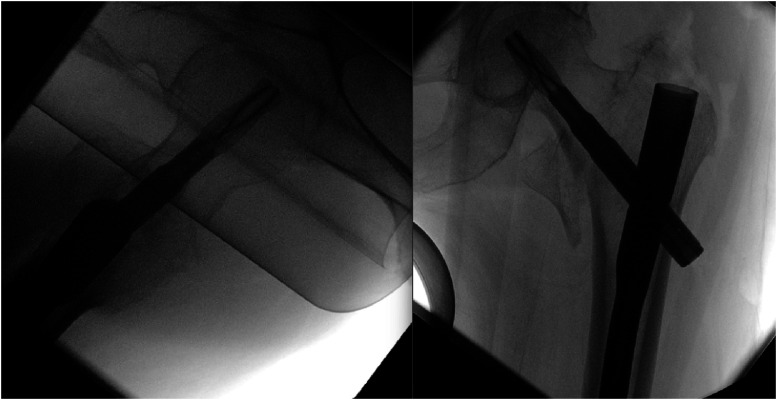
Case presentation: This report presents a rare case of delayed iatrogenic deep femoral artery (DFA) injury due to progressive displacement of the lesser trochanter following intertrochanteric fracture fixation. An 87-year-old female patient developed significant thigh swelling and pain 33 days postoperatively. Imaging confirmed migration of the lesser trochanter fragment, leading to DFA injury and active bleeding.
Discussion: Through a comprehensive literature review, we explore the incidence, diagnostic modalities, and management of vascular injuries associated with pertrochanteric fracture fixation. We emphasize the importance of identifying significantly displaced lesser trochanter fragments (>1 cm) preoperatively, as they markedly increase the risk of DFA injury. While debate continues over routine fragment fixation, our case suggests that surgical fixation may be beneficial in selected patients with large displacements to prevent vascular complications. Early CT angiography is highlighted as a crucial non-invasive diagnostic tool for timely detection and intervention in these high-risk cases.
Conclusion: This case underscores the need for careful postoperative monitoring and early intervention to optimize patient outcomes. As PFFs become more prevalent, further research is essential to improve geriatric orthopedic care.
Keywords: femoral artery injury; fracture fixation; hemorrhage; lesser trochanter migration; proximal femoral fractures; pseudoaneurysm.
© The Author(s) 2025.
Conflict of interest statement
The authors declare that there are no conflicts of interest related to this study.
Figures


Similar articles
-
[Ipsilateral femoral neck fracture after fixation of intertrochanteric fracture by InterTAN intramedullary nail: A case report].Beijing Da Xue Xue Bao Yi Xue Ban. 2025 Jun 18;57(3):610-613. doi: 10.19723/j.issn.1671-167X.2025.03.028. Beijing Da Xue Xue Bao Yi Xue Ban. 2025. PMID: 40509842 Free PMC article. Chinese.
-
Case Report: A case of femoral metastatic cancer misdiagnosed as isolated femoral lesser trochanter avulsion fracture.Front Oncol. 2025 Jun 17;15:1565771. doi: 10.3389/fonc.2025.1565771. eCollection 2025. Front Oncol. 2025. PMID: 40599861 Free PMC article.
-
High Risk of Venous Thromboembolism With Aspirin Prophylaxis After THA for High-riding Developmental Dysplasia of the Hip: A Retrospective, Comparative Study.Clin Orthop Relat Res. 2025 Jun 9. doi: 10.1097/CORR.0000000000003482. Online ahead of print. Clin Orthop Relat Res. 2025. PMID: 40536765
-
Has the documentation of chest injuries and the development of systemic complications in patients with long bone fractures changed over time?-A systematic literature review and meta-analysis by the IMPACT expert group.Injury. 2025 Mar;56(3):112182. doi: 10.1016/j.injury.2025.112182. Epub 2025 Jan 23. Injury. 2025. PMID: 39874866
-
Assessing the comparative effects of interventions in COPD: a tutorial on network meta-analysis for clinicians.Respir Res. 2024 Dec 21;25(1):438. doi: 10.1186/s12931-024-03056-x. Respir Res. 2024. PMID: 39709425 Free PMC article. Review.
References
-
- Xu H, Liu Y, Sezgin EA, et al. Comparative effectiveness research on proximal femoral nail versus dynamic hip screw in patients with trochanteric fractures: a systematic review and meta-analysis of randomized trials. J Orthop Surg Res. 2022;17(1):292. doi: 10.1186/s13018-022-03189-z - DOI - PMC - PubMed
-
- Gökmen MY, Uluöz M, Varmış HO, Çiçek H. Comparison of three methods of greater trochanter fixation in intertrochanteric femur fractures (AO type 31/A2) treated with cementless bipolar hemiarthroplasty. J Cukurova Anesth Surg Sci. 2024;7(3):195-199. doi: 10.36516/jocass.1534039 - DOI
Publication types
LinkOut - more resources
Full Text Sources
Research Materials
