. 2025 Jun 4:16:1550068.
doi: 10.3389/fendo.2025.1550068. eCollection 2025.

Extracellular vesicles in reproductive biology and disorders: a comprehensive review

Affiliations

Extracellular vesicles in reproductive biology and disorders: a comprehensive review

Jinguang Wang et al. Front Endocrinol (Lausanne). .

Abstract

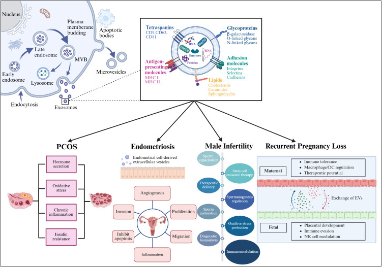
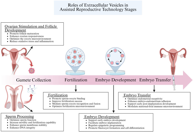
Extracellular vesicles (EVs) facilitate intercellular communication and the conveyance of bioactive substances, including proteins, lipids, and nucleic acids. They play a significant role in various reproductive biological processes, including gametogenesis, fertilization, early embryo development, and implantation. Dysfunctional EV activity is associated with various reproductive diseases, such as polycystic ovary syndrome (PCOS), endometriosis, male infertility, and recurrent pregnancy loss (RPL). This review systematically examines and categorizes current knowledge on EV functions in reproductive biology and disorders, and their potential as diagnostic and therapeutic tools. A systematic literature search from 2000 to 2024 identified studies showing EVs' influence on gamete maturation, fertilization, embryonic development, and implantation. They also play a role in reproductive disorders by affecting insulin resistance, androgen production, inflammation, angiogenesis, sperm quality, and maternal-fetal immune tolerance. The review concludes that EVs are integral to reproductive health, with further research needed to understand their mechanisms and clinical potential.

Keywords: embryo implantation; embryos; extracellular vesicles; fertility; oocyte; reproductive disorders; sperm.


Conflict of interest statement

The authors declare that the research was conducted in the absence of any commercial or financial relationships that could be construed as a potential conflict of interest.

Figures

Figure 1
Figure 1
The formation of EVs begins with endocytosis, which has two pathways: returning the cargo to the plasma membrane as “recycling endosomes” or transforming into “late endosomes,” or MVBs. MVBs will either merge with the lysosome or the plasma membrane, releasing their cargo outside the cell. Several RAB proteins, including Rab 27a and Rab 27b, as well as protein complexes, help transport MVBs to the plasma membrane and release EVs. In contrast, microvesicles are formed by the plasma membrane’s outward budding and scission, whereas apoptotic cellular membranes’ outward bubbling results in the production of apoptotic bodies. EVs and target cells interact in three ways (1): membrane proteins on EVs bind directly to receptors on target cells, activating an intracellular signaling cascade; (2) EVs transport their contents to target cells by fusing with the cell membrane; and (3) EVs are engulfed by endocytosis, releasing signaling molecules. Created with BioRender.com.
Figure 2
Figure 2
Structure and composition of EV. EV is a lipid bilayer structure that contains lipids, proteins and nucleic acids. Sphingomyelin, phosphatidylserine, cholesterol and ceramides are highly distributed on the membrane. In addition, EVs also contain a variety of proteins such as major histocompatibility complex I and II (MHC I and MHC II), proteins from the MVB machinery (ALIX, TSG101), heat shock proteins (HSP70, HSP90, HSP60), tetraspanins (CD9, CD63, CD81), receptors (FasL, TNF, TfR), adhesion molecules (Interins, Selectins, Cadherins) and cytosolic proteins, RNA and DNA. Created with BioRender.com.
Figure 3
Figure 3
This figure illustrates how EV-shuttled cargo, released from the epididymis (epididymosomes) and the prostate (prostasomes), affects various sperm functions. Epididymosomes enhance sperm motility, maturation, mediate cell-to-cell communication, protect sperm from oxidative damage, and participate in immune regulation. Similarly, prostasomes interact with sperm to improve motility, support maturation, facilitate cell-to-cell communication, protect against oxidative damage, and modulate immune responses. Both types of EVs play crucial roles in ensuring optimal sperm function and fertility. Created with BioRender.com.
Figure 4
Figure 4
This figure illustration highlights the critical roles of EVs in the female reproductive system. Part A, the ovary is depicted, showcasing the formation of the oocyte and emphasizing the functions of EVs within the follicular fluid, which include promoting oocyte maturation and enhancing fertility. Part B, the oviduct is illustrated, detailing how EVs are essential for oocyte maturation, fertility, and early embryo development. The image underscores the multifaceted impact of EVs throughout the reproductive process. Created with BioRender.com.
Figure 5
Figure 5
This figure illustrates the process of EV generation and their contents, including proteins, RNA, and lipids. The lower part of the image specifically details the role of EVs in four reproductive system-related diseases: Polycystic Ovary Syndrome (PCOS), endometriosis, male infertility, and recurrent pregnancy loss. In these conditions, EVs influence cellular communication by carrying specific biomolecules, thereby contributing to disease progression and pathogenesis. Created with BioRender.com.
Figure 6
Figure 6
This figure elucidates the crucial roles of EVs in Assisted Reproductive Technology (ART). EVs enhance the quality of sperm and oocytes by regulating the microenvironment of the reproductive tract and delivering signaling molecules and bioactive substances. During fertilization, they facilitate the recognition and binding between sperm and oocyte by transferring specific proteins and molecules, thereby increasing the success rate of fertilization. In the embryo development stage, EVs are vital in regulating gene expression and cell differentiation through intercellular communication, ensuring proper embryonic growth. Finally, during embryo transfer, EVs support the preparation of the uterine endometrium and enhance embryo-uterus interactions, which improves the implantation potential of the embryo. Collectively, these processes demonstrate the significant impact of EVs on the success of ART. Created with BioRender.com.

Similar articles

References

    1. Cox CM, Thoma ME, Tchangalova N, Mburu G, Bornstein MJ, Johnson CL. J Kiarie. Infertility prevalence and the methods of estimation from 1990 to 2021: a systematic review and meta-analysis. Hum Reprod Open. (2022) 2022:hoac051. doi: 10.1093/hropen/hoac051 - DOI - PMC - PubMed
    1. Liu Q, Kong L, Zhang J, Xu Q, Wang J, Xue Z, et al. Involvement of GJA1 and gap junctional intercellular communication between cumulus cells and oocytes from women with PCOS. BioMed Res Int. (2020) 2020:5403904. doi: 10.1155/2020/5403904 - DOI - PMC - PubMed
    1. Singh AB, Harris RC. Autocrine, paracrine and juxtacrine signaling by EGFR ligands. Cell Signal. (2005) 17:1183–93. doi: 10.1016/j.cellsig.2005.03.026 - DOI - PubMed
    1. Fyfe J, Casari I, Manfredi M, Falasca M. Role of lipid signalling in extracellular vesicles-mediated cell-to-cell communication. Cytokine Growth Factor Rev. (2023) 73:20–6. doi: 10.1016/j.cytogfr.2023.08.006 - DOI - PubMed
    1. Zhang Y, Liu Y, Liu H, Tang WH. Exosomes: biogenesis, biologic function and clinical potential. Cell Biosci. (2019) 9:19. doi: 10.1186/s13578-019-0282-2 - DOI - PMC - PubMed

Publication types

LinkOut - more resources