. 2025 Jun 4:13:1506206.
doi: 10.3389/fcell.2025.1506206. eCollection 2025.

Platinum(IV) anticancer therapies and cathepsin B: innovative strategies for overcoming resistance in glioblastoma cells

Affiliations

Platinum(IV) anticancer therapies and cathepsin B: innovative strategies for overcoming resistance in glioblastoma cells

Claudio Casali et al. Front Cell Dev Biol. .

Abstract

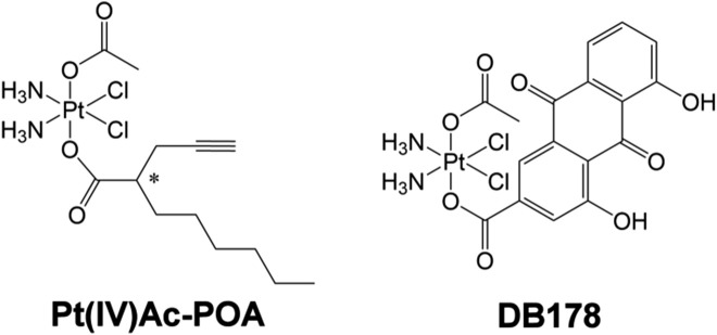
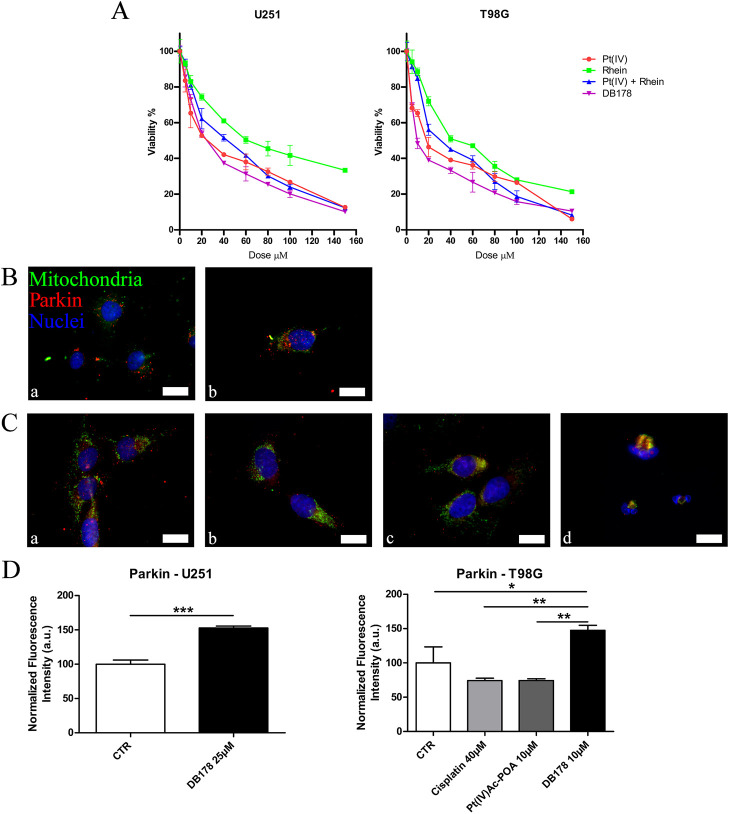
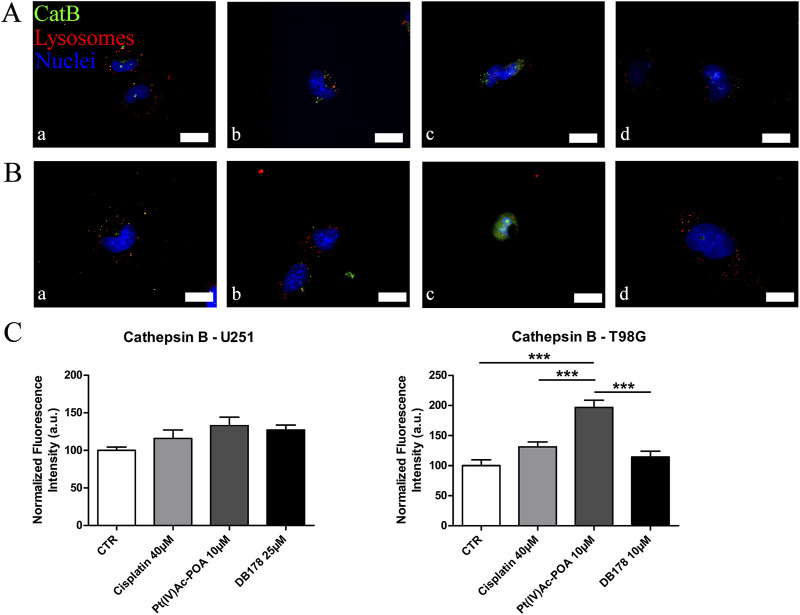
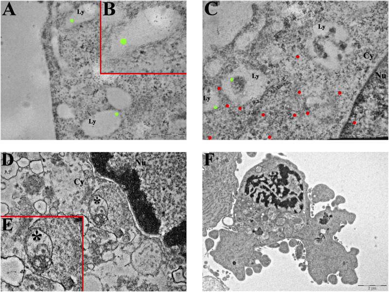
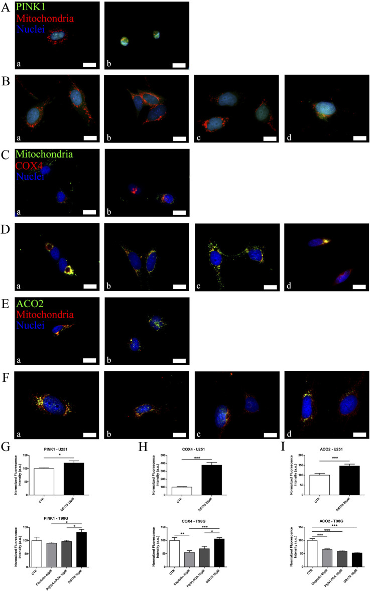
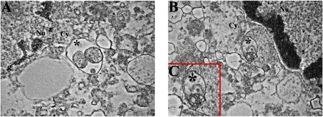
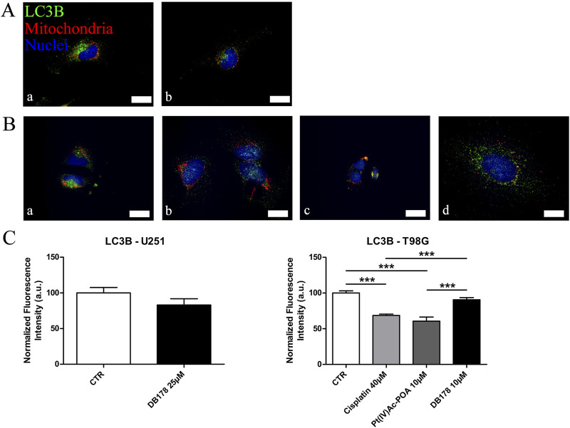
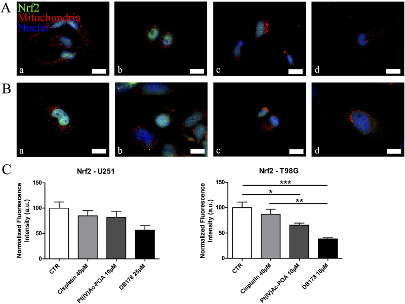
Glioblastoma (GBM) is the most frequent and aggressive brain tumor in adults. Due to its heterogeneity, the abundance of altered signaling pathways within the same tumoral mass, its low immunogenicity, and the presence of the blood-brain barrier, standard therapies based on surgical resection, radiotherapy, and chemotherapy result in ineffective tumor removal. For these reasons, the development of new drugs is mandatory to ameliorate patients' life expectancy and quality of life. Cathepsins are lysosomal proteases involved in several physiological and pathological processes, and they play key roles in modulating cell death and pharmacological resistance. In particular, cathepsin B is a crucial regulatory protein in different types of cell death, and its overexpression contributes to GBM angiogenesis and tumor progression. Octahedral platinum(IV) (Pt(IV))-based prodrugs have already demonstrated improved anticancer efficacy compared to routinely used cisplatin. This work aims to investigate the effects of two such prodrugs-Pt(IV)Ac-POA ((OC-6-44)-acetatodiamminedichlorido(2-(2-propynyl)octanoato)platinum(IV)) and DB178 ((OC-6-44)-acetatodiamminedichlorido(4,5-dihydroxy-9,10-dioxo-9,10-dihydroanthracene-2-carboxylato)platinum(IV))-on two different glioblastoma cell lines, U251 and T98G, with particular attention to their effects on cathepsin B. The immunocytochemical and biochemical results obtained on the two cell lines highlight the maintenance of basal levels of cathepsin B while efficiently activating programmed cell death mechanisms, as investigated by optical and electronic microscopy. These findings may serve as a valid starting point for further approaches that incorporate cathepsins' inhibitors to improve therapeutic efficacy and possibly reveal novel pharmacological targets.

Keywords: apoptosis; cathepsin B; drug resistance; glioblastoma; mitophagy; platinum(IV).


Conflict of interest statement

The authors declare that the research was conducted in the absence of any commercial or financial relationships that could be construed as a potential conflict of interest. The reviewer AP declared a shared affiliation with the author FDL to the handling editor at the time of review.

Figures

FIGURE 1
FIGURE 1
Pt(IV)Ac-POA and DB178. Structural formulas of the octahedral Pt(IV) complexes (OC-6-44)-acetatodiamminedichlorido(2-(2-propynyl)octanoato)platinum(IV) [Pt(IV)Ac-POA] and (OC-6-44)-acetatodiamminedichlorido(4,5-dihydroxy-9,10-dioxo-9,10-dihydroanthracene-2-carboxylato)platinum(IV) [DB178].
FIGURE 2
FIGURE 2
(A) MTT assay. Viability curves of U251 and T98G cell lines assessed by the MTT assay after a standard acute exposure of 48 h continuous treatment to increasing concentrations of Pt(IV), rhein, Pt(IV) and rhein, and DB178. Mean values ±SEM are reported; p < 0.0001. (B, C) Immunofluorescent labeling of mitochondria (green) and Parkin (red) in (B) U251 (a. CTR and b. DB178 25 µM) and (C) T98G cells (a. CTR, b. cisplatin 40 µM, c. Pt(IV)Ac-POA 10 µM, and d. DB178 10 µM). Nuclei were counterstained with Hoechst 33258 (blue). Scale bars, 25 µm. (D) Histograms report statistically significant increments in the normalized fluorescence intensity values following DB178 exposure both in U251 and T98G cell lines. *, p < 0.05; **, p < 0.01; ***, p < 0.001.
FIGURE 3
FIGURE 3
(A,B) Immunofluorescent labeling of cathepsin B (green) and lysosomes (red) in (A) U251 (a. CTR, b. cisplatin 40 µM, c. Pt(IV)Ac-POA 10 µM, and d. DB178 25 µM) and (B) T98G cells (a. CTR, b. cisplatin 40 µM, c. Pt(IV)Ac-POA 10 µM, and d. DB178 10 µM). Nuclei were counterstained with Hoechst 33258 (blue). Scale bars, 25 µm. (C) Histograms report maintenance of basal level in U251 cells and a statistically significant increase in the normalized fluorescence intensity values in T98G cells after exposure to Pt(IV)Ac-POA. ***, p < 0.001.
FIGURE 4
FIGURE 4
Ultrastructural analysis of T98G cells. (A–C) Transmission electron microscopy representative images of CatB immunolabeling in (A,B) control and (C) Pt(IV)Ac-POA-exposed T98G cells. CatB immunolabeling is confined to lysosomes in control cells ((B) inset of (A)), while the signal is mainly retrieved in the cytoplasm following treatment. Green dots, lysosomal CatB; red dots, extra-lysosomal CatB. Ly, lysosomes; Cy, cytoplasm; Nu, nucleus. Scale bars, 500 nm. (D,E) Representative morphological micrographs of T98G cells representing (D) the control cell and (E) apoptotic cell, characterized by the presence of a fragmented nucleus and apoptotic bodies, after treatment with Pt(IV)Ac-POA. Scale bars, 2 µm.
FIGURE 5
FIGURE 5
(A,B) Immunofluorescent labeling of PINK1 (green) and mitochondria (red) in (A) U251 (a. CTR and b. DB178 25 µM) and (B) T98G cells (a. CTR, b. cisplatin 40 µM, c. Pt(IV)Ac-POA 10 µM, and d. DB178 10 µM). (C, D) Immunofluorescent labeling of mitochondria (green) and COX4 (red) in (C) U251 (a. CTR and b. DB178 25 µM) and (D) T98G cells (a. CTR, b. cisplatin 40 µM, c. Pt(IV)Ac-POA10 µM, and d. DB178 10 µM). (E, F) Immunofluorescent labeling of ACO2 (green) and mitochondria (red) in (E) U251 (a. CTR and b. DB178 25 µM) and (F) T98G cells (a. CTR, b. cisplatin 40 µM, c. Pt(IV)Ac-POA 10 µM, and d. DB178 10 µM). Nuclei were counterstained with Hoechst 33258 (blue). Scale bars, 25 µm. (G) Histograms report trends of increasing normalized fluorescence intensity values of PINK1 following exposure to DB178 in both U251 and T98G cell lines. (H) Histograms report trends of increasing normalized fluorescence intensity values of COX4 following exposure to DB178 in both U251 and T98G cell lines. (I) Histograms report statistically significant alterations in normalized fluorescence intensity values of ACO2 in both U251 and T98G cell lines. *, p < 0.05; **, p < 0.01; ***, p < 0.001.
FIGURE 6
FIGURE 6
Mitophagy at the ultrastructural level. Representative TEM micrographs showing mitophagic vesicles (asterisk) in (A) U251 and (B, C) T98G cells following treatment with DB178. ((C) inset of (B)) Note the remains of the mitochondrial membranous system in the asterisk-labeled vesicle. Cy, cytoplasm; Nu, nucleus. Scale bars, 500 nm.
FIGURE 7
FIGURE 7
(A,B) Immunofluorescent labeling of LC3B (green) and mitochondria (red) in (A) U251 (a. CTR and b. DB178 25 µM) and (B) T98G cells (a. CTR, b. cisplatin 40 µM, c. Pt(IV)-POA 10 µM, and d. DB178 10 µM). Nuclei were counterstained with Hoechst 33258 (blue). Scale bars, 25 µm. (C) Histograms report maintaining low normalized fluorescence intensity values in U251 cells, while statistically significant reductions are evident in T98G cells exposed to Pt(IV)Ac-POA. ***, p < 0.001.
FIGURE 8
FIGURE 8
(A,B) Immunofluorescent labeling of Nrf2 (green) and mitochondria (red) in (A) U251 (a. CTR, b. cisplatin 40 µM, c. Pt(IV)Ac-POA 10 µM, and d. DB178 25 µM) and (B) T98G cells (a. CTR, b. cisplatin 40 µM, c. Pt(IV)Ac-POA 10 µM, and d. DB178 10 µM). Nuclei were counterstained with Hoechst 33258 (blue). Scale bars, 25 µm. (C) Histograms report trends of reduction in the normalized fluorescence intensity values following Pt(IV) prodrug exposure in both U251 and T98G cells. *: p < 0.05; **: p < 0.01; ***: p < 0.001.

Similar articles

References

    1. Alpini C., Lotzniker M., Valaperta S., Bottone M. G., Malatesta M., Montanelli A., et al. (2012). Characterization for anti-cytoplasmic antibodies specificity by morphological and molecular techniques. Auto. Immun. Highlights 3, 79–85. 10.1007/s13317-012-0033-4 - DOI - PMC - PubMed
    1. Alrouji M., Yasmin S., Alshammari M. S., Alhumaydhi F. A., Sharaf S. E., Shahwan M., et al. (2024). Unveiling Cathepsin B inhibition with repurposed drugs for anticancer and anti-Alzheimer’s drug discovery. PLoS One 19, e0316010. 10.1371/journal.pone.0316010 - DOI - PMC - PubMed
    1. Anes E., Pires D., Mandal M., Azevedo-Pereira J. M. (2022). Spatial localization of cathepsins: implications in immune activation and resolution during infections. Front. Immunol. 13, 955407. 10.3389/fimmu.2022.955407 - DOI - PMC - PubMed
    1. Astesana V., Faris P., Ferrari B., Siciliani S., Lim D., Biggiogera M., et al. (2021). [Pt(O,O’-acac)(γ-acac)(DMS)]: alternative strategies to overcome cisplatin-induced side effects and resistance in T98G glioma cells. Cell Mol. Neurobiol. 41, 563–587. 10.1007/s10571-020-00873-8 - DOI - PMC - PubMed
    1. Bairoch A. (2018). The Cellosaurus, a cell-line knowledge resource. J. Biomol. Tech. 29, 25–38. 10.7171/jbt.18-2902-002 - DOI - PMC - PubMed

LinkOut - more resources