. 2025 Jun 13:17:1127-1141.
doi: 10.2147/CMAR.S525738. eCollection 2025.

Integrating Machine Learning Algorithms to Construct a Triaptosis-Related Prognostic Model in Melanoma

Affiliations

Integrating Machine Learning Algorithms to Construct a Triaptosis-Related Prognostic Model in Melanoma

Jiaheng Xie et al. Cancer Manag Res. .

Abstract

Introduction: Melanoma is a highly aggressive skin cancer that accounts for a disproportionate number of skin cancer-related deaths due to early metastasis and therapy resistance. Programmed cell death (PCD), including ferroptosis and apoptosis, plays a crucial role in tumor progression and therapy response. Among these, triaptosis is a newly described form of PCD. It represents a novel mechanism of cell death with potential implications for cancer treatment. However, its role in melanoma remains largely unexplored.

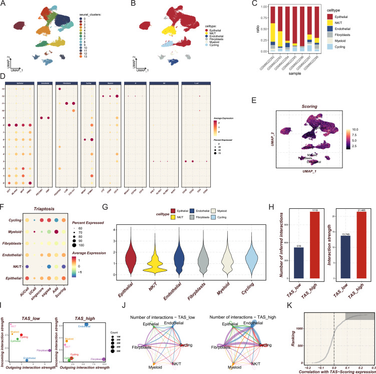
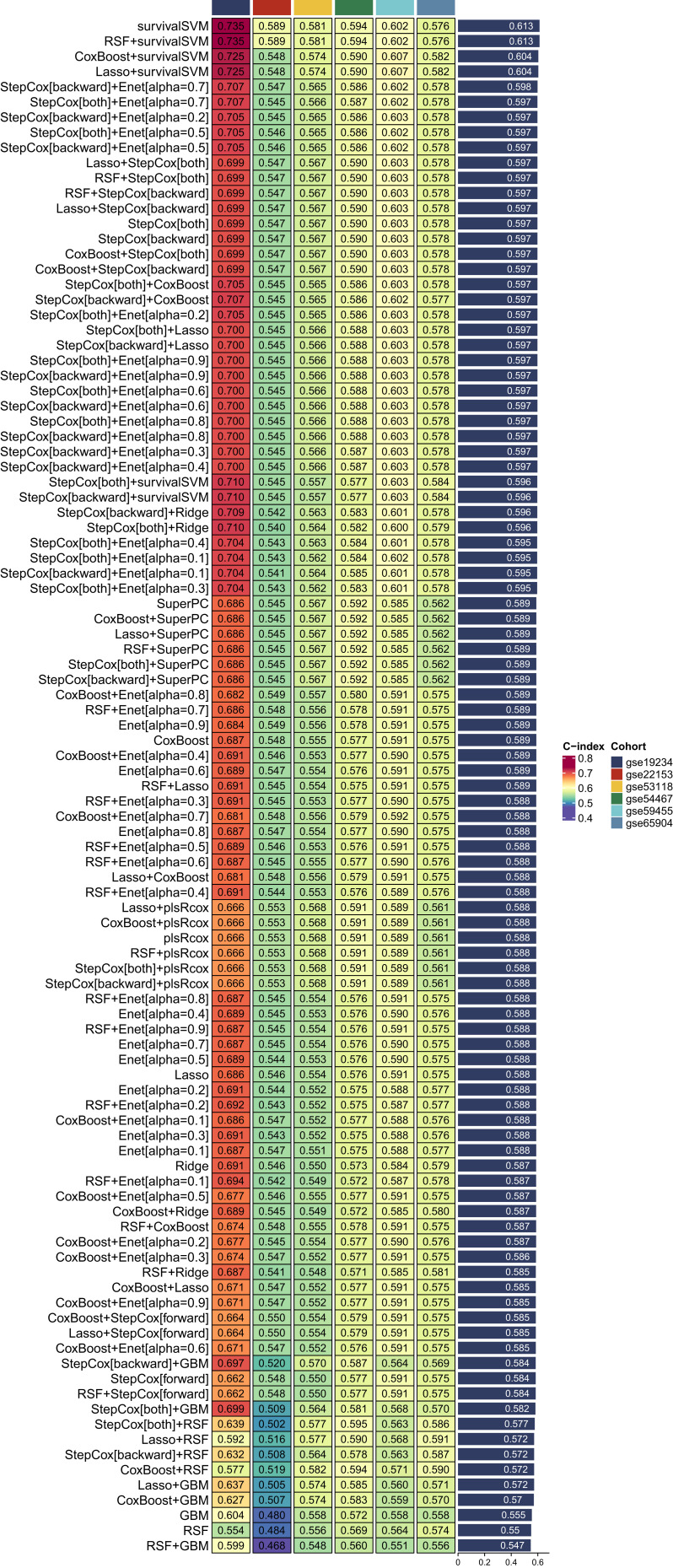
Methods: We explored the role of triaptosis in melanoma by integrating single-cell and bulk RNA sequencing data. Key triaptosis-related genes and pathways were identified and incorporated into machine learning models to construct a prognostic signature. The TCGA-SKCM cohort served as the training dataset, and GEO datasets were used for validation.

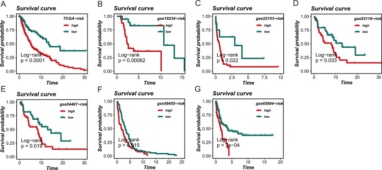
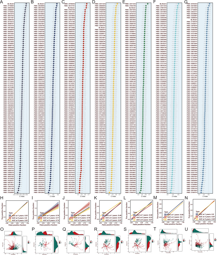
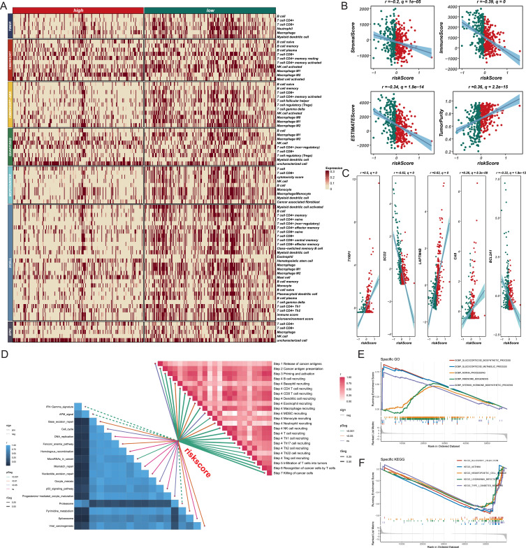
Results: A robust prognostic model based on triaptosis-associated signature (TAS) was established using the SurvivalSVM algorithm. This model showed superior predictive performance, with consistently high concordance index (C-index) values across independent validation datasets. Kaplan-Meier survival analysis indicated that high-risk patients had significantly worse overall survival than low-risk patients. The model's predictive accuracy was confirmed through receiver operating characteristic (ROC) curve analysis and principal component analysis (PCA). Moreover, immune infiltration and tumor microenvironment (TME) analyses revealed significant associations between TAS and immune cell populations.

Conclusion: Triaptosis-related gene expression patterns are closely linked with melanoma prognosis and immune infiltration. Our findings provide novel insights into triaptosis as a potential biomarker and therapeutic target, offering strategies to overcome treatment resistance in melanoma.

Keywords: cancer; cell death; melanoma; target; tumor microenvironment.


Conflict of interest statement

The authors declare no competing interests.

Figures

Figure 1
Figure 1
Cellular Heterogeneity and Triaptosis-Related Activity. (A) UMAP analysis identifies 14 distinct cell clusters across the dataset. (B) These clusters are annotated into six major cell types: epithelial, NKT, endothelial, fibroblasts, myeloid, and cycling cells. (C) The distribution of epithelial cells across different samples, showing their predominance. (D) Marker gene analysis confirming the identity of each cell type, with representative genes displayed. (E, F) Tas scores across cell populations, highlighting significant heterogeneity in triaptosis-related activity. (G) Epithelial cells exhibit the highest TAS scores, indicating their potential involvement in triaptosis within the tumor microenvironment. (HJ) Cell-cell communication analysis showing stronger intercellular interactions in the TAS-high group, with epithelial cells acting as central hubs in the interaction network. (K) Correlation analysis identifying the top 150 genes positively associated with TAS scores, providing insights into the molecular basis of triaptosis activity.
Figure 2
Figure 2
Performance of Prognostic Models Using SurvivalSVM. Performance of the SurvivalSVM model in the TCGA-SKCM training cohort and six independent validation cohorts (GSE19234, GSE22153, GSE53118, GSE54467, GSE59455, GSE65904). The model achieved the highest C-index in the training cohort and demonstrated robust predictive performance across the validation cohorts. Heatmaps summarize the C-index values, highlighting SurvivalSVM’s superior performance compared to other machine learning algorithms.
Figure 3
Figure 3
Survival Analysis of Risk Groups. Kaplan-Meier survival curves showing the prognostic stratification of the SurvivalSVM model in the TCGA training cohort (A) and validation cohorts (BG). Patients in the high-risk group exhibited significantly worse overall survival compared to the low-risk group in all cohorts, confirming the model’s effectiveness in stratifying risk groups and predicting survival outcomes.
Figure 4
Figure 4
Comparison of Prognostic Models with Published Bioinformatics Models. (AG) Comparison of the SurvivalSVM model with published bioinformatics models across the TCGA-SKCM training cohort and six validation cohorts. SurvivalSVM achieved the highest C-index in the TCGA-SKCM cohort and several validation cohorts, demonstrating its superior performance and robustness. (HN) Time-dependent ROC curves assessing the model’s predictive ability at 1-, 3-, and 5-year intervals across the validation cohorts. AUC values consistently support the model’s reliable predictive performance. (OU) PCA analysis showing distinct clustering of high- and low-risk groups, further validating the discriminative power of the SurvivalSVM model in both training and validation cohorts.
Figure 5
Figure 5
Immune and Tumor Microenvironment Analysis. (A) Heatmap of immune cell proportions between high- and low-risk groups, revealing reduced immune infiltration in high-risk samples. (B) Correlation analysis showing negative correlations between risk scores and immune/stromal/ESTIMATE scores, indicating immune suppression in high-risk samples. (C) Gene expression analysis of key model-associated genes (TYRP1, LAPTM4B, CA8, SCO2, BCL2A1), illustrating their correlation with risk scores. (D) GSVA pathway enrichment analysis showing associations between high-risk samples and immune suppression/tumor progression pathways. (E and F) GSEA results confirming the enrichment of immune-related pathways in the low-risk group, highlighting functional differences between risk groups.
Figure 6
Figure 6
Continued.
Figure 6
Figure 6
Functional Validation of LAPTM4B as a Key Model-Associated Gene. (AC) Expression analysis of LAPTM4B in tumor versus normal tissues, showing significantly higher expression in tumors. Stratification by BRAF mutation status reveals differential expression. (D) Cox regression analysis indicating that higher LAPTM4B expression correlates with poorer survival outcomes, including OS, DSS, RFS, and PFS. (EG) Functional enrichment analysis of LAPTM4B-associated genes, highlighting pathways related to cell migration, invasion, and immune suppression. (H and I) Experimental validation of LAPTM4B’s role in tumor aggressiveness through functional assays. Knockdown of LAPTM4B (si-LAPTM4B) in SK-MEL-48 cells significantly reduced cell migration and invasion (H) and impaired wound healing (I), confirming LAPTM4B’s role in promoting cellular migration and tumor progression.

Similar articles

References

    1. Arnold M, Singh D, Laversanne M, et al. Global burden of cutaneous melanoma in 2020 and projections to 2040. JAMA Dermatol. 2022;158(5):495–503. doi: 10.1001/jamadermatol.2022.0160 - DOI - PMC - PubMed
    1. Leonardi GC, Falzone L, Salemi R, et al. Cutaneous melanoma: from pathogenesis to therapy (Review). Int J Oncol. 2018;52(4):1071–1080. doi: 10.3892/ijo.2018.4287 - DOI - PMC - PubMed
    1. Belote RL, Le D, Maynard A, et al. Human melanocyte development and melanoma dedifferentiation at single-cell resolution. Nat Cell Biol. 2021;23(9):1035–1047. doi: 10.1038/s41556-021-00740-8 - DOI - PubMed
    1. Dhanyamraju PK, Schell TD, Amin S, Robertson GP. Drug-tolerant persister cells in cancer therapy resistance. Cancer Res. 2022;82(14):2503–2514. doi: 10.1158/0008-5472.CAN-21-3844 - DOI - PMC - PubMed
    1. Swetter SM, Tsao H, Bichakjian CK, et al. Guidelines of care for the management of primary cutaneous melanoma. J Am Acad Dermatol. 2019;80(1):208–250. doi: 10.1016/j.jaad.2018.08.055 - DOI - PubMed

LinkOut - more resources