. 2025 Jun 7:52:213-227.
doi: 10.1016/j.bioactmat.2025.06.002. eCollection 2025 Oct.

Bioengineered platelet nanoplatform enables renal-targeted dexamethasone delivery for chronic nephritis therapy with dual anti-inflammatory/anti-fibrotic effects and minimized systemic toxicity

Affiliations

Bioengineered platelet nanoplatform enables renal-targeted dexamethasone delivery for chronic nephritis therapy with dual anti-inflammatory/anti-fibrotic effects and minimized systemic toxicity

Lingling Zhang et al. Bioact Mater. .

Abstract

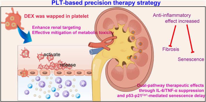
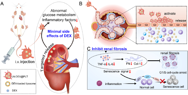
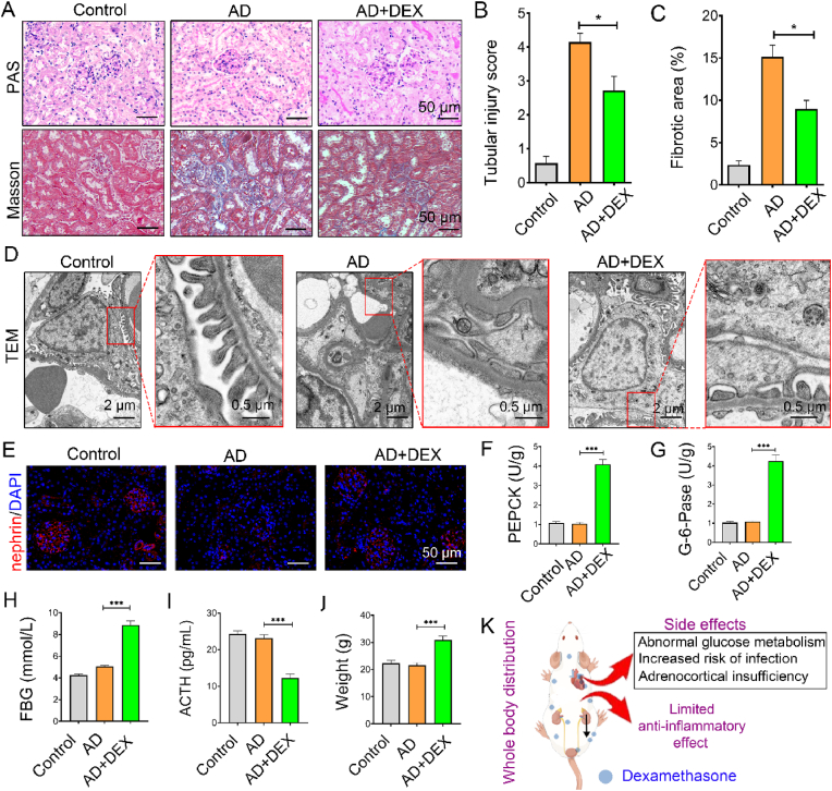
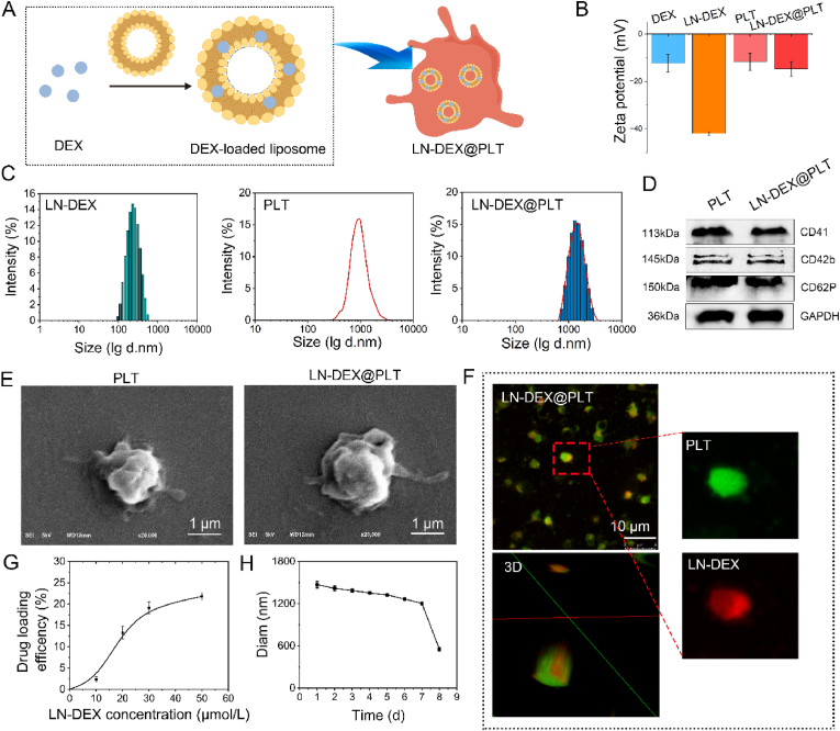
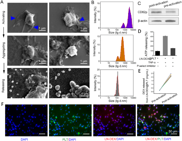
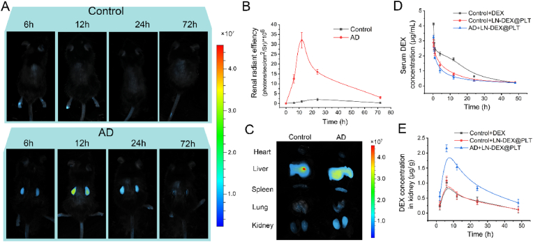
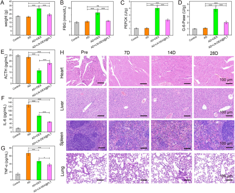
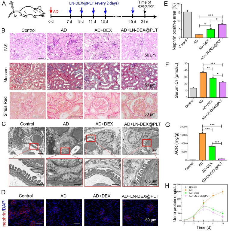
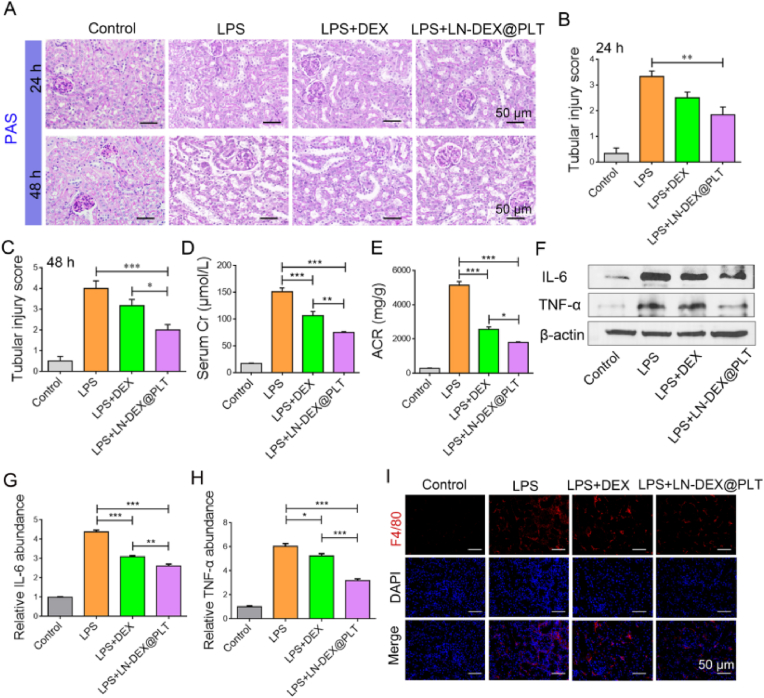
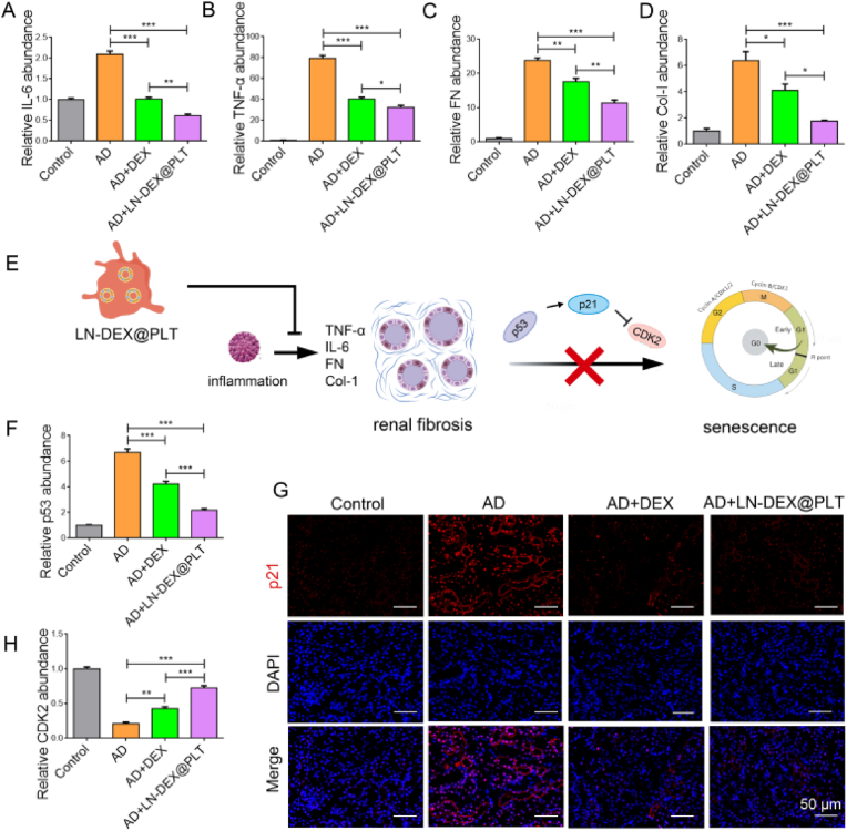
Chronic nephritis management remains challenging due to the compromised therapeutic efficacy and severe systemic complications of conventional glucocorticoid therapy. Here, we developed a bioinspired platelet-mediated delivery system (LN-DEX@PLT) that leverages platelet tropism toward injured glomeruli for precision drug delivery. This system integrates lipid nanoemulsion encapsulation with platelet-mediated hitchhiking delivery to achieve three key functionalities: (1) enhanced renal targeting demonstrated by 2.2-fold higher glomerular accumulation compared to free dexamethasone via In vivo imaging, (2) effective mitigation of glucocorticoid-induced metabolic toxicity evidenced by reduced fasting plasma glucose (5.2 ± 0.3 vs 8.3 ± 0.7 mmol/L in free DEX), suppression of hepatic gluconeogenic enzymes (PEPCK expression decreased by 43 %, G-6 Pase by 51 %, both p < 0.001), and suppressed body weight (-23.1 % versus free DEX group), and (3) dual-pathway therapeutic effects through IL-6/TNF-α suppression and p53-p21Cip1-mediated senescence delay. In Adriamycin-based chronic nephritis models, LN-DEX@PLT demonstrated superior renal protection with 81 % reduction in proteinuria (vs 33 % for free DEX). In LPS-induced and Adriamycin-based chronic nephritis models, LN-DEX@PLT demonstrated suppression of renal inflammation markers (IL-6 expression decreased to 68 %, TNF-α to 51 %) and macrophage infiltration (F4/80+ cells decreased 5.3-fold). This platelet-biohybrid system provides a clinically translatable paradigm for precision glucocorticoid therapy with reduced dosing frequency.

Keywords: Chronic nephritis; Dexamethasone; Minimized senescence effects; Platelets; Precision therapy.


Conflict of interest statement

The authors declare that they have no known competing financial interests or personal relationships that could have appeared to influence the work reported in this paper.

Figures

Image 1
Graphical abstract
Scheme 1
Scheme 1
Schematic of the platelet (PLT)-based precision therapy strategy (LN-DEX@PLT) as a targeted treatment of chronic nephritis with minimal side effects. (A) Schematic of LN-DEX@PLT in improving the efficacy and reducing the side effects of DEX. DEX was loaded in the PLT-based delivery system and exhibited long circulation time in vivo, reduced abnormal glucose metabolism, and inhibited inflammatory factors. LN-DEX@PLT was activated in the injured renal corpuscle and released LN-DEX with the help of PLTs. (B) Schematic illustration of PLT carrier activation at the site of inflamed renal corpuscles and DEX release. (C) Schematic illustrating the molecular mechanisms associated with LN-DEX@PLT, including inhibiting renal fibrosis (IL-6 and TNF-α) and the fibrosis index (fibronectin (FN) and collagen (Col)-I). The inhibition of senescence involved the p53-p21Cip1 pathway.
Fig. 1
Fig. 1
Effective treatment results and side effects of DEX in AD-treated chronic nephritis model mice. (A) Representative images of PAS staining, and Masson trichrome staining in control (normal mice), AD model mice, and DEX treated AD model mice. (B) The renal tubular injury score of PAS staining after different treatment. (C) Quantitative analysis of Masson staining in each group. (D) TEM results in assessing ultrastructural changes in podocyte morphology. (E) Immunofluorescence staining of the podocyte marker nephrin (red), and staining of the nuclei with DAPI (blue). (F–J) Comparison of PEPCK, G-6-Pase, FBG (fasting blood glucose), ACTH, and body weight in different groups. (K) Schematic diagram of the reason of DEX in clinical application. ∗P < 0.05, and ∗∗∗P < 0.001.
Fig. 2
Fig. 2
The synthesis and characterization of LN-DEX@PLT. (A) The preparation diagram of LN-DEX@PLT. (B) The zeta potential changes during preparation of LN-DEX@PLT. (C) The size distribution of DEX, PLT and LN-DEX@PLT. (D) The marker protein (CD41, CD42b, and CD62P) in LN-DEX@PLT membrane. (E) SEM images of raw PLT and LN-DEX@PLT. (F) Confocal laser scanning microscopy results of LN-DEX@PLT. Green, FITC-labeled PLTs. Red, Rhodamine B-labeled LN-DEX. (G) Drug loading efficiency of LN-DEX@PLT. (H) The stability results (particle size changes) of LN-DEX@PLT at room temperature.
Fig. 3
Fig. 3
Target behavior of LN-DEX@PLT. (A) Morphological changes of the activated LN-DEX@PLT. Activation: The characteristics of activation, specifically the tentacle-like structures on the surfaces, were indicated by the blue arrow. Aggregating: The tentacle-like structures interconnected with one another to form a larger aggregate. Releasing: LN-DEX@PLT disintegrated into smaller nanoparticles. (B) The size distribution of LN-DEX@PLT during the activation-aggregation phase and release-degradation phase, as well as the size distribution of particles released after final degradation. (C) The expression of CD62P in LN-DEX@PLT before and after activation. (D) Assay of ATP releasing for inactive and TNF-α activated LN-DEX@PLT. (E) DEX released behavior before and after LN-DEX@PLT activation. (F) The fluorescence microscopy images of the tubular epithelial cells after treated LN-DEX@PLT in the Transwell system.
Fig. 4
Fig. 4
Targeting delivery results of LN-DEX@PLT. (A) In vivo imaging of mice in the control group (normal mice) and AD-induced nephropathy mice (AD group). (B) Quantitative analysis of fluorescence intensity in the kidneys at 6, 12, 24 and 72 h after drug administration. (C) Ex vitro fluorescence images of vital organs 12 h post administration. (D) The plasma concentrations curves of DEX in the normal mice treated with free DEX (Control + DEX), the normal mice treated with LN-DEX@PLT (Control + LN-DEX@PLT), and AD-induced nephropathy mice treated with LN-DEX@PLT (AD + LN-DEX@PLT). (E) DEX concentration changes in AD model mice or normal mice renal after intravenous injection.
Fig. 5
Fig. 5
Limited DEX-associated adverse effects of LN-DEX@PLT. Comparison of (A) body weight, (B) FBG (Fasting Blood Glucose), (C) PEPCK, (D) G-6-Pase, (E) ACTH in different groups of mice, post 28 days treatments. Serum inflammatory indexes, (F) IL-6 and (G) TNF-α, in different groups of mice. (H) The results of HE staining of heart, liver, spleen and lung tissues of normal mice treated with LN-DEX@PLT for 28 days. ∗P < 0.05, ∗∗P < 0.01, and ∗∗∗P < 0.001.
Fig. 6
Fig. 6
Anti-inflammatory efficacy of LN-DEX@PLT in AD-treated chronic nephritis model mice. (A) Schematic diagram of drugs administration timeline in AD model mice. (B) Representative images of PAS staining, Masson trichrome staining, and Sirius red. (C) TEM results to assess ultrastructural changes in podocyte morphology. (D) Immunofluorescence staining of nephrin (red), while the nuclei were stained with DAPI (blue). (E) Quantification analyzes results of nephrin positive area%. (F–H) Changes of Cr, ACR, and urinary protein in normal mice, AD model mice, AD model mice treated with DEX or LN-DEX@PLT. ∗P < 0.05, ∗∗P < 0.01, and ∗∗∗P < 0.001.
Fig. 7
Fig. 7
The anti-inflammatory efficacy of LN-DEX@PLT in vivo. (A) Representative images of PAS staining of kidney samples. scale bars = 50 μm. (B–C) Quantification of the tubular injury score in PAS staining images above at 24 h and 48 h. (D–E) The concentration of serum creatinine and ACR after treatment changes in LPS-induced inflammation model mice. (F–H) The results of western blotting and quantitative analysis of TNF-α and IL-6 in LPS-induced inflammation model mice treated with LN-DEX@PLT. (I) Immunofluorescence staining of F4/80. ∗P < 0.05, ∗∗P < 0.01, and ∗∗∗P < 0.001.
Fig. 8
Fig. 8
LN-DEX@PLT ameliorates renal inflammation and reduce podocytes senescence in AD mice. (A–D) Expression of IL-6, TNF-α, FN, Col-I, in the kidney tissue 21 days post-treatment. (E) Schematic diagram of LN-DEX@PLT ameliorates renal inflammation and reduce podocytes senescence. (F) p53 value in the kidney tissue. (G) Immunofluorescence staining of p21. (H) CDK2 value in the kidney tissue. ∗P < 0.05, ∗∗P < 0.01, and ∗∗∗P < 0.001.

Similar articles

References

    1. Pofi R., Caratti G., Ray D.W., Tomlinson J.W. Treating the side effects of exogenous glucocorticoids; can we separate the good from the bad? Endocr. Rev. 2023;44:975–1011. doi: 10.1210/endrev/bnad016. - DOI - PMC - PubMed
    1. Liu H., Ji M., Xiao P., Gou J., Yin T., He H., Tang X., Zhang Y. Glucocorticoids-based prodrug design: current strategies and research progress. Asian J. Pharm. Sci. 2024;19 doi: 10.1016/j.ajps.2024.100922. - DOI - PMC - PubMed
    1. Mühlig A.K., Steingröver J., Heidelbach H.S., Wingerath M., Sachs W., Hermans-Borgmeyer I., Meyer-Schwesinger C., Choi H.Y., Lim B.J., Patry C., Hoffmann G.F., Endlich N., Bracke K., Weiß M., Guse A.H., Lassé M., Rinschen M.M., Braun F., Huber T.B., Puelles V.G., Schmitt C.P., Oh J. The calcium-sensing receptor stabilizes podocyte function in proteinuric humans and mice. Kidney Int. 2022;101:1186–1199. doi: 10.1016/j.kint.2022.01.036. - DOI - PubMed
    1. Liu H J.M., Xiao P., Gou J., Yin T., He H., Tang X., Zhang Y. Glucocorticoids-based prodrug design: current strategies and research progress. Asian J. Pharm. Sci. 2024;19 doi: 10.1016/j.ajps.2024.100922. - DOI - PMC - PubMed
    1. Cho Jh S.S. Glucocorticoid-induced hyperglycemia: a neglected problem. Endocrinol. Metab. 2024;39:222–238. doi: 10.3803/EnM.2024.1951. - DOI - PMC - PubMed

LinkOut - more resources