. 2025 May 27;16(8):2553-2566.
doi: 10.7150/jca.105863. eCollection 2025.

Ursolic Acid induces ferroptosis by affecting redox balance and FADS2-mediated unsaturated fatty acid synthesis in Non-Small Cell Lung Cancer

Affiliations

Ursolic Acid induces ferroptosis by affecting redox balance and FADS2-mediated unsaturated fatty acid synthesis in Non-Small Cell Lung Cancer

Lanlan Yang et al. J Cancer. .

Abstract

Ursolic Acid (UA) is a naturally occurring pentacyclic triterpenoid compound that is prevalent in various medicinal plants and fruits. It has garnered significant attention due to its broad spectrum of anticancer properties. In this study, we evaluated the antitumor effects of UA on Non-Small Cell Lung Cancer (NSCLC).UA significantly inhibited NSCLC viability and induced cell death in a time- and dose-dependent manner. Furthermore, the administration of UA resulted in an elevation of intracellular reactive oxygen species (ROS), lipid ROS, and ferrous iron levels, while concurrently suppressing the expression of SLC7A11, glutathione, and GPX4. Consequently, this led to an augmentation in the concentration of the lipid peroxidation substrate, malondialdehyde. All the changes were effectively attenuated by the ferroptosis inhibitor Ferrostatin-1(Fer-1) and Deferoxamine (DFO). Moreover, similar observations were made in animal experiments. The sequencing data indicate that UA influences ferroptosis by modulating Fatty Acid Desaturase-2 (FADS2). The reintroduction of FADS2 through ectopic expression restored the resistance to ferroptosis induced by UA in A549 cells, while the addition of exogenous oleic acid (OA) counteracted the impact of UA on the oxidative response. These results suggest that UA induces ferroptosis in NSCLC by affecting redox pathways and the FADS2-mediated synthesis of unsaturated fatty acids.These studies collectively underscore the promising role of UA in the development of effective anticancer therapies.

Keywords: FADS2; Ferroptosis; Redox Regulation; Unsaturated Fatty Acids; Ursolic Acid.


Conflict of interest statement

Competing Interests: The authors have declared that no competing interest exists.

Figures

Figure 1
Figure 1
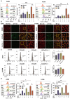
UA suppresses NSCLC growth by inducing ferroptosis. (A)Chemical structure of ursolic acid (UA). (B)The IC50 of A549 and H1299 was determined by the CCK-8 method after 24h of UA treatment. (C)A549/H1299 cells were observed at different concentrations of UA and combined with Fer-1 for 24h, SYTOX Green (100 nM) assay was performed by fluorescence microscopy. Scale bar:100μm. (D) A549/H1299 were treated with UA alone and in combination with Fer-1(1μM), DFO (60μM), Z-V(20μM), Nec(20μM), Baf-A1(50nM) for 24h, then the cell viability was detected by CCK8 assays.
Figure 2
Figure 2
UA increases ROS\lipid ROS\ferrous iron production in NSCLC. (A)Flow cytometry was performed to detect the changes of A549\H1299 ROS in UA, UA+Fer-1,and DMSO treatment at different times (0,6,12,24h). (B)Confocal images of lipid peroxidation by UA, UA+Fer-1, and DMSO action for 24h. The red color indicates non-oxidized state while the green color indicates oxidized state. Scale bar,100 μm. (C)Changes in lipid peroxidation were detected by flow cytometry. (D) FerroOrange probe to detect changes in ferrous iron (Bar graphs represent the proportion of changes)
Figure 3
Figure 3
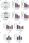
UA disrupted Redox Homeostasis. (A) The expression of SLC7A11 and GPX4 in A549\H1299 was analyzed by western blot analysis (GAPDH as an internal control). (B) Relative changes of GSH in A549\H1299 were treated with different concentrations of UA and combined with Fer-1 for 24h. (C) Relative changes of MDA in A549\H1299 exposed to UA and UA combined with Fer-1 for 48h.
Figure 4
Figure 4
mRNA sequence analyses. (A)Volcano plot showed differentially expressed genes between the UA and control (DMSO) groups. (B)Venn diagram illustrated the differentially expressed genes of UA VS Control, Erastin VS Control, and FerrDb database. (C)Hierarchical clustering heatmap of 8 common genes. (D)GO analysis. (E)KEGG analysis. (F)GSEA results of C5 reference gene (GO gene sets) set of UA VS control differential genes.
Figure 5
Figure 5
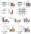
Overexpression of FADS2 renders A549 cells resistant to ferroptosis after UA treatment. (A)Western blot analysis of FADS2 and (B)SCD protein expression in UA-treated A549 cells. (C)FADS2 overexpression efficiency via Western blot analysis. (D) Changes in FADS2 and GPX4 protein levels after UA treatment of FADS2-OE A549 cell as compared to Vector. (E)FADS2-OE A549 and Vector A549 were treated with DMSO, UA (25μM), UA+Fer-1, Erastin(20μM) for 24h and cell viability was detected by CCK-8 assay. (F)Changes in GSH levels in FADS2-OE and Vector after UA and UA+Fer-1 for 24h. (G)Changes in MDA levels (histograms grouped as Fig 5 E). (H)DCFH-DA probe detected ROS changes after 24H of A549 after DMSO, OA (100μM), UA (25μM), and OA+UA combined were applied. (I)C11-Bodipy detected lipid ROS changes after the same treatments.
Figure 6
Figure 6
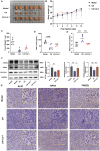
Effect of UA on subcutaneous xenograft tumors in vivo. (A)Tumor photo of each group. (B)Body weight. (C)Tumor weights (Data were plotted as mean ± SD, n = 6). (D)Serum levels of GSH and (E) MDA in each group of mice (n = 6). (F) GPX4, SLC7A11 and FADS2 protein expression levels in tumor tissues among different groups. The quantitative results of three replicate Western blot experiments. (G) Microscopic images of Ki-67 staining, and immunohistochemistry of GPX4 and FADS2 expression in different treated tumor tissues. (Scale bar, 50 μm).

Similar articles

References

    1. Herbst RS, Morgensztern D, Boshoff C. The biology and management of non-small cell lung cancer. Nature. 2018;553:446–54. - PubMed
    1. Dixon SJ, Lemberg KM, Lamprecht MR. et al. Ferroptosis: an iron-dependent form of nonapoptotic cell death. Cell. 2012;149:1060–72. - PMC - PubMed
    1. Dai C, Chen X, Li J, Comish P, Kang R, Tang D. Transcription factors in ferroptotic cell death. Cancer Gene Ther. 2020;27:645–56. - PubMed
    1. Liang D, Minikes AM, Jiang X. Ferroptosis at the intersection of lipid metabolism and cellular signaling. Mol Cell. 2022;82:2215–27. - PMC - PubMed
    1. Yu K, Chen Y, Zhang L. et al. Cancer-Erythrocyte Membrane-Mimicking Fe3O4 Nanoparticles and DHJS for Ferroptosis/Immunotherapy Synergism in Tumors. ACS Appl Mater Interfaces. 2023;15:44689–710. - PubMed

LinkOut - more resources