

Plasma-negative, Renal-limited Cryofibrinogen-associated Glomerulonephritis: A Unique Case Report
- PMID: 40535910
- PMCID: PMC12173047
- DOI: 10.1016/j.xkme.2025.101017
Plasma-negative, Renal-limited Cryofibrinogen-associated Glomerulonephritis: A Unique Case Report
Abstract
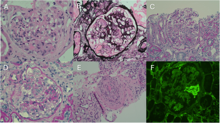
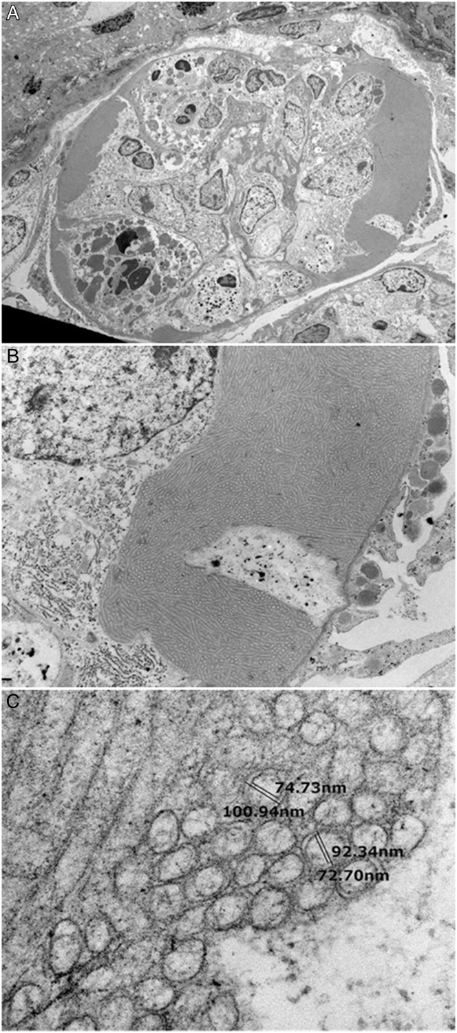
Cryofibrinogen-associated glomerulonephritis is characterized by membranoproliferative glomerulonephritis without immunoglobulin deposition and unique ultrastructural features. This case report presents a 63-year-old man with renal-limited cryofibrinogen-associated glomerulonephritis, with negative plasma cryofibrinogen levels. His medical history included metallic aortic valve replacement and long-term anticoagulation therapy. Clinical examination revealed no cutaneous manifestations or thrombotic events. Initial laboratory investigations showed severe kidney dysfunction, but negative results for plasma cryofibrinogen, serum cryoglobulin, and a comprehensive autoimmune, infective, and malignancy panel. Kidney biopsy revealed mesangiocapillary glomerulonephritis with focal vasculitis and significant interstitial fibrosis, and electron microscopy identified double-walled microtubules consistent with cryofibrinogen. Our patient was managed without immunosuppressive therapy due to significant kidney scarring and absence of extra-renal manifestations. To our knowledge, this case describes the first report of cryofibrinogen-associated glomerulonephritis in the absence of detectable cryofibrinogen in serum, with diagnosis relying on ultrastructural findings. Differential diagnoses such as immunotactoid glomerulonephritis were considered but ruled out based on morphological characteristics. This case adds to the limited literature on renal-limited cryofibrinogen and emphasizes the necessity for thorough investigation including electron microscopy assessment of kidney biopsies to ascertain the diagnosis.
Keywords: Biopsy-proven; cryofibrinogen; glomerulonephritis.
Crown Copyright © 2025 Published by Elsevier Inc. on behalf of the National Kidney Foundation, Inc.
Figures
Similar articles
-
Cryofibrinogen-associated glomerulonephritis with paraproteinemia.Front Immunol. 2025 Jul 15;16:1576917. doi: 10.3389/fimmu.2025.1576917. eCollection 2025. Front Immunol. 2025. PMID: 40735312 Free PMC article.
-
C3 glomerulopathy post kidney transplantation: A single center experience.World J Transplant. 2025 Jun 18;15(2):101517. doi: 10.5500/wjt.v15.i2.101517. World J Transplant. 2025. PMID: 40535493 Free PMC article.
-
Cryofibrinogen-Associated Glomerulonephritis.Am J Kidney Dis. 2017 Feb;69(2):302-308. doi: 10.1053/j.ajkd.2016.08.031. Epub 2016 Nov 17. Am J Kidney Dis. 2017. PMID: 27866967
-
Global Delphi consensus on treatment goals for generalized pustular psoriasis.Br J Dermatol. 2025 Mar 18;192(4):706-716. doi: 10.1093/bjd/ljae491. Br J Dermatol. 2025. PMID: 39844356
-
Detection methods and dynamic characteristics of specific antibodies in patients with COVID-19: A review of the early literature.Heliyon. 2024 Jan 24;10(3):e24580. doi: 10.1016/j.heliyon.2024.e24580. eCollection 2024 Feb 15. Heliyon. 2024. Retraction in: Heliyon. 2025 Mar 25;11(9):e43262. doi: 10.1016/j.heliyon.2025.e43262. PMID: 38317938 Free PMC article. Retracted. Review.
References
-
- Sethi S., Yachoui R., Murray D.L., Radhakrishnan J., Alexander M.P. Cryofibrinogen-associated glomerulonephritis. Am J Kidney Dis. 2017;69(2):302–308. - PubMed
-
- Beightler E., Diven D.G., Sanchez R.L., Solomon A.R. Thrombotic vasculopathy associated with cryofibrinogenemia. J Am Acad Dermatol. 1991;24(2, pt 2):342–345. - PubMed
-
- Jantunen E., Soppi E., Neittaanmäki H., Lahtinen R. Essential cryofibrinogenaemia, leukocytoclastic vasculitis and chronic purpura. J Intern Med. 1993;234(3):331–333. - PubMed
-
- Saadoun D., Elalamy I., Ghillani-Dalbin P., Sene D., Delluc A., Cacoub P. Cryofibrinogenemia: new insights into clinical and pathogenic features. Am J Med. 2009;122(12):1128–1135. - PubMed
-
- Terrier B., Izzedine H., Musset L., et al. Prevalence and clinical significance of cryofibrinogenaemia in patients with renal disorders. Nephrol Dial Transplant. 2011;26(11):3577–3581. - PubMed
Publication types
LinkOut - more resources
Full Text Sources
