

Ginsenoside Rh1 Alleviates Allergic Rhinitis by Mediating Mitochondrial Autophagy via Activation of the AMPK/ULK1/FUNDC1 Pathway
- PMID: 40535916
- PMCID: PMC12173953
- DOI: 10.1002/fsn3.70464
Ginsenoside Rh1 Alleviates Allergic Rhinitis by Mediating Mitochondrial Autophagy via Activation of the AMPK/ULK1/FUNDC1 Pathway
Abstract
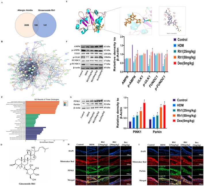
Ginsenoside Rh1, a bioactive compound derived from ginseng, exhibits notable anti-inflammatory and antioxidant effects and has shown promising therapeutic potential in the treatment of allergic diseases. However, its exact role in allergic rhinitis (AR) and the underlying molecular mechanisms remain inadequately understood. This study investigates whether Rh1 alleviates AR through AMPK/ULK1/FUNDC1-mediated mitochondrial autophagy. In this study, human nasal epithelial cells (HNEpCs) were stimulated with house dust mite (HDM) and treated with mitochondrial autophagy inhibitors or siRNA transfection techniques to assess the effects of Rh1. Network pharmacology and molecular docking (MD) were used to explore the interactions between Rh1 and AMPK, ULK1, and FUNDC1. To explore the effects of Rh1, enzyme-linked immunosorbent assay (ELISA) and flow cytometry (FC) were employed to measure IgE levels and various inflammatory mediators. Western blot (WB) analysis was conducted to assess protein expression related to mitochondrial autophagy, inflammation, and apoptosis in nasal tissues and HNEpCs. Immunofluorescence (IF) and transmission electron microscopy (TEM) provided further verification. The experimental data reveal that Rh1 effectively alleviates HDM-induced nasal mucosal epithelial thickening and eosinophil infiltration by modulating mitochondrial autophagy via the AMPK/ULK1/FUNDC1 signaling pathway. Additionally, Rh1 inhibits IL-4 secretion in nasal airway lavage fluid (NALF) and helps restore the Th1/Th2 immune balance. It also reduces mtROS production, inhibits NLRP3 inflammasome activation, and prevents apoptosis, thereby mitigating tissue damage associated with AR. Knockdown of AMPK or treatment with 3-Methyladenine (3-MA) further confirmed Rh1's inducing effect on mitophagy. In summary, Rh1 modulates mitophagy through the AMPK/ULK1/FUNDC1 pathway, reducing inflammatory responses and inhibiting apoptosis, thereby offering significant protection against AR.
Keywords: AMPK/ULK1/FUNDC1 pathway; Ginsenoside Rh1; allergic rhinitis; apoptosis; mitochondrial autophagy.
© 2025 The Author(s). Food Science & Nutrition published by Wiley Periodicals LLC.
Conflict of interest statement
The authors declare no conflicts of interest.
Figures


Similar articles
-
Ma Xing Shi Gan Decoction alleviates lipopolysaccharide-induced pneumonia by inhibiting NLRP3 inflammasome activation via AMPK/mTOR/ULK1-mediated autophagy.J Ethnopharmacol. 2025 Aug 14;353(Pt B):120418. doi: 10.1016/j.jep.2025.120418. Online ahead of print. J Ethnopharmacol. 2025. PMID: 40818519
-
Naringenin attenuates slow-transit constipation by regulating the AMPK/mTOR/ULK1 signaling pathway: In vivo and in vitro studies.J Nutr Biochem. 2025 Jun 24:110013. doi: 10.1016/j.jnutbio.2025.110013. Online ahead of print. J Nutr Biochem. 2025. PMID: 40571066
-
Ginsenoside Rd protects against acute liver injury by regulating the autophagy NLRP3 inflammasome pathway.Sci Rep. 2025 Jan 28;15(1):3569. doi: 10.1038/s41598-025-87991-9. Sci Rep. 2025. PMID: 39875579 Free PMC article.
-
Programmed cell death in allergic rhinitis: pathogenic mechanisms and therapeutic potential from a cellular perspective.Int Immunopharmacol. 2025 Aug 6;164:115319. doi: 10.1016/j.intimp.2025.115319. Online ahead of print. Int Immunopharmacol. 2025. PMID: 40773894 Review.
-
Assessing the comparative effects of interventions in COPD: a tutorial on network meta-analysis for clinicians.Respir Res. 2024 Dec 21;25(1):438. doi: 10.1186/s12931-024-03056-x. Respir Res. 2024. PMID: 39709425 Free PMC article. Review.
References
-
- Bai, X. , Liu P., Shen H., Zhang Q., Zhang T., and Jin X.. 2022. “Water‐Extracted <styled-content style="fixed-case"> Lonicera japonica </styled-content> Polysaccharide Attenuates Allergic Rhinitis by Regulating NLRP3‐IL‐17 Signaling Axis.” Carbohydrate Polymers 297: 120053. - PubMed
-
- Brożek, J. L. , Bousquet J., Agache I., et al. 2017. “Allergic Rhinitis and Its Impact on Asthma (ARIA) Guidelines‐2016 Revision.” Journal of Allergy and Clinical Immunology 140, no. 4: 950–958. - PubMed
LinkOut - more resources
Full Text Sources
Research Materials
Miscellaneous
