

Cerebral arterial lumens are enlarged in children and young adults with sickle cell disease compared to peers
- PMID: 40535957
- PMCID: PMC12172934
- DOI: 10.1016/j.ynirp.2025.100265
Cerebral arterial lumens are enlarged in children and young adults with sickle cell disease compared to peers
Abstract
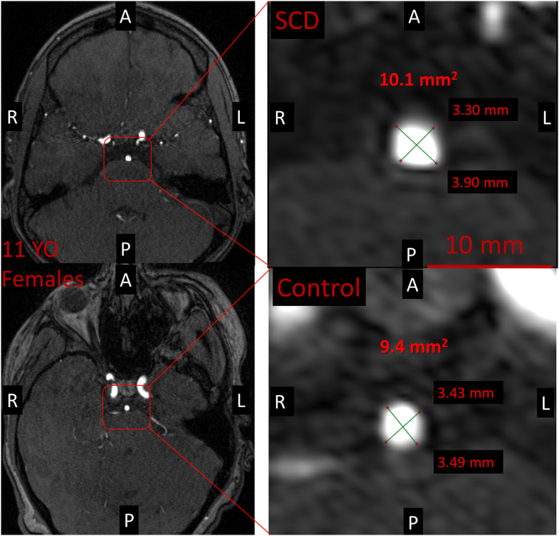
Children with sickle cell disease (SCD) may develop large vessel narrowing, but studies suggest vessels may also be enlarged, possibly related to increased cerebral blood flow (CBF). We used MRI to investigate whether the cross-sectional total inflow vessel luminal area (TIVLA) proximal to the circle of Willis (carotid arteries + basilar artery) would be increased in SCD compared to age- and sex-matched peers after adjusting for CBF. Across 36 children with SCD (19 female, median age 10.7 [8.0-14.5] years and 43 controls (26 female, median age 12.7 [9.2-18.2] years) matched by age (p = 0.13) and sex (p = 0.50), the median TIVLA in the SCD group (35.9 mm2 [30.7, 39.5]) was larger than controls (30.5 mm2 [27.8, 35.4], p = 0.002). In a mixed model including age, sex, hemoglobin, CBF, SCD status, and an interaction between hemoglobin and SCD status, CBF (β = 0.11, CI 0.02-0.20, p = 0.02), SCD (β = 28.02, CI 5.62-50.42, p = 0.015), and the interaction between SCD and hemoglobin (β = -2.48, CI -4.49 to -0.47, p = 0.018) were all significantly associated with increased TIVLA. Notably, TIVLA as a measure of arterial lumens is larger in children with SCD, even after adjusting for CBF in the mixed model. This implies disease-specific normative values may be needed to detect early vasculopathy.
Keywords: Arterial spin labeling; Brain development; Cerebral blood flow; Cerebrovascular disease; Hematology; MR angiography; MRI.
Conflict of interest statement
CRediT authorship contribution statement Josiah B. Lewis: Writing – review & editing, Writing – original draft, Visualization, Validation, Software, Methodology, Investigation, Formal analysis. Melanie E. Fields: Writing – review & editing, Writing – original draft, Investigation. Michael M. Binkley: Writing – review & editing, Formal analysis. Anita Zhou: Writing – review & editing, Validation, Investigation, Formal analysis. Amy Mirro: Writing – review & editing, Software, Investigation. Amy Ouyang: Writing – review & editing, Investigation. Niket Gupta: Writing – review & editing, Methodology, Investigation. Yasheng Chen: Writing – review & editing, Software, Methodology. Slim Fellah: Writing – review & editing, Software, Investigation. Alyssa E. Smith: Writing – review & editing, Investigation. Igor Dedkov: Writing – review & editing, Visualization, Investigation. Monica L. Hulbert: Writing – review & editing, Investigation. Andria L. Ford: Writing – review & editing, Investigation. Hongyu An: Writing – review & editing, Investigation. Jin-Moo Lee: Writing – review & editing, Conceptualization. Manu S. Goyal: Writing – review & editing, Writing – original draft, Methodology, Investigation, Conceptualization. Kristin P. Guilliams: Writing – review & editing, Writing – original draft, Supervision, Project administration, Methodology, Investigation, Conceptualization.
Figures


Similar articles
-
Association of Cerebral Hemodynamics and Anemia on Processing Speed in Adults with Sickle Cell Disease.J Neurol Exp Neural Sci. 2023;5(1):150. doi: 10.29011/2577-1442.100050. Epub 2023 Jul 24. J Neurol Exp Neural Sci. 2023. PMID: 37645351 Free PMC article.
-
Folate supplementation in people with sickle cell disease.Cochrane Database Syst Rev. 2018 Mar 16;3(3):CD011130. doi: 10.1002/14651858.CD011130.pub3. Cochrane Database Syst Rev. 2018. PMID: 29546732 Free PMC article.
-
Interventions for preventing silent cerebral infarcts in people with sickle cell disease.Cochrane Database Syst Rev. 2017 May 13;5(5):CD012389. doi: 10.1002/14651858.CD012389.pub2. Cochrane Database Syst Rev. 2017. Update in: Cochrane Database Syst Rev. 2020 Apr 6;4:CD012389. doi: 10.1002/14651858.CD012389.pub3. PMID: 28500860 Free PMC article. Updated.
-
Interventions for chronic kidney disease in people with sickle cell disease.Cochrane Database Syst Rev. 2023 Aug 4;8(8):CD012380. doi: 10.1002/14651858.CD012380.pub3. Cochrane Database Syst Rev. 2023. PMID: 37539955 Free PMC article.
-
Interventions for chronic kidney disease in people with sickle cell disease.Cochrane Database Syst Rev. 2017 Jul 3;7(7):CD012380. doi: 10.1002/14651858.CD012380.pub2. Cochrane Database Syst Rev. 2017. Update in: Cochrane Database Syst Rev. 2023 Aug 4;8:CD012380. doi: 10.1002/14651858.CD012380.pub3. PMID: 28672087 Free PMC article. Updated.
References
-
- Adams R.J., McKie V.C., Hsu L., Files B., Vichinsky E., Pegelow C., Abboud M., Gallagher D., Kutlar A., Nichols F.T., Bonds D.R., Brambilla D. Prevention of a first stroke by transfusions in children with sickle cell anemia and abnormal results on transcranial Doppler ultrasonography. N. Engl. J. Med. 1998;339:5–11. doi: 10.1056/NEJM199807023390102. - DOI - PubMed
-
- Adams Robert, Virgil McKie, Fenwick Nichols, Elizabeth Carl, Dao-Long Zhang, Kathy McKie, Figueroa Ramon, Mark Litaker, Thompson William, Hess David. The use of transcranial ultrasonography to predict stroke in sickle cell disease. N. Engl. J. Med. 1992;326:605–610. doi: 10.1056/NEJM199202273260905. - DOI - PubMed
-
- Alsop D.C., Detre J.A., Golay X., Günther M., Hendrikse J., Hernandez‐Garcia L., Lu H., MacIntosh B.J., Parkes L.M., Smits M., van Osch Matthias J.P., Wang Danny J.J., Wong E.C., Zaharchuk G. Recommended implementation of arterial spin-labeled perfusion MRI for clinical applications: a consensus of the ISMRM perfusion study group and the European consortium for ASL in dementia. Magn. Reson. Med. 2015;73:102–116. doi: 10.1002/mrm.25197. - DOI - PMC - PubMed
Grants and funding
LinkOut - more resources
Full Text Sources