. 2025 Jun 4:19:1551764.
doi: 10.3389/fnins.2025.1551764. eCollection 2025.

Cognitive and anti-inflammatory effects of Palmaria palmata in a schizophrenia mouse model: insights into CREB signaling, Iba-1 expression, and CD4+ cell modulation

Affiliations

Cognitive and anti-inflammatory effects of Palmaria palmata in a schizophrenia mouse model: insights into CREB signaling, Iba-1 expression, and CD4+ cell modulation

Shimaa Mohammad Yousof et al. Front Neurosci. .

Abstract

Background: Schizophrenia is a prevalent mental illness characterized by complex behavioral and emotional disturbances, with its underlying molecular mechanisms yet to be fully elucidated.

Aim: This study aims to examine the neuroprotective effects of Palmaria palmata (Palmaria p.) on cognitive function in a schizophrenia mouse model.

Methods: A total of 28 adult male SWR Swiss mice were used over a 30-day period. The animals were randomly divided into four groups (n = 7): control, cuprizone (CPZ) (0.2% CPZ in chow), CPZ + Palmaria p. (600 μg/kg bw/day via gavage), and Palmaria p. alone. The antioxidant activity of Palmaria p. was assessed using a radical scavenging assay. Behavioral assessments, hippocampal (HC) and frontal cortex (FC) gene expression analyses, and histopathological evaluations were conducted.

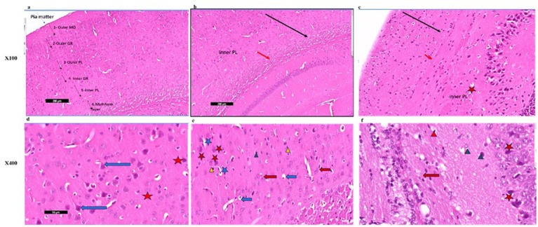
Results: Palmaria p. demonstrated remarkable antioxidant activity against CPZ-induced oxidative stress. No notable effects were observed in spatial memory, the novel object recognition test (NORT), or anxiety-related behaviors. In the CPZ-treated group, Iba1 and CREB expression levels increased in both the hippocampus (HC) and frontal cortex (FC). In the CPZ + Palmaria p. group, Iba1 expression was reduced by approximately one-fold in the HC and two-fold in the FC, while CREB expression was decreased by approximately two-fold in both regions compared to the CPZ group, indicating attenuation of neuroinflammation and restoration of neuroplasticity. Immunohistochemical analysis revealed a notable decline in CD4+ expression following Palmaria p. administration, suggesting a decrease in the immunological response induced by CPZ.

Conclusion: The results highlight the potential of Palmaria p. to enhance neuroplasticity and reduce neuronal inflammation associated with schizophrenia.

Keywords: Palmaria palmata; Y maze; anxiety; marble burying test; object recognition memory; red marine algae; schizophrenia; zero maze.


Conflict of interest statement

The authors declare that the research was conducted in the absence of any commercial or financial relationships that could be construed as a potential conflict of interest.

Figures

Figure 1
Figure 1
Timeline of the experiment with an overview of behavioral tasks. The presented graphical abstract illustrates the experimental methodology employed to examine the neuroprotective properties of Palmaria palmata in mitigating cognitive impairments and anxiety induced by CPZ in a mouse model of schizophrenia. The study included four distinct groups: control, Palmaria p. (Palmaria palmata treatment), CPZ (cuprizone treatment to cause cognitive deficits and anxiety), and CPZ + Palmaria p. (co-treatment with Palmaria palmata to evaluate its protective benefits). After the 30-day experiment, behavioral evaluations were administered to assess the participants’ memory and anxiety levels. In this study, the NORT and Y-Maze tests were utilized to measure memory, whereas the marble burying test and elevated plus maze (EPM) were implemented to assess anxiety (n = 7/group).
Figure 2
Figure 2
The upper figure (Aa,Ab) demonstrates the EZM. The lower part of the figure (Ba,Bb) demonstrates the MBT. Both tests are used for assessing anxiety-related behavior in mice. EZM, elevated zero maze; MBT, marble burying test (n = 7/group).
Figure 3
Figure 3
CPZ treatment impaired social recognition memory, as shown by the lack of preference for the novel mouse during the test stage (a) and the significantly reduced discrimination index (DI) (b). Control and P-only groups exhibited normal social memory, demonstrated by a preference for the novel over the familiar mouse and higher DI scores. Co-treatment with P (CPZ + P) showed a non-significant trend toward improvement, indicating that P may offer partial protection against CPZ-induced deficits (n = 7/group).
Figure 4
Figure 4
Showing the anxiety-related tests. (a) The number of spontaneous alterations (%) and the number of entries were assessed using the Y maze. (b) The number of closed arm entries and the time elapsed in the closed arm were compared using the elevated zero maze. The number of buried marbles was assessed via the marble burying test. p > 0.05 in the three tests. CPZ = cuprizone, P = Palmaria p. (n = 7/group).
Figure 5
Figure 5
Assessment of the anxiety-related behavior via the elevated zero mazes (a,b) and the marble burying test (c). The analysis of the number of closed arm entries and time spent in closed arms did not yield statistically significant differences between the groups (p > 0.05), suggesting a negligible effect on anxiety-related behavior. The number of buried marbles did not exhibit statistically significant changes across the groups (p > 0.05), implying an absence of a major impact of Palmaria palmata on anxiety-related compulsive behavior. CPZ = cuprizone, P = Palmaria p.
Figure 6
Figure 6
(a) A photomicrograph of sections (control –ve group) of adult male SWR Swiss mice showed the normal architecture of the different parts of the hippocampus: CA1, CA2, CA3, and dentate gyrus (DG), H&E; ×40. (b) A photomicrograph of sections (control +ve group) of adult male SWR Swiss mice showed the normal architecture of the different parts of the hippocampus: CA1, CA2, CA3, and dentate gyrus (DG), H&E; ×40. (c) A photomicrograph of Section (affected group) of adult male SWR Swiss mice showed the normal architecture of the different parts of the hippocampus: CA1, CA2, and CA3, with apparently increased thickness in the marked area of the dentate gyrus (DG), H&E; ×40. (d) A photomicrograph of sections (treated group) of adult male SWR Swiss mice showed the hippocampus, showing the different parts of the hippocampus: CA1, CA2, and CA3. Note the apparent decrease in the thickness of the dentate gyrus (G), H&E; ×40. (e) A photomicrograph of sections (control +ve group) of adult male SWR Swiss mice showed the normal architecture of the dentate region (black arrow D), formed mainly of an aggregation of granular cells (G), H&E; ×100. (f) A photomicrograph of sections (affected group) of adult male SWR Swiss mice showed higher magnification of the dentate gyrus (black arrow DG) with marked loss of its normal architecture (loss of granular layer) and marked thickness with apparently packed cells; note the shrunken cells (blue star) as well as the ruptured one (red star), H&E; ×100. (g) A photomicrograph of sections (treated group) of adult male SWR Swiss mice showed higher magnification of the dentate gyrus (black arrow DG), with a regain of its thickness with apparently moderate appearance of the granular cells (blue star), H&E; ×100. (h) A photomicrograph of sections (control +ve group) of adult male SWR Swiss mice showed the normal architecture of the dentate gyrus, formed mainly of an aggregation of granular cells (G) that appear rounded in shape with vesicular nuclei and prominent nucleoli (black star). Note the apparent presence of a pyramidal cell (red arrow), H&E; ×400. (i): A photomicrograph of sections (affected group) of adult male SWR Swiss mice showed higher magnification of the dentate gyrus. Note the complete loss of granular cells (G). Pyramidal cells showed open-face nuclei and prominent nucleoli (black arrow). Other cells appear markedly dark, shrunken, and detached from the surrounding (red arrow), H&E; ×400. (j) A photomicrograph of sections (treated group) of adult male SWR Swiss mice showed higher magnification of the dentate gyrus (DG). Note marked regain of granular cell layer (G). Other cells appear markedly dark, shrunken, and detached from the surrounding (red star), H&E; ×400.
Figure 7
Figure 7
A photomicrograph of sections (control –ve group) of adult male SWR Swiss mice showed the normal architecture of the different parts of the brain, showing frontal cortex (FC), corpus callosum (CC), and hippocampus (Hipo). H&E ×40.
Figure 8
Figure 8
(a) A photomicrograph of the Palmaria p. (control +ve) group of adult male SWR Swiss mice showed the normal architecture of the frontal cortex covered with pia matter and its six different layers (1—outer molecular layer, 2—outer granular layer, 3—outer pyramidal cell layer, 4—inner granular layer, 5—inner pyramidal cell layer, and 6—multiform layer), H&E ×100. (b) A photomicrograph of CPZ (affected group) of adult male SWR Swiss mice showing apparently amalgamated layers (black arrow) of frontal cortex and thickening of the multiform layer (red arrow). Apparently normal appearance of inner pyramidal cell layer, H&E ×100. (c) A photomicrograph of CPZ + Palmaria p. (treated group) of adult male SWR Swiss mice showing apparently amalgamated layers (black arrow) of frontal cortex, a mild decrease in the thickening of the multiform layer (red arrow), and a mild decrease in vacuolations. Note the open window cells of the pyramidal cell layers (red star), H&E ×100. (d) A photomicrograph of Palmaria p. (control +ve group) of adult male SWR Swiss mice showed the normal architecture of large pyramidal neurons with apical dendrites (blue arrow). Note the multiform layer of the frontal cortex shows pleomorphic nuclei of different cells (pyramidal and granular cells, red star, H&E; ×400). (e) A photomicrograph of the CPZ group shows a loss of shape in the pyramidal cell layer, with loss of its dendrites (red star) as well as ruptured cells with detached cytoplasm (blue star). Inner granular cell layer showing packed cytoplasmic cell layer (green triangle) as well as pyknotic nuclei (yellow triangle). Multiform cell layer showing multinucleated cells. Note vacuolated cells (blue arrow), apparently cells with eccentric nuclei, H&E ×400. (f) A photomicrograph of the CPZ+ Palmaria p. group in adult male SWR Swiss mice showing a normal pyramidal cell layer with regrowth of its dendrites (red star). The inner granular cell layer shows a normal pleomorphic cell layer (green triangle), but the appearance of pyknotic nuclei (red triangle) is still notable. Multiform cell layer showing multinucleated cells (red arrow). Note a decrease in the vacuolated cells, H&E ×400 (n = 7/group).
Figure 9
Figure 9
(a) A photomicrograph of the control –ve group of adult male SWR Swiss mice showed the normal architecture of the dentate gyrus of the hippocampus (DG) exhibiting CD4 mild immunoreaction of the nerve fibers, CD4 ×40. (b) A photomicrograph of the Palmaria p. only group of adult male SWR Swiss mice showed the normal architecture of the dentate gyrus of the hippocampus (DG) exhibiting CD4 Mild immunoreaction of the nerve fibers, CD4 ×40. (c) A photomicrograph of the CPZ group showed the normal architecture of the dentate gyrus of the hippocampus (DG) exhibiting CD4-marked immunoreaction of the nerve fibers, CD4 ×40. (d) A photomicrograph of CPZ + Palmaria p. showed the normal architecture of the dentate gyrus of the hippocampus (DG) exhibiting CD4 moderate immunoreaction of the nerve fibers, CD4 ×40 (n = 7/group).
Figure 10
Figure 10
(a) A photomicrograph of sections of the control –ve group of adult male SWR Swiss mice showed the normal architecture of the frontal cortex (FC) exhibiting CD4 Mild immunoreaction of the nerve fibers, CD4 ×40. (b) A photomicrograph of the Palmaria p. group showed the normal architecture of the frontal cortex (FC) exhibiting CD4 mild immunoreaction of the nerve fibers, D4 ×40. (c) A photomicrograph of CPZ of the adult male SWR group showed the frontal cortex (FC) exhibiting CD4-marked immunoreaction of the nerve fibers, CD4 ×40. (d) A photomicrograph of the Palmaria p. + CPZ group showed the frontal cortex (FC) exhibiting CD4 moderate immunoreaction of the nerve fibers, CD4 ×40. (n = 7/group).

Similar articles

References

    1. Alqurashi G. K., Hindi E. A., Zayed M. A., Abd El-Aziz G. S., Alturkistani H. A., Ibrahim R. F., et al. (2022). The impact of chronic unpredictable mild Stress-induced depression on spatial, recognition and reference memory tasks in mice: behavioral and histological study. Behav. Sci. 12:166. doi: 10.3390/bs12060166, PMID: - DOI - PMC - PubMed
    1. Angoa-Pérez M., Kane M. J., Briggs D. I., Francescutti D. M., Kuhn D. M. (2013). Marble burying and nestlet shredding as tests of repetitive, compulsive-like behaviors in mice. J. Vis. Exp. 82:50978. doi: 10.3791/50978 - DOI - PMC - PubMed
    1. Baliyan S., Mukherjee R. (2022). Determination of antioxidants by DPPH radical scavenging activity and quantitative phytochemical analysis of Ficus religiosa. Molecules 27:1326. doi: 10.3390/molecules27041326 - DOI - PMC - PubMed
    1. Bancroft J. D., Gamble M. (2013). Theory and practice of histological techniques. 7th Edn. Philadelphia: Churchill Livingstone of Elsevier, 172–186.
    1. Braun A. A., Skelton M. R., Vorhees C. V., Williams M. T. (2011). Comparison of the elevated plus and elevated zero mazes in treated and untreated male Sprague-Dawley rats: effects of anxiolytic and anxiogenic agents. Pharmacol. Biochem. Behav. 97, 406–415. doi: 10.1016/j.pbb.2010.09.013, PMID: - DOI - PMC - PubMed

LinkOut - more resources