Review
. 2025 Jul 1;64(13):2712-2726.
doi: 10.1021/acs.biochem.5c00234. Epub 2025 Jun 19.

A Tight-Knit Family: The Medium-Chain Dehydrogenase/Reductases of Monoterpene Indole Alkaloid Biosynthesis

Affiliations
Review

A Tight-Knit Family: The Medium-Chain Dehydrogenase/Reductases of Monoterpene Indole Alkaloid Biosynthesis

Samuel C Carr et al. Biochemistry. .

Abstract

Medium-chain dehydrogenases/reductases (MDRs) are enzymes that are well-known for catalyzing the reversible reduction of ketones or aldehydes or oxidation of alcohols. However, the biosynthetic pathways of the monoterpene indole alkaloids (MIAs), an important class of natural products derived from plants, highlight that MDRs can also catalyze 1,2- and 1,4-α,β-unsaturated iminium reductions, as well as 1,4-α,β-unsaturated carbonyl reduction. The noncanonical activities of these MDRs correlate with distinct catalytic architectures centered on amino acid substitutions that impact catalytic zinc coordination, acid/base catalysis, and proton relay. These noncanonical MDR catalytic architectures likely arose within the MDR subfamily of cinnamyl alcohol dehydrogenases (CADs). This review summarizes the currently characterized MIA biosynthetic MDRs along with an analysis of the catalytic mechanisms, structural underpinnings, and phylogeny.

Keywords: alcohol dehydrogenase; carbonyl reduction; catalytic architecture; iminium reduction; medium-chain dehydrogenase/reductase; monoterpene indole alkaloid; noncanonical activity; plant natural products.


Figures
1
1
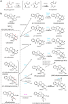
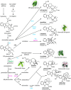
Roadmap to MDR-catalyzed reactions in MIA biosynthesis. A simplified diagram of the biosynthetic routes to major MIAs in various plants. MDRs conforming to the three major and one minor catalytic architectures described in the text are shown in gray (canonical), cyan (THAS-like), magenta (DPAS-like), and green (RedOx1-like). Additional biosynthetic enzymes are shown in black for reference. Abbreviations: 8-hydroxygeraniol oxidoreductase (8HGO), strictosidine synthase (STR), strictosidine β-d-glucosidase (SGD), geissoschizine synthase (GS), sarpagen bridge enzyme (SBE), dihydrocorynantheine synthases (DCS), tetrahydroalstonine synthase (THAS), heteroyohimbine synthase (HYS), yohimbine synthase (YOS), vomilenine reductase (VR), dihydrovomilenine reductase (DHVR), geissoschizine oxidase (GO), Weiland-Gumlich synthase (WS), reductive/oxidative enzyme 1 (RedOx1), reductive/oxidative enzyme 2 (RedOx2), precondylocarpine acetate synthase (PAS), dihydroprecondylocarpine acetate synthase (DPAS), coronaridine synthase (CorS), catharanthine synthase (CS), tabersonine synthase (TS), tabersonine 3-oxygenase (T3O), tabersonine 3-reductase (T3R).
2
2
The MDR superfamily: Overall structure, active site, and catalytic zinc binding. (a) The overall fold of Cr8HGOa (PDB 6K3G) with dimer protomers shown in gray and black. (b) Single protomer of Cr8HGOa. The homodimer interface is marked with a dotted line. (c) The active site of Cr8HGOa and schematic of tridentate catalytic zinc coordination. Protein backbones are shown in gray, key residue sidechains in green (carbon), red (oxygen), blue (nitrogen) and yellow (sulfur), cofactor is shown in magenta, and zinc ions as blue spheres. Active site hydrogen bonding network is drawn with dotted lines and the zinc coordinating water molecule is represented as a red star. Bond distances of zinc coordination were measured in Pymol.
3
3
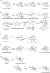
Early MIA pathway MDR-catalyzed reactions. (a) 8HGO catalyzed oxidation of 8-hydroxygeraniol. (b) MDR-catalyzed reductive trappings of strictosidine aglycone by DPAS, THAS/HYS, DCS, GS, and YOS. MDRs are highlighted based on their catalytic architectures described in the text: gray (canonical), cyan (THAS-like), magenta (DPAS-like), and green (RedOx1-like). Abbreviations: 8-hydroxygeraniol oxidoreductase (8HGO), strictosidine β-d-glucosidase (SGD), geissoschizine synthase (GS), dihydrocorynantheine synthases (DCS), tetrahydroalstonine synthase (THAS), heteroyohimbine synthase (HYS), yohimbine synthase (YOS), dihydroprecondylocarpine acetate synthase (DPAS).
4
4
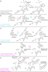
Downstream MDR-catalyzed reductions. (a) Concerted action of GO, RedOx1, and RedOx2 in the conversion of 19E-geissoschizine to stemmadenine. (b) DPAS catalyzed reductions of precondylocarpine acetate and downstream intermediates. (c) Concerted action of T3O and T3R in the conversion of tabersonine to 3-hydroxy-2,3-dihydrotabersonine. (d) WS catalyzed reductions of 18-OH norfluorocarine. (e) VR and DHVR catalyzed reductions of vomilenine and 1,2­(R)-dihydrovomilenine. MDRs are highlighted based on their catalytic architectures described in the text: gray (canonical), cyan (THAS-like), magenta (DPAS-like), and green (RedOx1-like). Abbreviations: vomilenine reductase (VR), dihydrovomilenine reductase (DHVR), geissoschizine oxidase (GO), Weiland-Gumlich synthase (WS), reductive/oxidative enzyme 1 (RedOx1), reductive/oxidative enzyme 2 (RedOx2), dihydroprecondylocarpine acetate synthase (DPAS), coronaridine synthase (CorS), tabersonine 3-oxygenase (T3O), tabersonine 3-reductase (T3R).
5
5
Proposed mechanisms of MDR-catalyzed reductions of 1,2-, 1,4-α,β-unsaturated iminium, 1,4-α,β-unsaturated aldehyde and 1,2-carbonyl moieties. (a) Canonical alcohol/aldehyde oxidation/reduction mechanism in Cr8HGOa. (b,c) 1,2-Iminium reduction mechanism in CrTHAS (b) and CrHYS (c). (d) 1,4-α,β-Unsaturated iminium reduction mechanism in CrDPAS. (e) 1,4-α,β-Unsaturated aldehyde reduction mechanism in CrDPAS. Abbreviations: 8-hydroxygeraniol oxidoreductase (8HGO), tetrahydroalstonine synthase (THAS), heteroyohimbine synthase (HYS), dihydroprecondylocarpine acetate synthase (DPAS).
6
6
The three major catalytic architectures of MIA MDRs. The active sites of representative MIA MDRs for canonical (Cr8HGOa; PDB 6K3G), THAS-like (CrGS; PDB 8A3N), and DPAS-like (TiDPAS2; PDB 8B25). Protein backbones are shown in gray, key residue sidechains in green (carbon), red (oxygen), blue (nitrogen) and yellow (sulfur), cofactor is shown in magenta, and zinc ions as blue spheres. Hydrogen bonding networks are marked by dotted lines and the zinc coordinating water molecule is represented by a red star. Corresponding schematics of zinc coordination for each respective architecture are shown below. Distances were measured in Pymol.
7
7
Sequence and phylogenetic analysis of characterized MIA MDRs. (a) Select positions from multiple sequence alignments of currently reported MIA MDRs (Table ) and related CADs. Amino acid sequences were aligned using the G-INS-i strategy. Residues pertaining to zinc coordination and the proton relay are shown. The numbering based on Cr8HGOa is shown on top of the alignment. (b) Phylogenetic analysis of MIA MDRs and related CADs. The maximum-likelihood phylogenetic tree was constructed using IQ-tree with the LG + I + G4 substitution model , from the multiple sequence alignment. The tree is rooted on ADH6 (KZV09178). Supported nodes are marked by black circles based on ultrafast bootstrap analysis using 1000 iterations (>95%) and branches were tested using SH-aLRT with 1000 replicates (>80%). Canonical catalytic architecture containing MDRs are highlighted in gray, THAS-like in cyan, DPAS-like in magenta, RedOx1-like in green, and MDRs not conforming to one of the described catalytic architectures in red. Their unique respective amino acid substitutions are highlighted in the multiple sequence alignment using the same coloring scheme.

References

    1. O’Connor S. E., Maresh J. J.. Chemistry and Biology of Monoterpene Indole Alkaloid Biosynthesis. Nat. Prod. Rep. 2006;23(4):532. doi: 10.1039/b512615k. - DOI - PubMed
    1. Himes R. H., Kersey R. N., Heller-Bettinger I., Samson F. E.. Action of the Vinca Alkaloids Vincristine, Vinblastine, and Desacetyl Vinblastine Amide on Microtubules in Vitro. Cancer Res. 1976;36:3798–3802. - PubMed
    1. Himes R. H.. Interactions of the Catharanthus (Vinca) Alkaloids with Tubulin and Microtubules. Pharmacol. Ther. 1991;51(2):257–267. doi: 10.1016/0163-7258(91)90081-V. - DOI - PubMed
    1. Gigant B., Wang C., Ravelli R. B. G., Roussi F., Steinmetz M. O., Curmi P. A., Sobel A., Knossow M.. Structural Basis for the Regulation of Tubulin by Vinblastine. Nature. 2005;435(7041):519–522. doi: 10.1038/nature03566. - DOI - PubMed
    1. Achan J., Talisuna A. O., Erhart A., Yeka A., Tibenderana J. K., Baliraine F. N., Rosenthal P. J., D’Alessandro U.. Quinine, an old anti-malarial drug in a modern world: role in the treatment of malaria. Malar. J. 2011;10(1):144. doi: 10.1186/1475-2875-10-144. - DOI - PMC - PubMed

MeSH terms

LinkOut - more resources