Review
. 2025 Aug;12(30):e17657.
doi: 10.1002/advs.202417657. Epub 2025 Jun 19.

Comparative Analysis of Clinical Outcomes and Financial Aspects of Phototherapies and Immunotherapy for Cancer

Affiliations
Review

Comparative Analysis of Clinical Outcomes and Financial Aspects of Phototherapies and Immunotherapy for Cancer

Deepak S Chauhan et al. Adv Sci (Weinh). 2025 Aug.

Abstract

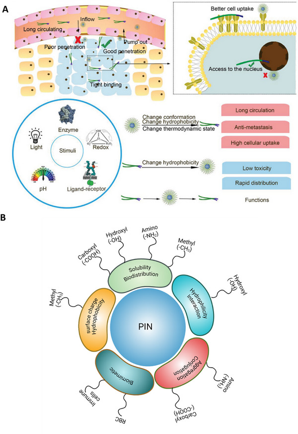
Photo-responsive/activable nanomedicine-driven therapies have emerged as a transformative approach to effectively target and eliminate tumors across different types of solid cancers. When synergistically combined with immunomodulators, these therapies significantly enhance anti-cancer efficacy. This comprehensive review examines the current advancements in photo-responsive/activable immunomodulator-loaded nanomedicines (PINs), providing a detailed analysis of their preclinical development, clinical translation, and associated research funding. The diverse PINs are systematically categorized, innovative design strategies are explored, and the underlying photo-responsive/activable and immunomodulatory mechanisms are elucidated. By integrating clinical advancements with technical innovations and financial perspectives, this review bridges the translational gap, underscoring the immense potential of PINs to revolutionize cancer therapy. This comprehensive resource is indispensable for researchers striving to bring phototherapies to clinical fruition in the battle against cancer.

Keywords: clinical trials; immunotherapy; photodynamic therapy; photoimmunotherapy; photothermal therapy.


Conflict of interest statement

The authors declare no conflict of interest.

Figures

Figure 1
Figure 1
Primary hurdles and valley of death in PIT development: Out of 530 studies analyzed, 394 studies have completed the in vitro validation, 136 completed the preclinical studies in mice, canine, pigs, rabbits, and primates, and 6 studies related to clinical trials have been published. The primary hurdles preventing successful transition from one stage to the next are indicated on the red color stop line. The figure on the right side depicts immunomodulators navigating through the “valley of death” under NIR light. Abbreviations: T: terminated, W: withdrawn, S: suspended clinical trials. Search engine: PubMed.
Figure 2
Figure 2
Different categories and types of interactions in PINs. A) Antibody‐dye conjugate, B) Liposome‐based PINs. C) Polymeric nanoparticle‐based PINs, D) Oncolytic virus‐based PINs, E) Inorganic–organic hybrid nanomaterials‐based PINs. A blue solid arrow indicates the transition of antibody‐based PINs from preclinical to clinical trials (Phase III). The numbers on each PIN type indicate the potential bonding for conjugating/loading NIR dye or immunomodulator to the nanocarriers. The sequence of numbers follows a hierarchical ranking, where the first number represents the most commonly used bonding type, followed by less frequent interactions (see interactions).
Figure 3
Figure 3
Influence of external stimuli and surface modifications on nanoparticle behavior and therapeutic efficacy. A) A concise depiction of how nanoparticle penetration, uptake, and stimuli‐responsive behavior influence delivery effectiveness. Light stimuli primarily exert effects via heat (photothermal response), enhancing permeability and cellular uptake. Other external stimuli, including enzymes, redox conditions, pH, and ligand–receptor interactions, can trigger conformational or hydrophobicity changes, improving circulation, tumor penetration, and targeted drug release. B) Surface functionalization of PINs and their role in biodistribution and targeting. Surface modifications such as carboxylation, hydroxylation, amination, and methylation influence solubility, biodistribution, cellular interactions, and immune targeting. Additionally, biomimetic coatings derived from immune cells or RBCs enhance tumor targeting and prolong circulation time. Adapted with permission[ 38 ] Copyright2011, Wiley.
Figure 4
Figure 4
Combinatorial photoimmunotherapeutic approach for cancer treatment. A) Schematic showing the synthesis of macrophage membrane‐coated PIN and laser‐triggered release of an immunomodulator (IND), a photosensitizer (Ce6), and cytotoxic nanoparticles. B‐i, ii) Nanogel for the photoimmunotherapy of cancer. The assembly of the combined treatment plan and the release mechanism of CpG‐P‐ss‐M (i). A simplified explanation of CpG‐P‐ss‐M‐induced DCs maturation for cancer immunotherapy (ii). The letters LDIMP, highlighted in orange, symbolize the loading of tumor‐specific antigens through drug delivery systems (DDS), their transport to lymph nodes (LNs), internalization by DCs, maturation of DCs leading to the expression of costimulatory molecules, and the presentation of peptide‐MHC‐I complexes to T cells, respectively. C‐i–iii) TEM images of CpG‐PP (i), CpG‐PM (ii), and CpG‐P‐ss‐M (iii). D‐i–iv) Photograph showing the gelation process following ultrasonication (i), scanning electron microscopy images of PP/CD gel (ii, iii), rheological properties of gels as a function of frequency (iv). E‐i, ii) Tumor growth curves for primary (i) and distant (ii) tumors (n = 6). Reproduced with permission.[ 4f,g ] Copyright 2020, Science; Copyright 2020, Elsevier. Abbreviations: M1m: M1 macrophage membrane, DOX: doxorubicin, BRPEG3400: PEGylated bilirubin, LPS: lipopolysaccharide, Ce6: chlorin e6, IND: indoximode, B‐A(D): DOX inserted and bovine serum albumin‐protected gold nanoclusters (B–AuNCs), (C/I)BP: PEGylated bilirubin to co‐deliver hydrophobic Ce6 and IND, (C/I)BP@B‐A(D): physically adsorbed (C/I)BP on B‐A(D), (C/I)BP@B‐A(D)&M1m: macrophage membrane coated (C/I)BP@B‐A(D), α‐CD: α‐cyclodextrin, MAN: reductive‐cleavable 4‐aminophenyl‐α‐d‐mannopyranoside, PAMAM: polyamidoamine dendrimer, pMHC: Peptide major histocompatibility complex.
Figure 5
Figure 5
Photothermally controlled synthetic gene switches for controlling the activity of CAR‐T cells. A) Thermal and luminescence imaging of wells containing TS‐Fluc T cells post‐irradiation with a NIR laser. The thermal images on the left were taken using a FLIR thermal camera, while the luminescence images on the right were recorded 6 h after heating with an IVIS Spectrum CT system. B) A diagram showing the TS‐Fluc αCD19 CAR construct transduced into primary human T cells before their transfer into NSG mice with two flank tumors (either K562 or Raji), followed by photothermal heating of one of the tumors. C) Thermal images of a mouse captured during laser irradiation of the tumor site at 0 and 3 min. D) Schematic of TS‐BiTE and TS‐Rluc αHER2 CAR vectors. E) Flow cytometry quantification of activation markers CD69, PD‐1, and CD107a by TS‐BiTE αHER2 CAR T cells after 30 min heating and co‐culture with HER2–MDA‐MB‐468 target cells. F) Tumor growth curves of MDA‐MB‐468 tumors inoculated at a 3:1 HER2+ to HER2− ratio and treated with TS‐BiTE or TS‐Rluc αHER2 CAR T cells. Heat treatments were carried out on days 45, 47, 52, 59, 66, and 72. G) Spider plots of individual tumors, with vertical dotted lines indicating heat treatments. H) In vivo luminescence imaging time course. I) Individual spider plots acquired by an IVIS Spectrum CT system representative of the HER2−/Fluc+ cell population in the mixed MDA‐MB‐468 tumor model. Adapted with permission.[ 80 ] Copyright 2021, Nature. Abbreviations: TS‐BiTE αHER2 CAR: CAR‐T cell construct designed for targeting HER2 positive cancers, TS‐BiTE αHER2 CAR + NIR: NIR light driven photothermal activation of TS‐BiTE αHER2 CAR, TS‐Rluc αHER2 CAR: tumor‐specific receptor‐luciferase with a CAR targeting HER2‐positive tumor cells, TS‐Rluc αHER2 CAR + NIR: NIR light driven photothermal activation of TS‐Rluc αHER2 CAR, UTD: untransduced groups, ACT: adoptive cell transfer, BiTEs: bispecific T cell engagers, IVIS: in vivo imaging system, CT: computed tomography.
Figure 6
Figure 6
Mechanism of PINs driven photoimmunotherapy. 1) After intravenous administration of PINs, it reaches the primary tumor via the EPR effect. 2) It's actively taken up by the cancer cells with the help of targeting moiety. 3) Laser irradiation causes a) ROS generation or b) hyperthermia. 4) The ICD is due to ROS generation or hyperthermia, which causes the release of tumor antigens and immunomodulators loaded in PINs. 5) Immunomodulators help in enhancing the antigen presentation by the APC, which migrates to the lymph node. 6) Anti‐tumor immune response eliminates the secondary tumor and left out cancer cells in the body.
Figure 7
Figure 7
Number of clinical trials and their phases in different cancer therapies. A) Number of different clinical trials (CT) under nanomedicines driven phototherapies, i.e., photothermal, photodynamic, and photoimmunotherapy. B) Comparison of clinical trials in phototherapies to immunotherapy. C) Number of clinical trials under the head of different clinically viable nanomaterials. D) Number of clinical trials under the head of different clinically viable immunomodulators. Source: Generated with the information from clinicaltrials.gov.
Figure 8
Figure 8
Clinical trial distribution by administration routes, targeting agents, and funding dynamics. A) The number of clinical trials following different routes of administration. IV administration is the most common route for PTT, immunotherapy, and photoimmunotherapy, while topical administration predominates in PDT. B) The use of clinically used targeting agents. Anti‐PD1 and anti‐PD‐L1 are the most frequently used targeting agents, with most trials in phase II. GM‐CSF and folate follow as the second most utilized immunomodulators, with over 100 clinical trials in phase II. C) The financial landscape of photoimmunotherapy, including the distribution of funds to different light‐driven cancer therapies. D) Comparison of funding for phototherapies versus immunotherapy. E) Funding allocation under major heads of clinically viable immunomodulators. F) Funding allocation under major heads of clinically viable nanomaterials for photoimmunotherapy. Source: Data generated using information from clinicaltrials.gov and NIH Reporter. Abbreviations: MUC1: mucin‐type 1 glycoprotein, AS1411: nucleic acid against nucleolin, GM‐CSF: cytokine adjuvant, NY‐ESO‐1: cancer‐testis antigens, MAGE A3: Melanoma‐associated antigen A3.

Similar articles

References

    1. a) Siegel R. L., Miller K. D., Wagle N. S., Jemal A., Ca‐Cancer J. Clin. 2023, 73, 17; - PubMed
    2. b) Pucci C., Martinelli C., Ciofani G., ecancermedicalscience 2019, 13, 961. - PMC - PubMed
    1. a) Fan D., Cao Y., Cao M., Wang Y., Cao Y., Gong T., Signal Transduction Targeted Ther. 2023, 8, 293; - PMC - PubMed
    2. b) Howard D., Garcia‐Parra J., Healey G. D., Amakiri C., Margarit L., Francis L. W., Gonzalez D., Conlan R. S., Interface Focus 2016, 6, 20160054. - PMC - PubMed
    1. a) Riehemann K., Schneider S. W., Luger T. A., Godin B., Ferrari M., Fuchs H., Angew. Chem., Int. Ed. Engl. 2009, 48, 872; - PMC - PubMed
    2. b) Kim H., Yang M., Kwon N., Cho M., Han J., Wang R., Qi S., Li H., Nguyen V. N., Li X., Cheng H. B., Yoon J., Bull. Korean Chem. Soc. 2022, 44, 236.
    1. a) Vats M., Mishra S. K., Baghini M. S., Chauhan D. S., Srivastava R., De A., Int. J. Mol. Sci. 18, 924; - PMC - PubMed
    2. b) Chauhan D. S., Kumawat M. K., Prasad R., Reddy P. K., Dhanka M., Mishra S. K., Bahadur R., Neekhra S., De A., Srivastava R., Colloids Surf., B 2018, 172, 430; - PubMed
    3. c) Chauhan D. S., Bukhari A. B., Ravichandran G., Gupta R., George L., Poojari R., Ingle A., Rengan A. K., Shanavas A., Srivastava R., De A., Sci. Rep. 2018, 8, 16673; - PMC - PubMed
    4. d) Chauhan D. S., Arunkumar P., Prasad R., Mishra S. K., BP K. R., De A., Srivastava R., Mater. Sci. Eng., C 2018, 90, 539; - PubMed
    5. e) Fan F., Hou Y., Zhang Y., Zeng Y., Zhang Y., Zhang S., Meng X., Wang X., Front. Oncol. 2022, 12, 987491; - PMC - PubMed
    6. f) Qin L., Cao J., Shao K., Tong F., Yang Z., Lei T., Wang Y., Hu C., Umeshappa C. S., Gao H., Peppas N. A., Sci. Adv. 2020, 6, 3116; - PMC - PubMed
    7. g) Hu C., Lei T., Wang Y., Cao J., Yang X., Qin L., Liu R., Zhou Y., Tong F., Umeshappa C. S., Gao H., Biomaterials 2020, 255, 120159. - PubMed
    1. a) Zhang J. Y., Gao W. D., Lin J. Y., Xu S., Zhang L. J., Lu X. C., Luan X., Peng J. Q., Chen Y., J. Drug Targeting 2023, 31, 555; - PubMed
    2. b) Deng X., Shao Z., Zhao Y., Adv. Sci. 2021, 8, 2100950. - PMC - PubMed

LinkOut - more resources