. 2025 Aug;120(4):707-725.
doi: 10.1007/s00395-025-01124-x. Epub 2025 Jun 19.

The innate immune receptor NLRX1 is a novel required modulator for mPTP opening: implications for cardioprotection

Affiliations

The innate immune receptor NLRX1 is a novel required modulator for mPTP opening: implications for cardioprotection

Y Xiao et al. Basic Res Cardiol. 2025 Aug.

Abstract

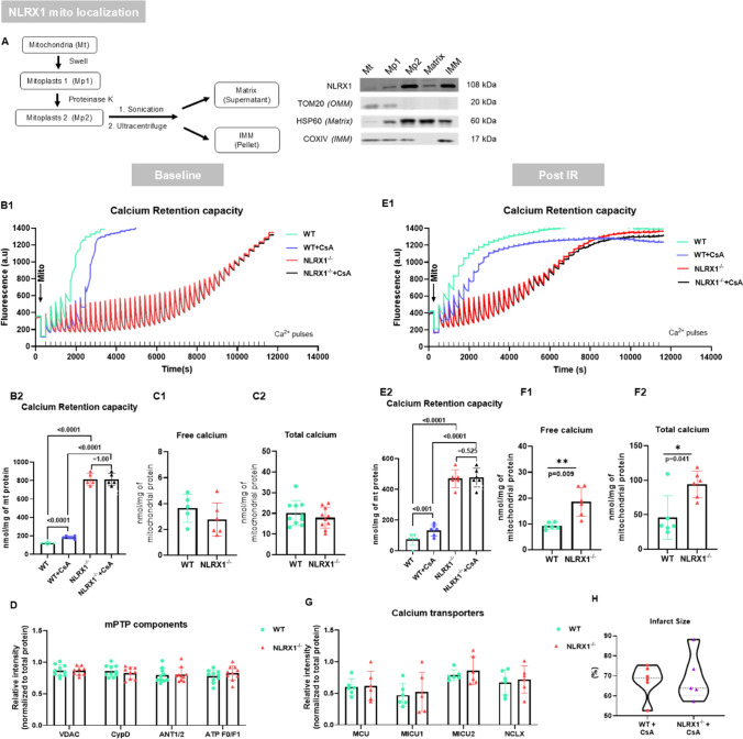
NLRX1 is the only NOD-like innate immune receptor that localises to mitochondria. We previously demonstrated that NLRX1 deletion increased infarct size in isolated mouse hearts subjected to ischemia-reperfusion injury (IRI); however, underlying mechanisms are yet to be identified. Given the crucial role played by mitochondria in cardiac IRI, we here hypothesise that NLRX1 affects key mechanisms of cardiac IRI. Cardiac IRI was evaluated in isolated C57BL/6J (WT) and NLRX1 knock out (KO) mouse hearts. The following known modulators of IRI were explored in isolated hearts, isolated mitochondria; or permeabilised cardiac fibres: 1) mTOR/RISK/autophagy regulation, 2) AMPK and mitochondrial energy production, and 3) mitochondrial permeability transition pore (mPTP) opening. NLRX1 deletion increased IRI, and cardiac NLRX1 was decreased after IRI in mouse and pig hearts. NLRX1 ablation caused decreased mTOR and RISK pathway (Akt, ERK, and S6K) activation following IR, without affecting autophagy/inflammation/oxidative stress markers. The RISK activator Urocortin dissipated NLRX1 effects on mTOR, RISK pathway and IRI, indicating that increased cardiac IRI with NLRX1 deletion is, at least partly, due to impaired RISK activation. The energy sensor AMPK was activated in NLRX1 KO hearts, possibly due to slowed mitochondrial respiratory responses (impaired mitochondrial permeability) towards palmitoylcarnitine in permeabilised cardiac fibres. NLRX1 deletion completely abolished calcium-induced mPTP opening, and cyclosporine A (CsA) effects on mPTP, both before and after IR, and was associated with increased mitochondrial calcium content after IR. Mitochondrial sub-fractionation studies localised NLRX1 to the inner mitochondrial membrane. NLRX1 deletion associated with decreased phosphorylation of mitochondrial Got2, Cx43, Myl2, Ndufb7 and MICOS10. The mPTP inhibitor CsA abolished IRI differences between KO and WT hearts, suggesting that the permanent closure of mPTP due to NLRX1 deletion contributed to the increased IR sensitivity of NLRX1 KO hearts. This is the first demonstration that the mitochondrial NLRX1 is a novel factor required for mPTP opening and contributes to cardioprotection against acute IRI through RISK pathway activation and prevention of permanent mPTP closure.

Keywords: AMPK; I/R injury; Mitochondria; Mitochondrial transition pore opening; NLRX1; RISK pathway.


Conflict of interest statement

Declarations. Conflict of interest: PF is the founder and CEO of Pharmahungary Group, a group of R&D companies www.pharmahungary.com . All other authors report no conflict of interest.

Figures

Fig. 1
Fig. 1
Experimental series and hypotheses being tested (I) NLRX1 and cardiac IRI and mTOR/RISK/autophagy pathways: Cardiac NLRX1 levels were determined in post-ischemic mouse and pig hearts, WT and NLRX1−/− isolated mouse hearts were subjected to 20 min baseline normoxic perfusion or to 20 min baseline perfusion followed by 35 min global ischemia (I) and 90 min reperfusion (R) and cardiac IR injury and signalling pathways evaluated; (II) NLRX1 and the role of RISK pathway activation in cardiac IRI: WT and NLRX1−/− hearts were pretreated with 2 nM urocortin during 15min baseline, followed by I/R as series I; (III) NLRX1 and cardiac energy regulation: cardiac AMPK was determined from experimental series I, cardiac fibres permeabilised for mitochondrial respiratory measurements in Oxygraph-2k, hearts fixated for EM analysis of mitochondrial density, and OXPHOS components determined in isolated mitochondria from series IV; (IV) NLRX1 and mPTP regulation: Mitochondria were isolated from WT and KO hearts for determination of NLRX1 localisation and calcium retention capacity (CRC) to determine Ca2+-sensitivity of mPTP opening, mitochondrial calcium and mPTP components and phosphoproteome analysis for hearts immediately sacrificed or subjected to 20 min baseline perfusion followed by 35 min I and 7 R, and mitochondrial ROS production at early reperfusion; (V) NLRX1 and the role of mPTP inhibition in cardiac IRI: WT and NLRX1−/− hearts were treated with 500 nM cyclosporine A during 20min baseline and the first 60min of reperfusion, I/R procedure as series I
Fig. 2
Fig. 2
NLRX1 protects against cardiac IRI and is associated with mTOR and RISK pathway activation. Hearts from WT and NLRX1−/− mice were subjected to 20 min normoxic perfusion (Baseline) followed by 35 min ischemia and 90 min reperfusion (Post IR). There was less protein expression of NLRX1 in WT mouse heart in post IR group than in baseline group (A, n = 8 per group), similar findings were observed after 3 h (n = 7 sham, n = 6 IR) and 3 d (n = 7 sham, n = 8 IR) reperfusion for NLRX1 in the ischemic regions of post IR pig hearts. (B1-B4). Data shown are mean ± SD (A-B).More severe IRI in NLRX1−/− than in WT evident from increased infarct size (C, n = 13 WT, n = 11 KO). Data shown are median ± IQ (C). D1 and D2, Representative expression analysis of phospho-mTOR/total mTOR at baseline and after IR in heart lysate (n = 8 per group). E, Co-immunoprecipitation (IP) was performed with hearts tissues from WT and NLRX1−/− mice after IR procedure (n = 6 per group). mTOR complexes proteins (Raptor, Rictor, mTOR, mLST8) and NLRX1 protein levels were shown in. F and G, Reperfusion injury salvage kinase (RISK) survival signalling pathway at baseline and post IR were investigated (n = 8 per group). Expression analysis of phospho-Akt/total Akt, phospho-ERK/total ERK, and phospho-S6K/total S6K at baseline (F) and after IR (G) (n = 8 per group). Expression analysis of autophagic parameters (p62, LC3, LAMP1, LAMP2, CTSB) at baseline (H) condition or after IRI (I) (n = 8 per group). Data shown are mean ± SD (D-I). Statistical significance was evaluated by nonpaired t test (normally distributed data) or Mann–Whitney test (non-normally distributed data). *P < 0.05, **P < 0.01, ***P < 0.001, and ****P < 0.0001 between the groups indicated by the solid lines
Fig. 3
Fig. 3
RISK activator urocortin negates genotype effects on post IR RISK pathway activation and thereby abrogates genotype effects on IRI. Each heart was perfused with 2 nM urocortin during baseline, then subjected to IR procedure (n = 9 per group). A, Representative protein expression analysis of phospho-mTOR /total mTOR, phospho-Akt/total Akt, phospho-ERK/total ERK, and phospho-S6K/total S6K (data shown are mean ± SD). B, Infarct size (% of area at risk) of 2, 3, 5-triphenyltetrazolium chloride (TTC) staining in the hearts after 90 min reperfusion. Data shown are median ± IQ (B). Statistical significance was evaluated by nonpaired t test (normally distributed data) or Mann–Whitney test (non-normally distributed data)
Fig. 4
Fig. 4
NLRX1 deletion increased capacity of complex 1 and delayed respiratory responses to FA carnitines. A1 and A2, Hearts from WT and NLRX1−/− mice were subjected to 20min normoxic perfusion (Baseline) followed by 35min ischemia and 90 min reperfusion (Post IR). Representative immunoblots and analysis of phospho-AMPKα/total AMPKα at baseline (A1, A2) and after IR (A3, A4) in heart lysate (n = 8 per group). B1-D2, Mitochondrial respiration was detected in isolated heart fibers (n = 5 per group): B1, Rates of mitochondrial oxygen consumption during substrates-uncoupler-inhibitor Titration (SUIT) protocol. B2, Excess capacity calculated as the ratio of uncoupled respiration to OXPHOS. C1, Rates of mitochondrial oxygen consumption during palmitoylcarnitine oxidation. C2, The timing to reach maximal palmitoylcarnitine oxidation. C3, The final concentration to reach plateau of palmitoylcarnitine oxidation. D1, Rates of mitochondrial oxygen consumption during NADH saturate gradually. D2, Rates of mitochondrial oxygen consumption upon NADH saturation. E, Immunoblots’ analysis of OXPHOS complex I, II, III, IV, and V measured in isolated mitochondria lysate (n = 10 per group). Data shown are mean ± SD. Statistical significance was evaluated by nonpaired t test (normally distributed data) or Mann–Whitney test (non-normally distributed data). NADH curve (C1) was analysed by two-way repeated measures ANOVA with Bonferroni adjustment for multiple comparisons. *P < 0.05, **P < 0.01, ***P < 0.001, and ****P < 0.0001 between WT and NLRX1−/− group
Fig. 5
Fig. 5
NLRX1 deletion blocks mPTP opening, and NLRX1’s effect on IR injury is dependent on mPTP opening A, Mitochondria were isolated from hearts excised directly after in-chest cannulation and further separated into different enrichment fractions, NLRX1 protein was analysed in all sub-fractions by western blot (n = 5). B1-D, Mitochondria were isolated from the hearts excised directly after in-chest cannulation (Baseline). The sensitivity of mPTP opening was measured by calcium retention capacity assay (B1) and analysed (B2) in baseline mitochondria (n = 6 [WT] and [WT + CsA], 5 [NLRX1−/−] and [NLRX1−/− + CsA]). Free calcium content (C1, n = 5 [WT], 6 [NLRX1−/−]) and total calcium content (C2, n = 10 per group) were measured in baseline mitochondria. D, Protein expression analysis of mPTP components in basal mitochondria (n = 9–10 per group), following components were examined: voltage-dependent anion channel (VDAC), cyclophilin D (Cyp D), adenine nucleotide translocator (ANT), and ATP F0/F1 proteins (one value missing in KO group for VDAC and Cyp D due to air bubble in lane blot). E1-G, Mitochondria were isolated from the hearts after 35 min ischemia and 7min reperfusion (Post IR, n = 6 for each group). The sensitivity of the mPTP opening was measured by calcium retention capacity assay (E1) and analysis (E2) in Post IR mitochondria. Free calcium content (F1) and total calcium content (F2) were measured in Post IR mitochondria. G, Representative analysis of important mitochondrial calcium transporters expression in Post IR mitochondria. The following calcium transporter proteins were examined: mitochondrial calcium uniporter (MCU), mitochondrial calcium uptake 1 and 2 (MICU1 and MICU2), and mitochondrial sodium calcium exchanger (NCLX) proteins. Data shown are mean ± SD. Hearts from WT and NLRX1-/- mice were subjected to IR procedure with cyclosporine A (CsA, mPTP inhibitor) administrated during 20 min baseline and first 60 min reperfusion (n = 6 [WT + CsA], 5 [NLRX1-/- + CsA]). CsA equalized IRI between WT and KO, as indicated by infarct size (H, % of area at risk) of 2, 3, 5-triphenyltetrazolium chloride (TTC) staining in the hearts after 90 min reperfusion. Data shown are median ± IQ. Statistical significance was evaluated by nonpaired t test (normally distributed data) or Mann–Whitney test (non-normally distributed data). Calcium retention capacity data (B2 and E2) were analysed by two-way repeated measures ANOVA with Bonferroni adjustment for multiple comparisons. *P < 0.05, **P < 0.01 between the groups indicated by the solid lines
Fig. 6
Fig. 6
Summary of NLRX1 working scheme. A, In physiological conditions, NLRX1 is needed for mPTP regulation and opening, maintaining a proper level of mitochondrial respiratory activity by regulating complex I activity and mitochondrial response to fatty acid (FA) carnitine. B, Under pathological conditions (IR [41, 72], Traumatic brain injury (TBI) [73], Rheumatoid arthritis (RA) [32], Chronic obstructive pulmonary disease (COPD) [30], Cancer [12, 77], and HIV [51]), NLRX1 is downregulated. The loss of NLRX1 results in the inhibition of the mPTP, while inducing higher complex I respiratory capacity and slower mitochondrial responses to FA substrates possibly due to impaired mitochondrial permeability of FA. This mitochondrial dysregulation then, possibly due to the impaired inner membrane permeability, activates AMPKα which impairs stress-induced activation of mTOR and the cardioprotective RISK pathway. Further studies are still needed to explore in detail how mitochondrial dysfunction induced by NLRX1 deletion activates AMPKα

References

    1. Alavian KN, Beutner G, Lazrove E, Sacchetti S, Park HA, Licznerski P, Li H, Nabili P, Hockensmith K, Graham M, Porter GA Jr, Jonas EA (2014) An uncoupling channel within the c-subunit ring of the F1FO ATP synthase is the mitochondrial permeability transition pore. Proc Natl Acad Sci U S A 111:10580–10585. 10.1073/pnas.1401591111 - PMC - PubMed
    1. Altschuld RA, Hohl CM, Castillo LC, Garleb AA, Starling RC, Brierley GP (1992) Cyclosporin inhibits mitochondrial calcium efflux in isolated adult rat ventricular cardiomyocytes. Am J Physiol 262:H1699-1704. 10.1152/ajpheart.1992.262.6.H1699 - PubMed
    1. Arslan F, Houtgraaf JH, Keogh B, Kazemi K, de Jong R, McCormack WJ, O’Neill LA, McGuirk P, Timmers L, Smeets MB, Akeroyd L, Reilly M, Pasterkamp G, de Kleijn DP (2012) Treatment with OPN-305, a humanized anti-Toll-Like receptor-2 antibody, reduces myocardial ischemia/reperfusion injury in pigs. Circ Cardiovasc Interv 5:279–287. 10.1161/CIRCINTERVENTIONS.111.967596 - PubMed
    1. Arslan F, Smeets MB, O’Neill LA, Keogh B, McGuirk P, Timmers L, Tersteeg C, Hoefer IE, Doevendans PA, Pasterkamp G, de Kleijn DP (2010) Myocardial ischemia/reperfusion injury is mediated by leukocytic toll-like receptor-2 and reduced by systemic administration of a novel anti-toll-like receptor-2 antibody. Circulation 121:80–90. 10.1161/CIRCULATIONAHA.109.880187 - PubMed
    1. Baines CP, Kaiser RA, Purcell NH, Blair NS, Osinska H, Hambleton MA, Brunskill EW, Sayen MR, Gottlieb RA, Dorn GW, Robbins J, Molkentin JD (2005) Loss of cyclophilin D reveals a critical role for mitochondrial permeability transition in cell death. Nature 434:658–662. 10.1038/nature03434 - PubMed

MeSH terms

Substances