. 2025 Jun 19;149(1):64.
doi: 10.1007/s00401-025-02902-6.

The olfactory epithelium: a critical gateway for pathological tau propagation and a target for mitigating tauopathy in the central nervous system

Affiliations

The olfactory epithelium: a critical gateway for pathological tau propagation and a target for mitigating tauopathy in the central nervous system

Marion Dourte et al. Acta Neuropathol. .

Abstract

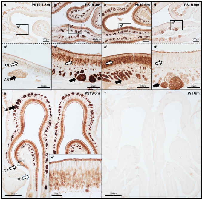
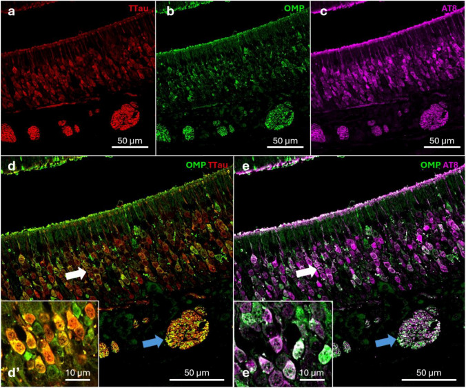
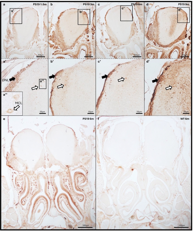
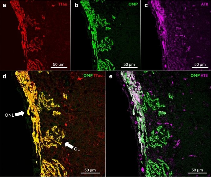
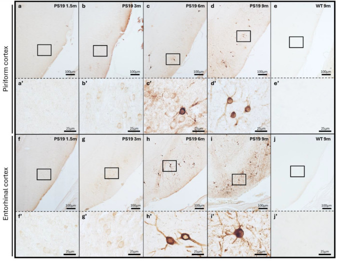
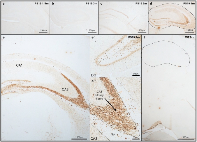
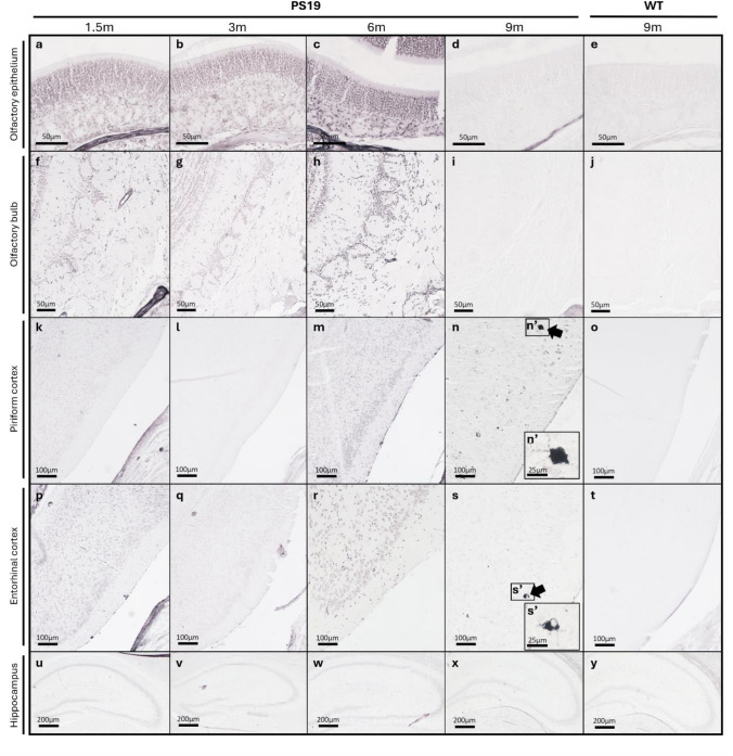
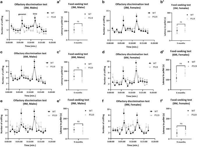
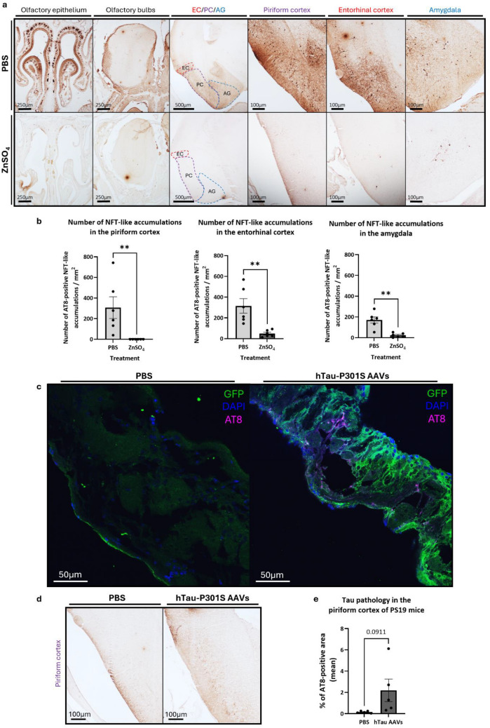
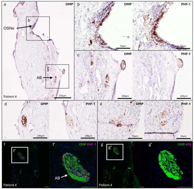
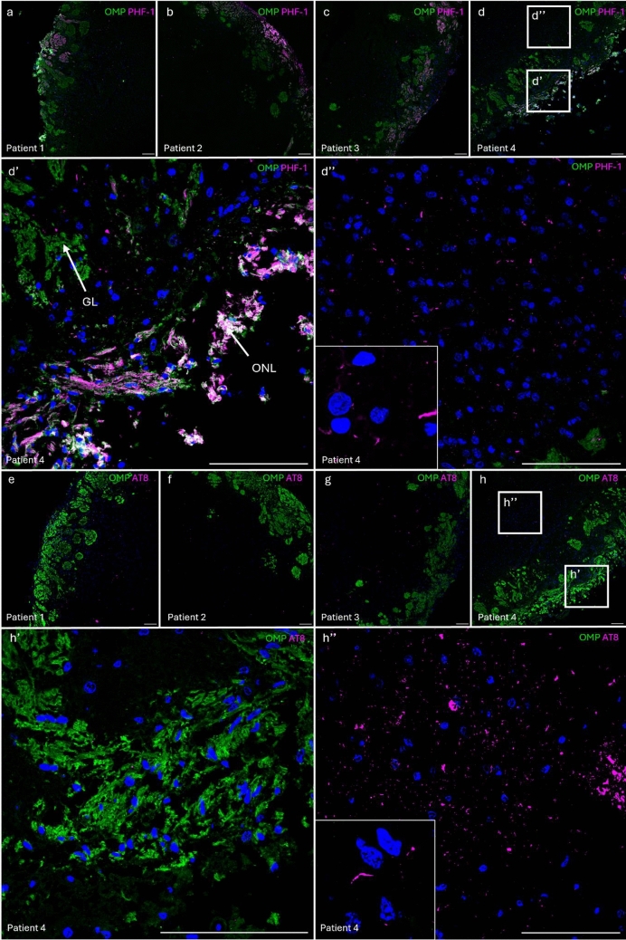
Olfactory impairment is a recognized early indicator of neurodegenerative diseases (NDs), such as Alzheimer's disease (AD). Intracellular aggregates of hyperphosphorylated tau protein, referred to as neurofibrillary tangles (NFTs), are a hallmark of AD. NFTs are found in the olfactory bulb (OB) and entorhinal cortex (EC), both crucial for processing olfactory information. We explored the hypothesis that typical tau lesions could appear early and progress along olfactory regions to reach connected areas critically affected in AD (e.g., EC and hippocampal formation). To that end, we used transgenic PS19 mice expressing mutated human tau protein (1N4R isoform, P301S mutation). They recapitulate major phenotypes of AD, such as accumulation of NFTs, synaptic dysfunction, cognitive impairment, and neuronal loss. The presence of pathological hyperphosphorylated human tau protein (pTau) was monitored in olfactory regions: olfactory epithelium (OE), OB, piriform cortex (PC), and in connected regions of the hippocampal formation (hippocampus and EC). pTau was detected in the OE's middle stratum and in the OB's olfactory nerve layer (ONL) at 1.5 months. At 6 months of age, tau accumulations were found in the PC and EC, along with the CA3 region and dentate gyrus of the hippocampus. We found that olfactory function remained unaffected in PS19 mice, despite the presence of tau pathology in key regions of the olfactory system. Targeted treatments (ZnSO4 and AAVs) were applied at the OE level to assess the impact on tau pathology in the CNS. Complete stripping of the OE by intranasal administration of ZnSO4 led to a significant reduction in pretangle-like tau pathology within the PC, amygdala, and EC of 6-month-old PS19 mice. Finally, we observed in human postmortem samples that pTau signal was present in the olfactory regions (OE and OB) of patients at early Braak stages (I/II). Based on these observations, we propose that pTau could appear, due to aging or environmental agents, in the OE and subsequently spread in a prion-like manner to the hippocampal formation along neuroanatomical connections. These findings also indicate the interest of the OE as a target for intervention aimed at mitigating the progression of tauopathy in the CNS.

Keywords: Olfaction; Olfactory system; Tau pathology; Tau spreading.


Conflict of interest statement

Declarations. Conflicts of interest: DRT and SOT received consultant honoraria from Muna Therapeutics. DRT collaborated with Novartis Pharma AG (Switzerland), and GE Healthcare (UK). The other authors have no competing interests to declare that are relevant to the content of this article. Ethical approval: All animal procedures were conducted in accordance with institutional and European guidelines and approved by the UCLouvain Ethical Committee for Animal Welfare (2021/UCL/MD/018). Human postmortem brain tissue was collected in accordance with the applicable legislation in Belgium. The recruitment protocols for the collection of human brains were approved by the ethical committee (2020/02JUL/355).

Figures

Fig. 1
Fig. 1
pTau expression in the OE of PS19 mice. The OE from 1.5-month (a, a’), 3-month (b, b’), 6-month (c, c’, e), 9-month-old PS19 mice (d, d’), and 6-month-old WT mice (f) were fixed and stained with the AT8 antibody (pTau (Ser202,Thr205)). pTau recognized by the AT8 antibody is found in the middle stratum of the OE (white arrows) and in the ABs (black arrows) as soon as 1.5 months in PS19 mice (a, a’). The signal increases up to 6 months (c, c’, e) and appears to decrease at 9 months (d, d’). No signal is detected in WT mice (f). All the mice used were males. The immunostainings shown are representative of n = 4 mice per time point. High magnification of the OE (white arrows) is shown in the inset (e’). ABs: axon bundles, OE: olfactory epithelium, RE: respiratory epithelium
Fig. 2
Fig. 2
pTau expression in the OSNs and ABs of PS19 mice. Paraffin sections immunostained for total tau (TTau, red) (a, d), olfactory marker protein (OMP, green) (b, d, e), and pTau (AT8, purple) (c, e) show colocalization between TTau-OMP (yellow) (d) and AT8-OMP (white) (e) indicating tau hyperphosphorylation in the OSNs (white arrows) and in the ABs (blue arrows) of PS19 mice at 3 months. High magnifications are shown in the insets (d’, e’). OSNs: olfactory sensory neurons, ABs: axon bundles
Fig. 3
Fig. 3
pTau expression in the OB of PS19 mice. The OB from 1.5-month (a, a’), 3-month (b, b’), 6-month (c, c’, e), 9-month-old PS19 mice (d, d’), and 6-month-old WT mice (f) were immunohistochemically stained with AT8 (ad, a’–d’). pTau recognized by the AT8 antibody is found in the external layer of the OB (black arrows) and in the MCL (white arrows) from 1.5 months (ad, a’–d’). The pTau signal seems stable over time (a’–d’). No signal is detected in WT mice (f). All the mice used for immunostainings were males. The immunostainings shown are representative of n = 4 mice per time point. High magnification of the MCL (white arrows) is shown in the inset (a’’). ONL: olfactory nerve layer, MCL: mitral cells layer
Fig. 4
Fig. 4
pTau expression in the ONL of PS19 mice. Paraffin sections immunostained for total tau (a, d) (TTau, red), OMP (b, d, e) (green), and AT8 (c, e) (purple) show colocalization between TTau-OMP (yellow) (d) and AT8-OMP (white) (e) indicating tau protein expression both in ONL and GL as well as pTau expression in the ONL and inner layers of the OB from 3-month-old PS19 mice. ONL: olfactory nerve layer, GL: glomerular layer
Fig. 5
Fig. 5
pTau expression in the PC and EC of PS19 mice. The PC (Bregma − 1.94 mm) from 1.5-month (a, a’), 3-month (b, b’), 6-month (c, c’), 9-month-old PS19 mice (d, d’), and 9-month-old WT mice (e, e’) were immunohistochemically stained with AT8 (ae). pTau recognized by the AT8 antibody is found in the PC as soon as 3 months (b, b’) with an accumulation up to 9 months (bd, b’–d’). The first NFT-like tau accumulations appear at 6 months (c, c’). There is no AT8 signal in the PC of WT mice (e, e’). The EC (Bregma − 3.08 mm) from 1.5-month (f, f’), 3-month (g, g’), 6-month (h, h’), 9-month-old PS19 mice (i, i’), and 9-month-old WT mice (j, j’) were immunohistochemically stained with AT8 (fj). pTau recognized by the AT8 antibody is found in the EC as soon as 1.5 months (f, f’) with an accumulation up to 9 months (fi, f’–i’). PS19 mice develop NFT-like tau accumulations in this region at 6 months (h, h’). No AT8 signal is detected in the EC of WT mice (j, j’). All the mice used were males. The immunostainings shown are representative of n = 4 mice per time point. Insets were taken from the PC area (a’–e’) and in the lateral EC (f’–j’)
Fig. 6
Fig. 6
pTau expression in the hippocampus of PS19 mice. The hippocampus (Bregma − 1.82 to − 2.18 mm) from 1.5-month (a), 3-month (b), 6-month (c), 9-month-old PS19 mice (d, e), and 9-month-old WT mice (f) were immunohistochemically stained with AT8 (af). pTau recognized by the AT8 antibody is found in specific hippocampal subregions like the CA3 (e’’) and DG (e’) as soon as 6 months with an increased signal at 9 months (ce). No AT8 signal is found in the hippocampus of WT mice (f). All the mice used for immunostainings were males. The immunostainings shown are representative of n = 4 mice per time point. DG: Dentate gyrus, CA: Cornu Ammonis, Sp: Stratum pyramidale
Fig. 7
Fig. 7
Gallyas staining in the OE, OB, PC, EC, and hippocampus of PS19 mice. The OE (ae), OB (fj), PC (ko), EC (pt), and hippocampus (uy) of PS19 mice at 1.5, 3, 6, and 9 months, as well as 9-month-old WT mice were stained with Gallyas silver staining to highlight NFT inclusions. A few Gallyas-positive inclusions (black arrows) are only observed in the PC (n) and EC (s) from 9 months PS19 mice. No NFTs are detected in the OE, OB, or hippocampus at any time point, nor in the PC or EC before 9 months. No signal is detected in WT mice (e, j, o, t, y). High magnifications of the Gallyas-positive aggregates (black arrows) are shown in the inset (n’) and (s’). All the mice used for Gallyas silver staining were males. The Gallyas stainings shown are representative of n = 3 mice per time point
Fig. 8
Fig. 8
Olfactory function assessment in PS19 mice. The olfactory discrimination (af) and food-seeking tests (a’–f’) show no significant effect of genotype (PS19) on olfaction at either 3, 6, or 9 months, both in males and females (af, a’–f’). For PS19 males; n = 9 (3 months), n = 9 (6 months), and n = 7 (9 months). For WT males; n = 9 (3 months), n = 9 (6 months), and n = 9 (9 months). For PS19 females; n = 8 (3 months), n = 8 (6 months), and n = 5 (9 months). For WT females; n = 8 (3 months), n = 8 (6 months), and n = 8 (9 months). Data were analyzed using two-way ANOVA with Šídák’s multiple comparisons test (olfactory discrimination test) or using independent t test (food-seeking test)
Fig. 9
Fig. 9
Modulation of tau pathology in the CNS via intranasal interventions. Sections of the OE, OB, PC, EC, and amygdala from 6-month-old PS19 males were fixed and stained with the AT8 antibody. After intranasal ZnSO4 irrigation, the OE and ONL from the OB are stripped, no more pTau signal is found in these regions (a). Quantification of the number of NFT-like tau accumulations/mm2 was performed in the PC, EC, and amygdala. In the PC, there are no NFT-like tau accumulations compared to the PBS-treated mice. In the EC and amygdala, there is a significant decrease in NFT-like accumulations compared to the control condition. **P < 0.01 (Mann–Whitney test, n = 6 mice/group) (b). OE sections immunostained for GFP (green), AT8 (purple), and DAPI showed efficient transduction following intranasal AAVs administration (c). In the PC, AT8 immunoreactivity appeared increased in AAV-treated mice compared to PBS controls (d). Quantification of the AT8-positive area in the PC showed a trend toward increased pTau signal following nasal delivery of hTau-P301S AAVs (Mann–Whitney, n = 5 mice per group) (e). ONL: olfactory nerve layer, AG: amygdala
Fig. 10
Fig. 10
pTau expression in human OE. The OE was obtained from a Braak stage II patient (patient 4). The sections were immunohistochemically stained with PHF-1 or with OMP (ae). PHF-1 signal is observed in the OMP-positive OSNs (b) and ABs (ce) of the OE. Paraffin sections immunostained for OMP (green) and PHF-1 (purple) show colocalization between OMP-PHF-1 (white) in the AB (f, f’). PHF-1 signal is also observed inside the tissue (f). Paraffin sections immunostained for OMP (green) and AT8 (purple) show no colocalization between OMP-AT8 (g, g’). No signal is detected in the negative controls without primary antibodies. Scale bar: 100 μm. OSNs: olfactory sensory neurons, ABs: axon bundles
Fig. 11
Fig. 11
pTau expression in human OB. The OB were obtained either from patients 1 and 2 (Braak stage I) (a, b, e, f) or from patients 3 and 4 (Braak stage II) (c, d, g, h). Paraffin sections immunostained for OMP (green) and PHF-1 (purple) show colocalization between OMP-PHF-1 (white) in the ONL of all patients (ad, d’). PHF-1 signal is also found in the inner layers of the OB (ad, d’’). Paraffin sections immunostained for OMP (green) and AT8 (purple) show no colocalization between OMP-AT8 (eh, h’). AT8 signal is only found in the inner layers of the OB, similarly to PHF-1 (eh, h’’). No signal is detected in the negative controls without primary antibodies. Scale bar: 100 μm. ONL: Olfactory nerve layer, GL: Glomerular layer

Similar articles

References

    1. Ahmad F, Mein H, Jing Y, Zhang H, Liu P (2021) Behavioural functions and cerebral blood flow in a P301S Tauopathy mouse model: a time-course study. Int J Mol Sci. 10.3390/ijms22189727 - PMC - PubMed
    1. Ahnaou A, Rodriguez-Manrique D, Biermans R, Embrechts S, Manyakov NV, Drinkenburg WH (2020) Functional alterations in the olfactory neuronal circuit occur before hippocampal plasticity deficits in the P301S mouse model of tauopathy: implications for early diagnosis and translational research in Alzheimer’s disease. Int J Mol Sci. 10.3390/ijms21155431 - PMC - PubMed
    1. Amani M, Lauterborn JC, Le AA, Cox BM, Wang W, Quintanilla J et al (2021) Rapid aging in the Perforant path projections to the rodent dentate gyrus. J Neurosci 41(10):2301–2312. 10.1523/jneurosci.2376-20.2021 - PMC - PubMed
    1. Aragão Gomes L, Uytterhoeven V, Lopez-Sanmartin D, Tomé SO, Tousseyn T, Vandenberghe R et al (2021) Maturation of neuronal AD-tau pathology involves site-specific phosphorylation of cytoplasmic and synaptic tau preceding conformational change and fibril formation. Acta Neuropathol 141(2):173–192. 10.1007/s00401-020-02251-6 - PubMed
    1. Arnold SE, Smutzer GS, Trojanowski JQ, Moberg PJ (1998) Cellular and molecular neuropathology of the olfactory epithelium and central olfactory pathways in Alzheimer’s disease and schizophrenia. Ann N Y Acad Sci 855:762–775. 10.1111/j.1749-6632.1998.tb10656.x - PubMed

Publication types

LinkOut - more resources