

Deep learning detects retropharyngeal edema on MRI in patients with acute neck infections
- PMID: 40536731
- PMCID: PMC12179047
- DOI: 10.1186/s41747-025-00599-6
Deep learning detects retropharyngeal edema on MRI in patients with acute neck infections
Abstract
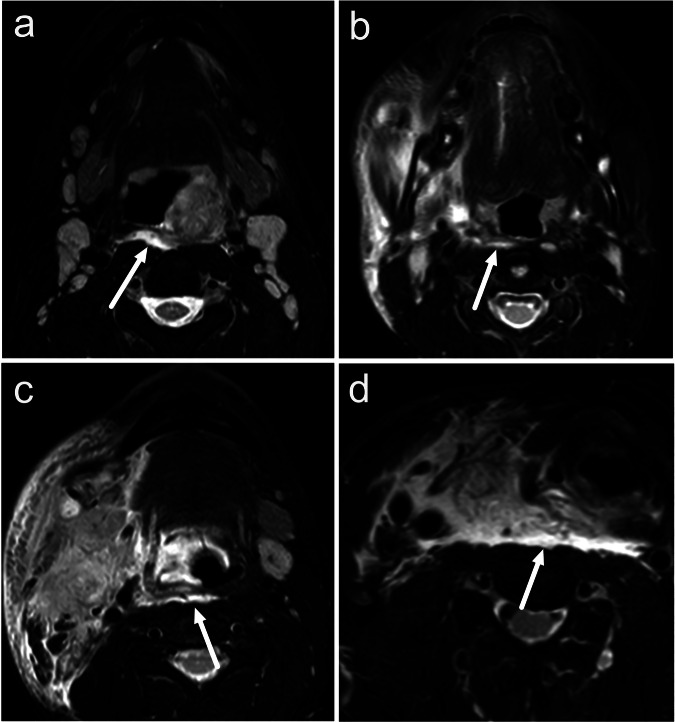
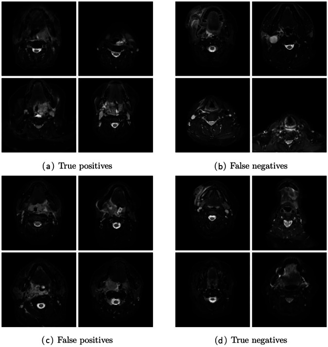
Background: In acute neck infections, magnetic resonance imaging (MRI) shows retropharyngeal edema (RPE), which is a prognostic imaging biomarker for a severe course of illness. This study aimed to develop a deep learning-based algorithm for the automated detection of RPE.
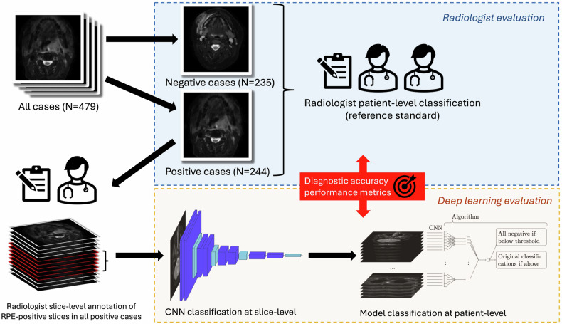
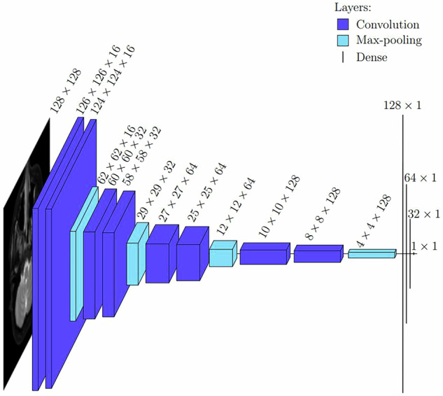
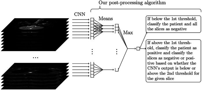
Methods: We developed a deep neural network consisting of two parts using axial T2-weighted water-only Dixon MRI images from 479 patients with acute neck infections annotated by radiologists at both slice and patient levels. First, a convolutional neural network (CNN) classified individual slices; second, an algorithm classified patients based on a stack of slices. Model performance was compared with the radiologists' assessment as a reference standard. Accuracy, sensitivity, specificity, and area under receiver operating characteristic curve (AUROC) were calculated. The proposed CNN was compared with InceptionV3, and the patient-level classification algorithm was compared with traditional machine learning models.
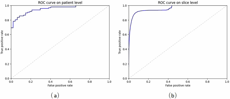
Results: Of the 479 patients, 244 (51%) were positive and 235 (49%) negative for RPE. Our model achieved accuracy, sensitivity, specificity, and AUROC of 94.6%, 83.3%, 96.2%, and 94.1% at the slice level, and 87.4%, 86.5%, 88.2%, and 94.8% at the patient level, respectively. The proposed CNN was faster than InceptionV3 but equally accurate. Our patient classification algorithm outperformed traditional machine learning models.
Conclusion: A deep learning model, based on weakly annotated data and computationally manageable training, achieved high accuracy for automatically detecting RPE on MRI in patients with acute neck infections.
Relevance statement: Our automated method for detecting relevant MRI findings was efficiently trained and might be easily deployed in practice to study clinical applicability. This approach might improve early detection of patients at high risk for a severe course of acute neck infections.
Key points: Deep learning automatically detected retropharyngeal edema on MRI in acute neck infections. Areas under the receiver operating characteristic curve were 94.1% at the slice level and 94.8% at the patient level. The proposed convolutional neural network was lightweight and required only weakly annotated data.
Keywords: Artificial intelligence; Magnetic resonance imaging; Neural networks (computer); Respiratory tract infections; Retropharyngeal abscess.
© 2025. The Author(s).
Conflict of interest statement
Declarations. Ethical approval and consent to participate: According to the national legislation, no separate ethics committee approval is needed for retrospective studies that involve a secondary use of registry or archival data. Consent for publication: According to the national legislation, no separate ethics committee approval is needed for retrospective studies that involve a secondary use of registry or archival data. Competing interests: The authors declare that they have no competing interests.
Figures
Similar articles
-
A deep learning approach to direct immunofluorescence pattern recognition in autoimmune bullous diseases.Br J Dermatol. 2024 Jul 16;191(2):261-266. doi: 10.1093/bjd/ljae142. Br J Dermatol. 2024. PMID: 38581445
-
Identifying Primary Sites of Spinal Metastases: Expert-Derived Features vs. ResNet50 Model Using Nonenhanced MRI.J Magn Reson Imaging. 2025 Jul;62(1):176-186. doi: 10.1002/jmri.29720. Epub 2025 Jan 27. J Magn Reson Imaging. 2025. PMID: 39868626
-
Diagnosis of Sacroiliitis Through Semi-Supervised Segmentation and Radiomics Feature Analysis of MRI Images.J Magn Reson Imaging. 2025 Aug;62(2):563-572. doi: 10.1002/jmri.29731. Epub 2025 Feb 6. J Magn Reson Imaging. 2025. PMID: 39912494
-
Pharmacological and electronic cigarette interventions for smoking cessation in adults: component network meta-analyses.Cochrane Database Syst Rev. 2023 Sep 12;9(9):CD015226. doi: 10.1002/14651858.CD015226.pub2. Cochrane Database Syst Rev. 2023. PMID: 37696529 Free PMC article.
-
Transformers for Neuroimage Segmentation: Scoping Review.J Med Internet Res. 2025 Jan 29;27:e57723. doi: 10.2196/57723. J Med Internet Res. 2025. PMID: 39879621 Free PMC article.
References
-
- Boscolo-Rizzo P, Stellin M, Muzzi E et al (2012) Deep neck infections: a study of 365 cases highlighting recommendations for management and treatment. Eur Arch Otorhinolaryngol 269:1241–1249. 10.1007/s00405-011-1761-1 - PubMed
-
- Kamalian S, Avery L, Lev MH et al (2019) Nontraumatic head and neck emergencies. Radiographics 39:1808–1823. 10.1148/rg.2019190159 - PubMed
MeSH terms
LinkOut - more resources
Full Text Sources
Medical