. 2025 Sep;39(9):701-714.
doi: 10.1177/15459683251345434. Epub 2025 Jun 19.

Diffusion Tensor Imaging Findings in Cerebral Sensorimotor Areas in Patients After Spinal Cord Injury Correlate With Neurophysiological Deficits

Affiliations

Diffusion Tensor Imaging Findings in Cerebral Sensorimotor Areas in Patients After Spinal Cord Injury Correlate With Neurophysiological Deficits

Anna Zimny et al. Neurorehabil Neural Repair. 2025 Sep.

Abstract

ObjectivesAssessment of sensorimotor cortex and tracts degeneration using novel diffusion tensor imaging (DTI) templates in patients with chronic spinal cord injury (SCI) and its correlation with clinical and neurophysiological findings.MethodsSex and age-matched 29 patients with chronic SCI (paraplegic: p-SCI; tetraplegic: t-SCI) and 29 healthy controls underwent neurophysiological assessment including motor evoked potentials (MEP). DTI was performed on 3 T magnetic resonance imaging scanner and postprocessed using Human Motor Area and Sensorimotor Area Tract Templates. DTI parameters were compared using analysis of covariance with post hoc Scheffé and Bonferroni corrections. Spearman's rank test was used for correlations with P < .05 considered significant.ResultsCompared to controls, all SCI patients showed significantly lower fractional anisotropy (FA) in several tracts (primary motor [M1], somatosensory [S1], pre-supplementary motor area [preSMA], and dorsal premotor [PMd]) and cortices (M1, pre-SMA, and S1). There were no differences in DTI parameters between p-SCI and t-SCI or p-SCI and controls. Compared to controls, t-SCI showed significantly decreased FA within M1 and S1 tracts. In t-SCI higher motor scores were associated with higher FA from ventral premotor area (PMv) tracts and cortex; higher sensory scores were associated with higher FA from S1 tracts. Positive correlations were found between MEP amplitudes from rectus femoris muscles and FA for M1, PMd, PMv, pre-SMA, SMA tracts, and PMv cortex.ConclusionsDTI shows remote degeneration of sensorimotor cortex and supraspinal tracts in SCI correlating with several clinical motor and sensory scores, and MEP parameters. DTI metrics have the potential to become biomarkers of remote degeneration.

Keywords: chronic spinal cord injury; clinical metrics; diffusion tensor imaging; motor evoked potentials; sensorimotor cortex; sensorimotor tracts.


Conflict of interest statement

Declaration of Conflicting InterestsThe author(s) declared no potential conflicts of interest with respect to the research, authorship, and/or publication of this article.

Figures

Figure 1.
Figure 1.
(A) Examples of the motor evoked potentials recordings from abductor pollicis brevis and rectus femoris muscles bilaterally following the transcranial magnetic stimulation in the healthy volunteer (a), a tetraplegic patient after SCI (b), and a paraplegic patient after SCI (c). Calibration bars for amplification (vertical) and the time base (horizontal) of MEP recordings set during neurophysiological tests are shown in the left part of the figure. Note that amplifications of recordings in (b) and (c) are greater than in (a). (B) Sensorimotor tract parcellation using Sensorimotor Area Tract Template (SMATT) and sensorimotor cortical areas parcellation using Human Motor Area Template (HMAT)—planar multislice and 3D views. Abbreviations: M1, primary motor area, PMd, dorsal premotor area, PMv, ventral premotor area, preSMA, pre-supplementary motor area, S1, primary somatosensory area, SMA, supplementary motor area, Thenar, Abductor pollicis brevis muscle, Rect fem, Rectus femoris muscle, R, Right, L, Left.
Figure 2.
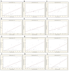
Figure 2.
The graphical presentations of correlations between clinical motor and sensory scores and mean fractional anisotropy (FA) values from different cortical and white matter locations in t-SCI patients: (A) LEMS versus mean FA from PMv tracts, (B) SCIM Mobility Score versus mean FA from PMv tracts, (C) Light Touch Score versus mean FA from S1 tracts, (D) LEMS versus mean FA from PMv cortices, (E) SCIM Mobility Score versus mean FA from PMv cortices, and (F) Total Touch Score versus mean FA from S1 tracts as well as graphical presentations of correlations between motor evoked potential (MEP) amplitudes recorded from rectus femoris muscles (RFM) and mean fractional anisotropy (FA) values from different cortical and white matter locations in t-SCI patients: (G) mean MEP amplitude recorded from RFM versus mean FA from M1 tracts, (H) mean MEP amplitude recorded from RFM versus mean FA from PMv tracts, (I) mean MEP amplitude recorded from RFM versus mean FA from SMA tracts, (J) mean MEP amplitude recorded from RFM versus mean FA from PMd tracts, (K) mean MEP amplitude recorded from RFM versus mean FA from Pre-SMA tracts and (L) mean MEP amplitude recorded from RFM versus mean FA from PMv cortices. Abbreviations: FA, fractional anisotropy; t-SCI, tetraplegic patients after spinal cord injury; LEMS, Lower Extremity Motor Score; SCIM, Spinal Cord Independence Measure; PMv, ventral premotor area; S1, primary somatosensory area; MEP, motor evoked potential; RFM, rectus femoris muscle; M1, primary motor area; PMd, dorsal premotor area; preSMA, pre-supplementary motor area; SMA, supplementary motor area.

Similar articles

References

    1. Ahuja CS, Wilson JR, Nori S, et al. Traumatic spinal cord injury. Nat Rev Dis Primers. 2017;3:17018. doi: 10.1038/nrdp.2017.18 - DOI - PubMed
    1. Alizadeh A, Dyck SM, Karimi-Abdolrezaee S. Traumatic spinal cord injury: an overview of pathophysiology, models, and acute injury mechanisms. Front Neurol. 2019;10:282. doi: 10.3389/fneur.2019.00282 - DOI - PMC - PubMed
    1. Freund P, Weiskopf N, Ashburner J, et al. MR investigation of the sensorimotor cortex and the corticospinal tract after acute spinal cord injury: a prospective longitudinal study. Lancet Neurol. 2013;12:873-881. doi: 10.1016/S1474-4422(13)70146-7 - DOI - PMC - PubMed
    1. Huber J, Leszczyńska K, Wincek A, et al. The role of peripheral nerve electrotherapy in functional recovery of muscle motor units in patients after incomplete spinal cord injury. Appl Sci. 2021;11(20):9764. doi: 10.3390/app11209764 - DOI
    1. Kim BG, Dai HN, McAtee M, Vicini S, Bregman BS. Remodeling of synaptic structures in the motor cortex following spinal cord injury. Exp Neurol. 2006;198(2):401-415. doi: 10.1016/j.expneurol.2005.12.010 - DOI - PubMed

LinkOut - more resources