. 2025 Jun 19;16(1):5329.
doi: 10.1038/s41467-025-60839-6.

ELD1 mediates photoperiodic flowering via OsCCA1 alternative splicing and interacts with phytochrome signaling in rice

Affiliations

ELD1 mediates photoperiodic flowering via OsCCA1 alternative splicing and interacts with phytochrome signaling in rice

Liang Cai et al. Nat Commun. .

Abstract

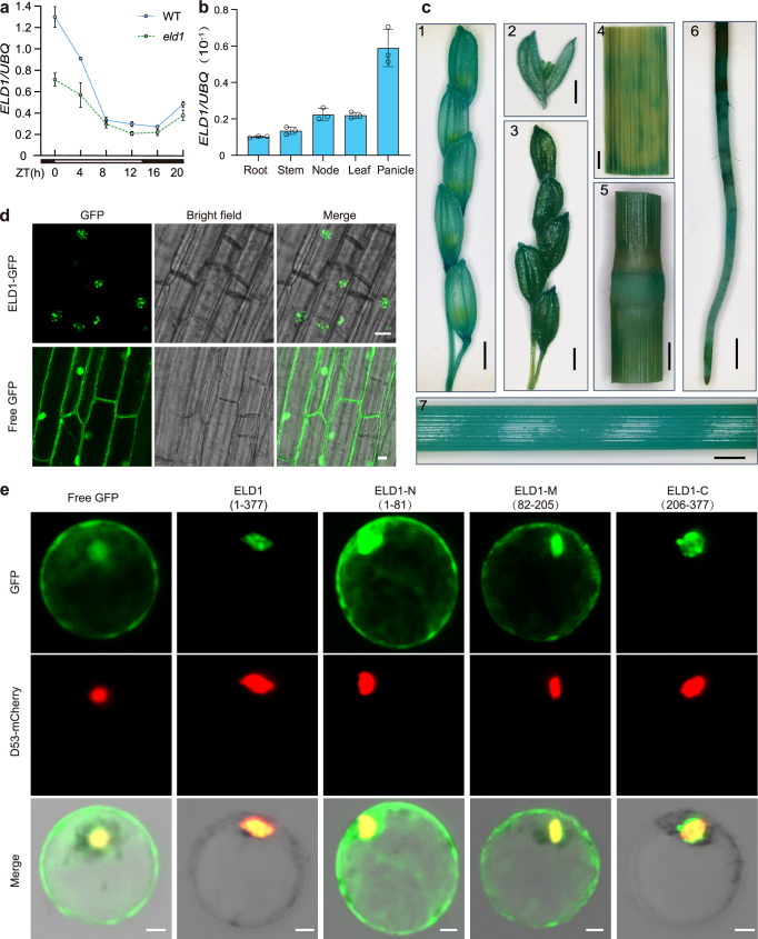
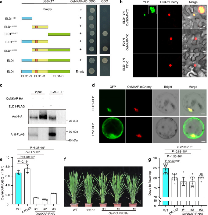
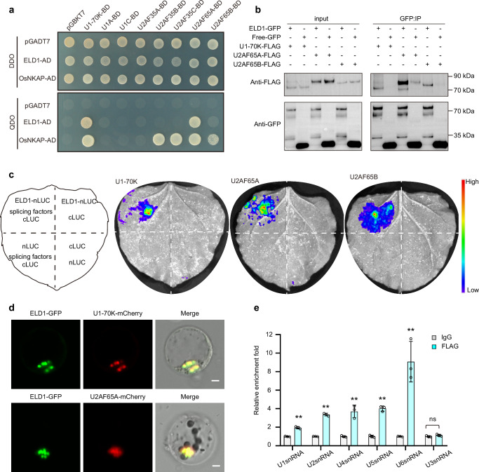
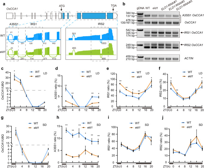
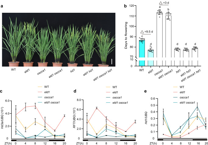
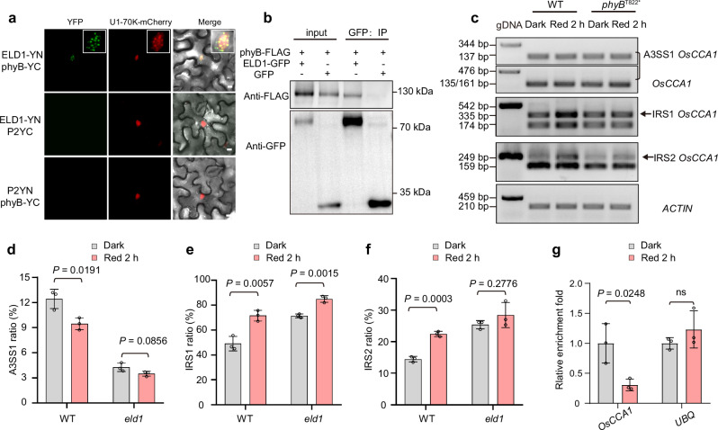
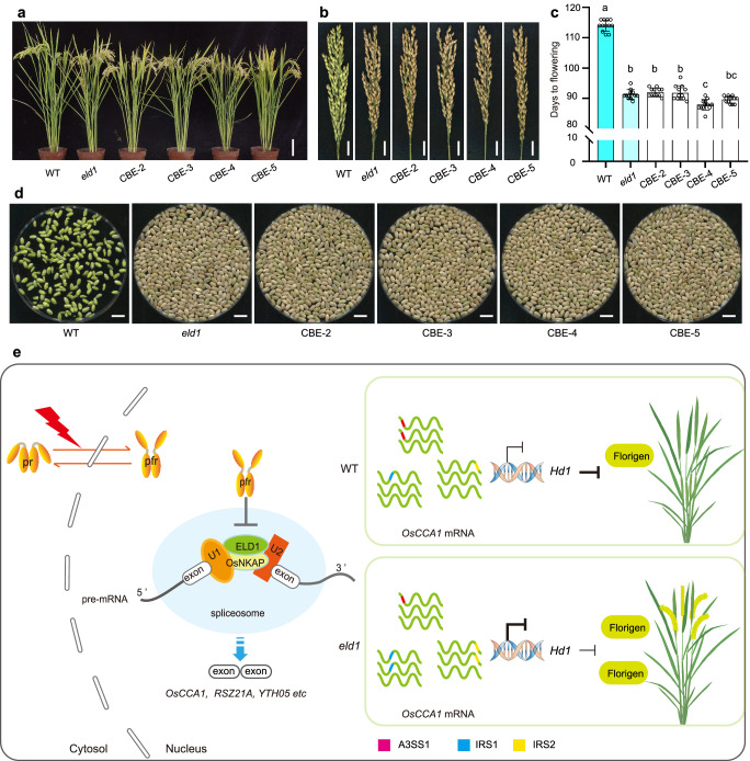
Photoperiodic flowering in plants is orchestrated by the dynamic interaction between light signals and the endogenous circadian clock, but how light signals integrate into the clock remains to be fully elucidated. Here, we identify ELD1, a CCHC-type zinc finger protein that is essential for rice embryo survival. Notably, partial loss of ELD1 function results in early flowering under long-day conditions. Further investigations demonstrate that ELD1 physically interacts with OsNKAP, an orthologue of mammal NF-κB activating protein, as well as core splicing factors to regulate the splicing profile of OsCCA1, a core oscillator of the circadian clock. Molecular and genetic evidence indicate that OsCCA1 is the primary target of ELD1 in controlling flowering time. Additionally, ELD1 interacts with photoactivated phyB, mediating red-light-regulated alternative splicing of OsCCA1. Collectively, our findings establish a molecular connection between light signaling and the circadian clock, with ELD1 modulating OsCCA1 alternative splicing to control photoperiodic flowering.


Conflict of interest statement

Competing interests: The authors declare no competing interests.

Figures

Fig. 1
Fig. 1. Phenotype analysis of eld1 mutant and cloning of the corresponding gene.
a, b Phenotypic comparison of WT and eld1 plants at heading stage under Nanjing natural long-day conditions (NLD, ~14.25 h of daylight at transplanting) and Hainan natural short-day conditions (NSD, ~11 h of daylight at transplanting). White arrows indicate rice panicles. Scale bar, 10 cm. c Days to flowering of WT and eld1 mutant under Beijing NLD conditions (~ 15 h of daylight at transplanting), Nanjing NLD conditions, and Hainan NSD condition. Values are presented as means ± SD (n = 10 biological replicates), two-sided Student’s t-test was used to calculate P values. d Gene structure diagram of ELD1. The white and blue boxes represent UTR and CDS. The sequence and trace file of WT, eld1, and CRISPR-based adenine base editor (ABE) recover edit lines are indicated. e Phenotypic comparison of WT, eld1, and two independent ABE recover edit lines at the heading stage in Nanjing. Scale bars, 10 cm. f Days to flowering of WT, eld1, and two independent ABE recover edit lines at the heading stage in Nanjing. Values are presented as means ± SD (n = 10 biological replicates). g Phenotypic comparison of WT, eld1, and three independent transgenic complemented lines (C1 to C3) at heading stage under Nanjing NLD conditions. Scale bars, 10 cm. h Days to flowering of WT, eld1, and three independent transgenic complemented lines (C1 to C3) grown under Nanjing NLD conditions. Values are presented as means ± SD (n = 10 biological replicates). For (f and h), different letters were used to denote statistically significant differences, which were determined using one-way ANOVA with Tukey’s multiple comparisons test (P < 0.05).
Fig. 2
Fig. 2. ELD1 is broadly expressed and encodes a nuclear speckle localized protein.
a Rhythmic expression of ELD1 over 24 h in WT and eld1 mutant, detected by RT-qPCR in the leaves of 40-day-old plants grown under controlled long-day conditions (CLD, 14 h light/10 h dark). Values are means ± SD (n = 3 biological replicates). b RT-qPCR analysis of ELD1 expression in various tissues at heading stage. Values are means ± SD (n = 3 biological replicates). c GUS staining of the tissues collected from pELD1:GUS transgenic plants at the heading stage. (1) young panicle, (2) (3) panicle, (4) flag leaf, (5) stem node, (6) root, and (7) stem. Scale bars, 2.5 mm. d Subcellular localization of ELD1-GFP and free GFP in root cells of 7-day-old transgenic plants. Scale bars, 10 μm. e Subcellular localization of full-length ELD1 and various truncated forms in rice protoplasts. ELD1 (1–377) represents the full-length ELD1, ELD1-N (1–81) includes the conserved N-terminal, ELD1-M (82–205) contains the conserved ZCCHC domain, and ELD1-C (206–377) comprises the RS-enriched C-terminal region. D53-mCherry was used as a nuclear marker. Scale bars, 5 μm.
Fig. 3
Fig. 3. OsNKAP interacts with ELD1 and suppresses flowering in rice.
a The interaction between ELD1 and OsNKAP in yeast cells. The ELD1-N (1–81), ELD1-M (82–205), and ELD1-C regions are indicated; The ZCCHC domain was marked in red. DDO refers to SD/-Trp/-Leu, and QDO refers to SD/-Trp/-Leu/-His/-Ade. The symbol “+” indicates the presence the corresponding construct, whereas “empty” represents the empty construct. b Bimolecular fluorescence complementation (BiFC) analysis revealed the interaction between ELD1 and OsNKAP in N. benthamiana cells. The inset images show the magnified portions of recombinant YFP signal. D53-mCherry serving as nuclear marker. Scale bars, 10 μm. c Co-immunoprecipitation (Co-IP) assays in rice protoplasts show that ELD1 can interact with OsNKAP. Immunoprecipitated samples were detected using anti-HA and anti-FLAG antibodies, respectively. The symbols “+” represent presence corresponding construct; symbols “−” represent free construct. Three independent experiments were performed with similar results. d ELD1-GFP co-localizes with OsNKAP-mCherry in rice protoplasts, and exhibited nuclear speckles localization. Free GFP was used as the control, Scale bars, 5 μm. e RT-qPCR analysis OsNKAP expression level in WT, OsNKAP-CR162, and three OsNKAP-RNAi lines. Samples were collected from 20-day-old seedlings grown under CLD conditions at ZT0. Values are presented as means ± SD (n = 3 biological replicates), two-sided Student’s t-test was used to calculate P values. f Phenotypes of WT, OsNKAP-CR162, and three distinct OsNKAP-RNAi lines at heading stage growth in Nanjing. Scale bars, 10 cm. g Days to flowering of WT, OsNKAP-CR162, and three independent OsNKAP-RNAi lines in Nanjing. Values are presented as means ± SD (n = 10 biological replicates), two-sided Student’s t-test was used to calculate P values.
Fig. 4
Fig. 4. ELD1 interacts with snRNPs and snRNA.
a A yeast-two hybrid assay shows ELD1 and OsNKAP interact with core splicing factors. DDO refers to SD/-Trp/-Leu, and QDO refers to SD/-Trp/-Leu/-His/-Ade. b The in vivo Co-IP assay confirms that ELD1 interacts with U1-70K, U2AF65A, and U2AF65B in N. benthamiana. The immunoprecipitated samples were detected using the anti-GFP and anti-FLAG antibodies. The symbol “+” indicates presence of the corresponding protein, while the symbol “−” represents the empty construct. Three independent experiments were performed with similar results. c The luciferase complementation imaging (LCI) assay confirms that ELD1 interacts with U1-70K, U2AF65A, and U2AF65B. d Co-localization of ELD1-GFP with U1-70K-mCherry and U2AF65A-mCherry in rice protoplasts. Scale bars, 5 μm. e RNA immunoprecipitation followed by qPCR (RIP-qPCR) assay demonstrating the binding affinity of ELD1 protein to snRNAs in vivo. The pUBI:ELD1-FLAG/eld1 transgenic seedlings grown for 10 days under CLD conditions were used. Samples were immunoprecipitated with anti-FLAG Magnetic Beads or the IgG control. UBQ gene serving as an internal control. Values are presented as means ± SD (n = 3 biological replicates). The asterisks indicate significant differences from IgG control using the two-sided student’s t-test (**P < 0.01).
Fig. 5
Fig. 5. ELD1 regulates AS of OsCCA1.
a The Sashimi plot from the Integrative Genomics Viewer (IGV) illustrates three distinct alternative splicing sites of OsCCA1, highlighting differences between the WT and eld1 mutant. b sqRT-PCR analysis of A3SS1, IRS1 and IRS2 splicing pattern of OsCCA1 in WT, eld1 and three ELD1-RNAi lines. Using cDNA as a template, the sqRT-PCR primers (ACTIN, A3SS1, and IRS1) amplify the target regions along with adjacent constitutive introns, with genomic DNA (gDNA) serving as a control. All the sqRT-PCR reactions run for 27 cycles, except for A3SS1, which runs for 28 cycles due to its relatively low isoform expression. Samples were collected from 20-day-old seedlings grown under CLD conditions at ZT0. More than three independent experiments were performed with similar results. c Rhythmic expression of OsCCA1 over 24 h in WT and eld1 mutant, detected by RT-qPCR in the leaves of 40-day-old plants grown under CLD conditions. Values are means ± SD (n = 3 biological replicates). d–f The rhythmic splicing pattern of A3SS1 (d), IRS1 (e), and IRS2 (f) was detected by RT-qPCR in both WT and eld1. Total RNA was extracted from 40-day-old plants under CLD conditions. Values are presented as means ± SD (n = 3 biological replicates). g Rhythmic expression of OsCCA1 over 24 h in WT and eld1 mutant, detected by RT-qPCR in the leaves of 27-day-old plants grown under CSD conditions. Values are means ± SD (n = 3 biological replicates). h–j The rhythmic splicing pattern of A3SS1 (h), IRS1 (i) and IRS2 (j) detected by RT-qPCR in WT and eld1. Total RNA was extracted from 27-day-old plants under CSD conditions. Values are presented as means ± SD (n = 3 biological replicates). For (cf and h), Asterisks indicate significant differences from WT using the two-sided Student’s t-test (*P < 0.01, *P < 0.05).
Fig. 6
Fig. 6. ELD1 regulates flowering time through OsCCA1-Hd1 pathway.
a Phenotypes of WT, eld1, oscca1, eld1 oscca1, hd1, eld1 hd1 and eld1 hd1 oscca1 at heading stage growth in Nanjing NLD conditions. Scale bars, 10 cm. b Days to flowering of WT, eld1, oscca1, eld1 oscca1, hd1, eld1 hd1 and eld1 hd1 oscca1 in Nanjing NLD conditions. Values are presented as means ± SD (n = 12 biological replicates). Different letters were used to denote statistically significant differences, which were determined using one-way ANOVA with Tukey’s multiple comparisons test (P < 0.05). c–e Expression of Hd1 and two florigens genes in WT, eld1, oscca1, and eld1 oscca1 detected by RT-qPCR. Samples were collected from 40-day plants grown under CLD conditions. Values are presented as means ± SD (n = 3 biological replicates).
Fig. 7
Fig. 7. ELD1 interacts with phyB to regulate OsCCA1 AS.
a BiFC analysis revealed the interaction between ELD1 and phyB in N. benthamiana cells. The inset images display the magnified portions of YFP signal. U1-70K mCherry serving as nuclear speckles marker, Scale bars, 10 μm. b The in vivo Co-IP assay confirms the interaction between ELD1 and phyB in N. benthamiana. The immunoprecipitated samples were detected using the anti-GFP and anti-FLAG antibodies. The symbols “+” represent presence corresponding protein; symbols “−” represent empty construct. Three independent experiments were performed with similar results. c The AS pattern of A3SS1, IRS1, and IRS2 detected by sqRT-PCR after 2 h of red-light treatment in WT and phyBT822*. Continuous dark treatment serves as control. More than three independent experiments were performed with similar results. d–f The AS pattern of A3SS1 (d), IRS1 (e), and IRS2 (f) detected by RT-qPCR after 2 h of red-light treatment in WT and eld1 mutant. The indicated genotypes of 10-day-old seedlings were grown under CLD conditions, pretreated with darkness for 48 h, and then exposed to red light for 2 h. Continuous dark treatment serves as a control. Values are presented as means ± SD (n = 3 biological replicates). g RIP-qPCR assay shows the in vivo binding affinity of ELD1 to OsCCA1 mRNA. pUBI:ELD1-FLAG/eld1 transgenic seedlings grown for 10 days under CLD conditions were pretreated with darkness for 48 h, and then exposed to red light for 2 h. Continuous dark treatment serves as the control. Samples were immunoprecipitated with anti-FLAG Magnetic Beads. UBQ gene serves as an internal control. Values are presented as means ± SD (n = 3 biological replicates). For (dg), two-sided Student’s t-test was used to calculate P values.
Fig. 8
Fig. 8. Breeding utilization of ELD1 through CRISPR-based cytosine base editors (CBE) and a proposed working model of ELD1.
a Phenotypes of WT, eld1 and four independent CBE editing lines at heading stage during growth in Beijing NLD conditions. Scale bars, 10 cm. b Panicle maturity of WT, eld1, and four independent CBE editing lines at the time of harvest in Beijing. Scale bars, 2 cm. c Days to flowering of WT, eld1 and the CBE editing lines in Beijing NLD conditions. Values are presented as means ± SD (n = 12 biological replicates). Different letters were used to denote statistically significant differences, which were determined using one-way ANOVA with Tukey’s multiple comparisons test (P < 0.05). d Brown rice of WT, eld1, and CBE editing lines at the time of harvest in Beijing. The WT failed to mature and nearly no harvest, while eld1 and four CBE lines were able to mature and be harvested normally. e The working model of ELD1 in regulating photoperiodic flowering. ELD1 interacts OsNKAP and core splicing factors to regulate global AS. The ELD1-containing spliceosome complex directly binds and regulates the rhythmic AS of OsCCA1. Additionally, phyB physically interacts with ELD1 in a light dependent manner, inhibiting its binding to OsCCA1 pre-mRNA. This regulation ensures appropriate OsCCA1 activity and a properly tuned circadian clock, resulting in high Hd1 transcription and prolonged flowering at LD. In the eld1 mutant, the OsCCA1 has aberrant splicing pattern at A3SS1, IRS1, and IRS2 sites. These aberrantly spliced forms of OsCCA1 strongly suppress Hd1 transcription, leading to the activation of downstream florigen genes and ultimately causing early flowering.

Similar articles

References

    1. Yano, M. et al. Hd1, a major photoperiod sensitivity quantitative trait locus in rice, is closely related to the Arabidopsis flowering time gene CONSTANS. Plant Cell12, 2473–2483 (2000). - PMC - PubMed
    1. Hu, Z. et al. Autophagy targets Hd1 for vacuolar degradation to regulate rice flowering. Mol. Plant15, 1137–1156 (2022). - PubMed
    1. Doi, K. et al. Ehd1, a B-type response regulator in rice, confers short-day promotion of flowering and controls FT-like gene expression independently of Hd1. Gene Dev.18, 926–936 (2004). - PMC - PubMed
    1. Zhou, S. et al. Transcriptional and post-transcriptional regulation of heading date in rice. N. Phytol.230, 943–956 (2021). - PMC - PubMed
    1. Lee, S. J., Kang, K., Lim, J. H. & Paek, N. C. Natural alleles of CIRCADIAN CLOCK ASSOCIATED1 contribute to rice cultivation by fine-tuning flowering time. Plant Physiol.190, 640–656 (2022). - PMC - PubMed

LinkOut - more resources