. 2025 Jun 19;16(1):131.
doi: 10.1186/s13244-025-02005-6.

Pelvic nerve endometriosis: MRI features and key findings for surgical decision

Affiliations

Pelvic nerve endometriosis: MRI features and key findings for surgical decision

Justine Bourg et al. Insights Imaging. .

Abstract

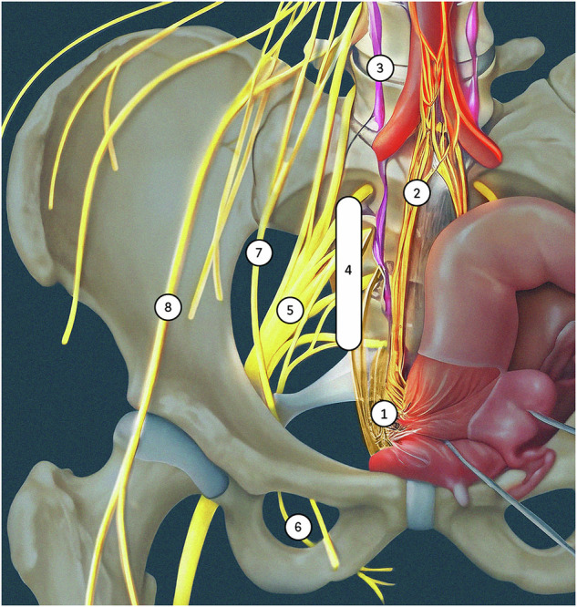
Endometriosis is a prevalent gynecological disorder in women of reproductive age. It is the leading cause of chronic pelvic pain. While the mechanisms underlying this pain remain elusive, rare cases of pelvic nerve involvement can result in severe, debilitating symptoms, adding complexity to the clinical landscape. Nerve involvement typically results from the direct extension of deep infiltrating endometriosis, though it may also occur in isolation. The nerves most commonly affected include the inferior hypogastric and lumbosacral plexuses, as well as the sciatic, pudendal, obturator, and femoral nerves. Early and accurate diagnosis is essential for the effective management of the pain and the prevention of irreversible nerve damage. Given the limitations of transvaginal ultrasonography in visualizing the lateral compartment, MRI is considered the gold standard for detecting and evaluating pelvic nerve involvement. Through the use of optimized protocols to enhance the visualization of nerves and their anatomical landmarks, radiologists play a key role in the identification of endometriotic lesions. A comprehensive and structured radiology report is essential for surgical planning, as nerve involvement often requires precise interventions to alleviate symptoms and restore quality of life. CRITICAL RELEVANCE STATEMENT: Accurate identification and a structured reporting of pelvic nerve endometriosis in the lateral compartment are pivotal to guide surgical decision-making and optimize patient outcomes. KEY POINTS: Pelvic nerve endometriosis is often overlooked, underestimated by clinicians, and underdiagnosed on imaging. Timely nerve involvement diagnosis prevents permanent damage in pelvic pain with neurological symptoms. Deep endometriosis in the lateral compartment may extend to the pelvic nerves. The inferior hypogastric plexus, sacral plexus, sciatic, and pudendal nerves are commonly affected. A dedicated MRI protocol with 3D T2-weighted sequence ensures accurate pelvic nerve assessment.

Keywords: Endometriosis; Magnetic resonance imaging; Pelvic pain; Pelvic plexus.


Conflict of interest statement

Declarations. Ethics approval and consent to participate: All procedures performed in studies involving human participants were in accordance with the ethical standards of the institutional and/or national research committee and with the 1964 Helsinki Declaration and its later amendments or comparable ethical standards. Consent for publication: Written informed consent was not required for this educational review because of its retrospective design and nature. No patients can be identified in the images or clinical information presented in this educational review. Competing interests: Pascal Rousset reported consultant fees from Ziwig and EDAP TMS France, reported receiving lecture fees from Bracco, General Electric Health care, and compensation for serving on the board from Guerbet. Isabelle Thomassin-Naggara reported receiving lecture fees from General Electric, Siemens, Canon, and GSK; lecture fees and compensation for serving on the board from Guerbet; compensation for serving on the board from Bayer; lecture fees from Incepto, ICAD, Fujifilm, and Hologic; and lecture fees and compensation for serving on the board from Bracco. François Golfier reported consultant fees from ZIWIG.

Figures

Fig. 1
Fig. 1
Anatomical illustration of somatic and autonomic pelvic nerves in a female pelvis (frontal view, adapted from Alkatout et al [37]). 1: inferior hypogastric plexus, 2: hypogastric nerve, 3: sympathetic trunk with chain of ganglia, 4: sacral roots (S1–S4), 5: sciatic nerve, 6: pudendal nerve, 7: obturator nerve, and 8: femoral nerve
Fig. 2
Fig. 2
DE in a 29-year-old woman with dysmenorrhea, deep dyspareunia, and dyschezia. A Axial and (B) sagittal T2W MR images show a right subperitoneal infiltrative lesion (dotted lines) involving the anterolateral rectal wall and extending to the mesorectal fascia (A, white arrowhead) and beyond into the right posterolateral parametrium. The infiltration of the inferior hypogastric plexus shows spiculated margins (B, black arrows) but no extension to the sacral roots or the pelvic wall (A, dashed arrow). Note the safety fat line between the lesion and the iliococcygeus muscle of the levator ani muscle (B, white arrows). Laparoscopic surgery confirms the involvement of the inferior hypogastric plexus and a cleavage plane with the levator ani muscle and sacral roots
Fig. 3
Fig. 3
DE in a 42-year-old woman, with a history of rectal shaving 10 years ago, who presented with a recurrence of symptoms, in particular deep dyspareunia and right sciatica. A, B Axial T2 and (C) sagittal T2W MR images show right subperitoneal infiltrative lesion involving the anterolateral rectal wall (B, arrowhead), extending to the right posterolateral parametrium with inferior hypogastric plexus involvement (stars), up to the pelvic wall with encasement of hypogastric vessels (A, B, dashed arrows) and contact with the piriformis muscle. The posterior spiculated margins come into contact with the S3 and S4 sacral roots (C, arrows) and the posterior part of the right sciatic nerve (B, circle). D Axial fat-suppressed T1W MR image reveals T1-hyperintense endometriotic hemorrhagic microcysts (arrows). The surgical procedure confirmed extrinsic involvement and included a shaving of the right sacral roots and right sciatic nerve, as well as a section of the hypogastric vessels
Fig. 4
Fig. 4
Severe endometriosis in a 31-year-old woman with dysmenorrhea, dyspareunia, and dysesthesia in the right lower extremity. AC Axial 3D T2W images from top to bottom show endometriotic infiltration of the rectal wall (C, arrowhead) and posterior vaginal fornix (C, star), with complete extension to the right posterolateral parametrium including the inferior hypogastric plexus (black dashed arrows) with posterior attraction of S1 and circumferential involvement of S2, S3 and S4. Note the absence of extension of the ureter (B, C, white dashed arrow), the piriformis muscle, or internal iliac vessels. D Sagittal 3D reconstruction T2W image at the level of the right posterolateral parametrium showing involvement of the inferior hypogastric plexus (star) extending inferiorly to the levator ani muscle (dashed arrow), and posteriorly to sacral roots S2, S3, and S4, and with spiculation up to S1 but no involvement. E The robotic laparoscopic view shows a complete adhesion (white arrows) between the anterior rectal wall (star), the uterus and vagina, the peritoneal infiltration is being subperitoneal (dotted line). Note the hypogastric nerve (black arrow) passing through the inferior hypogastric plexus affected by the endometriosis and the course of the ureter (dashed arrows) distant from the endometriotic lesion. F The robotic laparoscopic photography view shows, after careful dissection, the upper part of the endometriotic lesion (dotted line) involving the rectal wall (star), S2, and partially attracting S1. Note the course of the ureter (dashed arrow). G Robotic laparoscopic view showing, after dissection of the endometriotic lesion (dotted line) from the rectal wall, involvement of the posterior vaginal fornix with a submucous hemorrhagic cystic component (arrow) after colpotomy, of S2, S3, and S4 and retraction of S1. Note the absence of involvement of the piriformis muscle (star). A complete nerve-sparing resection was performed, with S1 being retracted but removable from the lesion, and S2–S4 being circumscribed but without macroscopic intrinsic infiltration
Fig. 5
Fig. 5
DE in a 31-year-old woman with right catamenial sciatica. A Axial T2W MR image shows fibrotic thickening in the right sciatic notch involving the right sciatic nerve (circle) with loss of its spaghetti aspect compared to the left sciatic nerve (arrow). This lesion is isolated without involvement of the ovarian fossa or the sacrorectal septum, and could illustrate the potential existence of a peritoneal diverticulum. Note the hypertrophy of the piriformis muscle (star). B Axial fat-suppressed T1W MR image shows diffuse T1-hyperintense endometriotic hemorrhagic microcysts (dashed arrow). C Axial T2W MR image shows fatty atrophy of the right obturator internus muscle (black arrow), indicating a concomitant involvement of the nerve to the obturator internus muscle passing through the sciatic notch. The patient was medically treated with an LHRH analog
Fig. 6
Fig. 6
DE in a 31-year-old woman with right sciatica initially cyclical, then became chronic with walking difficulties. A Axial and (B) coronal T2W MR images show an infiltrative endometriotic mass of 5 cm (measure not shown) (dotted line) centered on the right sciatic notch with involvement of the sciatic nerve. Note the hypertrophy of the piriformis muscle (A, star). C Axial fat-suppressed T2W MR image shows denervation of the gluteus medius and minimus muscles (stars), indicating the involvement of the superior gluteal nerve in the sciatic notch, as well as the piriformis muscle. D Axial fat-suppressed T1W MR image shows a right-sided endometrioma (arrow) and hemorrhagic implants of the left ovary (dashed arrow). Surgical intervention was not possible due to the intrinsic involvement of the sciatic nerve, which could risk nerve damage. The patient was treated medically with an LHRH analog
Fig. 7
Fig. 7
DE in a 29-year-old woman with deep dyspareunia and left pudendalgia. AC Axial and (D) sagittal T2W MR images show fibrotic nodular infiltration of the left posterolateral parametrium (stars) extending to the pelvic wall and to the iliococcygeus muscle of the levator ani muscle (D, black arrows), involving the left inferior hypogastric plexus, the left sacral nerves courses S3 and S4, and the level of origin of the left proximal pudendal nerve (C, circle). The patient was treated medically with hormone therapy
Fig. 8
Fig. 8
DE in a 43-year-old woman with dyspareunia and perineal pain. A Axial and (B) coronal T2W MR images show an endometriotic lesion (circle) of the right distal mediolateral parametrium extending to the pelvic wall in contact with the pudendal vascular-nerve bundle at the level of arcus tendineus of the levator ani muscle (B, arrowhead). The right ureter passes quite widely inside this lesion (A, arrow). This lesion is located in front of the right ischial spine mark (A, dashed arrow) and the sciatic nerve. This lesion is isolated without involvement of the ovarian fossa or the posterolateral parametrium. C Axial fat-suppressed T1W MR image showing diffuse T1-hyperintense endometriotic hemorrhagic microcysts (arrows). The patient was treated medically with hormone therapy
Fig. 9
Fig. 9
DE in a 34-year-old woman with pudendalgia after vaginal delivery with forceps extraction and bilateral mediolateral episiotomy. A Axial and (B) coronal T2W MR images show a bilateral fibrotic infiltration (arrows) of the levator ani muscle at the level of the distal course of bilateral pudendal nerves, as well as infiltration of the left part of the vagina (A, arrowhead). C Axial fat-suppressed T1W MR image reveals some T1-hyperintense endometriotic hemorrhagic microcysts (arrows). D Axial T1W post-contrast MR image shows a better delineation of the left vaginal (sub) mucosal involvement (arrow). E Axial T2W and (F) axial fat-suppressed T1W post-contrast MR images show a right vulvar lesion (arrows) correlating with clinical photographs (G). The patient was treated medically with hormone therapy
Fig. 10
Fig. 10
DE in a 29-year-old woman with deep dyspareunia, right sciatica, pain in the right inner thigh, and difficulty in walking. AC Axial and (D) coronal T2W MR images show subperitoneal endometriosis lesion along and from the right adnexa (A, B, white arrows), extending to the right distal mediolateral parametrium (C, D, dashed arrows) and to the right obturator nerve (B, C, black arrows). The lesion is in contact with the right sciatic nerve posteriorly (B, circle), with an extrinsic traction effect but no involvement. After the failure of the medical treatment, the patient underwent surgery, including hysterectomy, right adnexectomy, right parametrectomy, ureterolysis, and dissection of the obturator nerve, which was entrapped by the mass
Fig. 11
Fig. 11
Femoral nerve endometriosis in a 34-year-old woman. A, B Axial 3D reconstruction T2W MR images show a fibrotic and retractile infiltration (circles) in contact with the right iliacus muscle (stars), on the course of the right femoral nerve (arrow). C Axial 3D reconstruction fat-suppressed T1W MR image reveals some T1-hyperintense endometriotic hemorrhagic microcysts (arrow), not to be confused with the external iliac artery (dashed arrow). D Axial T1W post-contrast MR image shows persistent enhancement of the lesion (arrows)

Similar articles

References

    1. Buck Louis GM, Hediger ML, Peterson CM et al (2011) Incidence of endometriosis by study population and diagnostic method: the ENDO study. Fertil Steril 96:360–365. 10.1016/j.fertnstert.2011.05.087 - PMC - PubMed
    1. Bourgioti C, Preza O, Panourgias E et al (2017) MR imaging of endometriosis: spectrum of disease. Diagn Interv Imaging 98:751–767. 10.1016/j.diii.2017.05.009 - PubMed
    1. Tomassetti C, Johnson NP, Petrozza J et al (2021) An international terminology for endometriosis, 2021. J Minim Invasive Gynecol 28:1849–1859. 10.1016/j.jmig.2021.08.032 - PubMed
    1. Siquara De Sousa AC, Capek S, Amrami KK, Spinner RJ (2015) Neural involvement in endometriosis: review of anatomic distribution and mechanisms: neural involvement in endometriosis: review. Clin Anat 28:1029–1038. 10.1002/ca.22617 - PubMed
    1. Possover M, Chiantera V (2007) Isolated infiltrative endometriosis of the sciatic nerve: a report of three patients. Fertil Steril 87:417.e17–417.e19. 10.1016/j.fertnstert.2006.05.084 - PubMed

LinkOut - more resources