

A rare variation of five major vessels arising from the aortic arch with an absence of brachiocephalic trunk
- PMID: 40537711
- PMCID: PMC12178958
- DOI: 10.1007/s00276-025-03674-0
A rare variation of five major vessels arising from the aortic arch with an absence of brachiocephalic trunk
Abstract
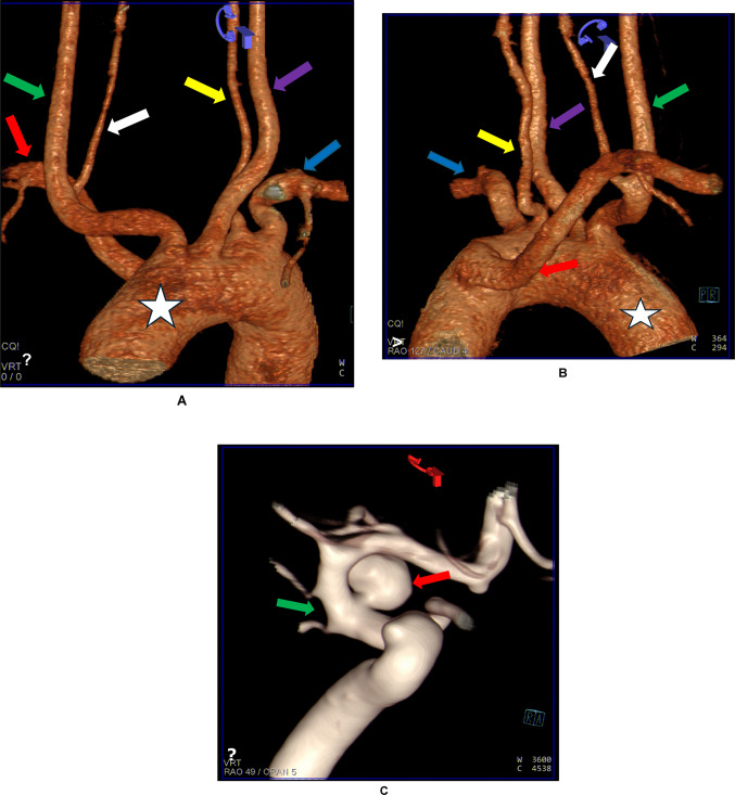
The most common variations of the aortic arch branching pattern usually involve the distance between the vessels arising from it and their dimensions. Changes in the number of vessels originating from the aortic arch, ranging from one to four or more major vessels instead of the classical three vessels as independent branches, are uncommon. Incidents of branching patterns involving four independent vessels arising from the aortic arch are rare, and reports of five or six independent vessels are extremely rare. We report on a case of an absent brachiocephalic trunk associated with an aberrant right subclavian artery and five distinct major vessels arising directly from the aortic arch in a South African male. Although most congenital vascular variations are incidental findings on angiographic images, some have also been associated with cerebrovascular diseases such as cerebral aneurysms. In addition, knowledge of these rare variations is of diagnostic importance as their presence may increase the difficulty and alter the specificity of vascular procedures performed using endovascular and open techniques.
Keywords: Aberrant right subclavian artery; Aortic arch; Brachiocephalic trunk; Computed tomography angiography; Vertebral artery.
© 2025. The Author(s).
Conflict of interest statement
Declarations. Conflict of interest: The authors declare no competing interests. Consent for publication: Not applicable.
Figures
Similar articles
-
A systematic review and meta-analysis of variations in branching patterns of the adult aortic arch.J Vasc Surg. 2018 Jul;68(1):298-306.e10. doi: 10.1016/j.jvs.2017.06.097. Epub 2017 Aug 31. J Vasc Surg. 2018. PMID: 28865978
-
A systematic classification of the vertebral artery variable origin: clinical and surgical implications.Surg Radiol Anat. 2018 Jul;40(7):779-797. doi: 10.1007/s00276-018-1987-3. Epub 2018 Feb 19. Surg Radiol Anat. 2018. PMID: 29459992
-
Anatomy, Thorax, Subclavian Arteries.2023 Jul 24. In: StatPearls [Internet]. Treasure Island (FL): StatPearls Publishing; 2025 Jan–. 2023 Jul 24. In: StatPearls [Internet]. Treasure Island (FL): StatPearls Publishing; 2025 Jan–. PMID: 30969558 Free Books & Documents.
-
Embryology, Aortic Arch.2023 Mar 6. In: StatPearls [Internet]. Treasure Island (FL): StatPearls Publishing; 2025 Jan–. 2023 Mar 6. In: StatPearls [Internet]. Treasure Island (FL): StatPearls Publishing; 2025 Jan–. PMID: 31985966 Free Books & Documents.
-
Dysplastic aberrant left subclavian artery originating from a thoracic intersegmental artery associated with a right aortic arch.Surg Radiol Anat. 2024 Apr;46(4):519-522. doi: 10.1007/s00276-024-03333-w. Epub 2024 Mar 13. Surg Radiol Anat. 2024. PMID: 38480591
References
-
- Abdelmotalab M, Abdelaziz O, Abdalla I, Ali AE, Abdalla SEM, Karti IAA, Saleh HE (2025) Anatomical variation of the aortic arch branches: a case report and literature review. Iraqi Natl J Med 7:153–156
-
- Bernardi L, Dettori P (1975) Angiographic study of a rare anomalous origin of the vertebral artery. Neuroradiology 9:43–47
-
- Keet K, Gunston GD, Alexander RL (2019) Variations in the branching pattern of the aortic arch: an African perspective. Eur J Anat 23:91–102
Publication types
MeSH terms
Supplementary concepts
LinkOut - more resources
Full Text Sources
