. 2025 Jun 19;23(1):291.
doi: 10.1186/s12964-025-02298-6.

Evidence for a JAK2/STAT3 proinflammatory and vasculogenic mimicry interrelated molecular signature in adipocyte-derived mesenchymal stromal/stem cells

Affiliations

Evidence for a JAK2/STAT3 proinflammatory and vasculogenic mimicry interrelated molecular signature in adipocyte-derived mesenchymal stromal/stem cells

Carolane Veilleux et al. Cell Commun Signal. .

Abstract

Background: During obesity, the excessive accumulation of fat in tissue promotes dysregulated hormonal and cytokine homeostasis that triggers chronic inflammation, which is, in part, associated with an increased incidence of some cancers. This protumoral inflammatory environment is further exacerbated through the secretome of mature adipocytes, which promotes tumor angiogenesis. Emerging studies suggest that human adipocyte-derived mesenchymal stromal/stem cells (ADMSCs) may contribute to a complementary process supporting local angiogenesis termed vasculogenic mimicry (VM). The molecular mechanisms linking ADMSCs to VM and inflammation remain poorly understood.

Methods: ADMSC 3D capillary-like structures were generated upon seeding on Cultrex. Structure analysis was performed using WIMASIS. Total RNA was extracted using TRIzol and RT-qPCR was performed to assess gene expression or screen RT2 PCR arrays. Transient gene silencing was performed by transfecting cells with a specific siRNA against STAT3. Protein lysates were harvested and used for Western blotting. Realtime cell migration was performed with the xCELLigence system.

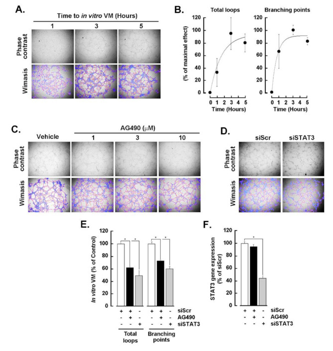
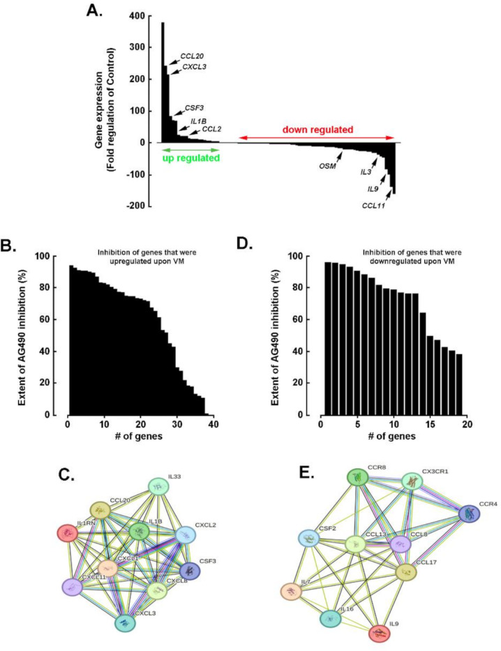
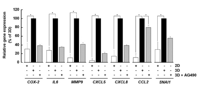
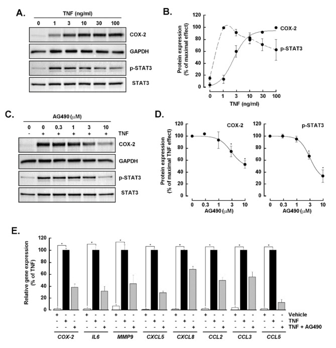
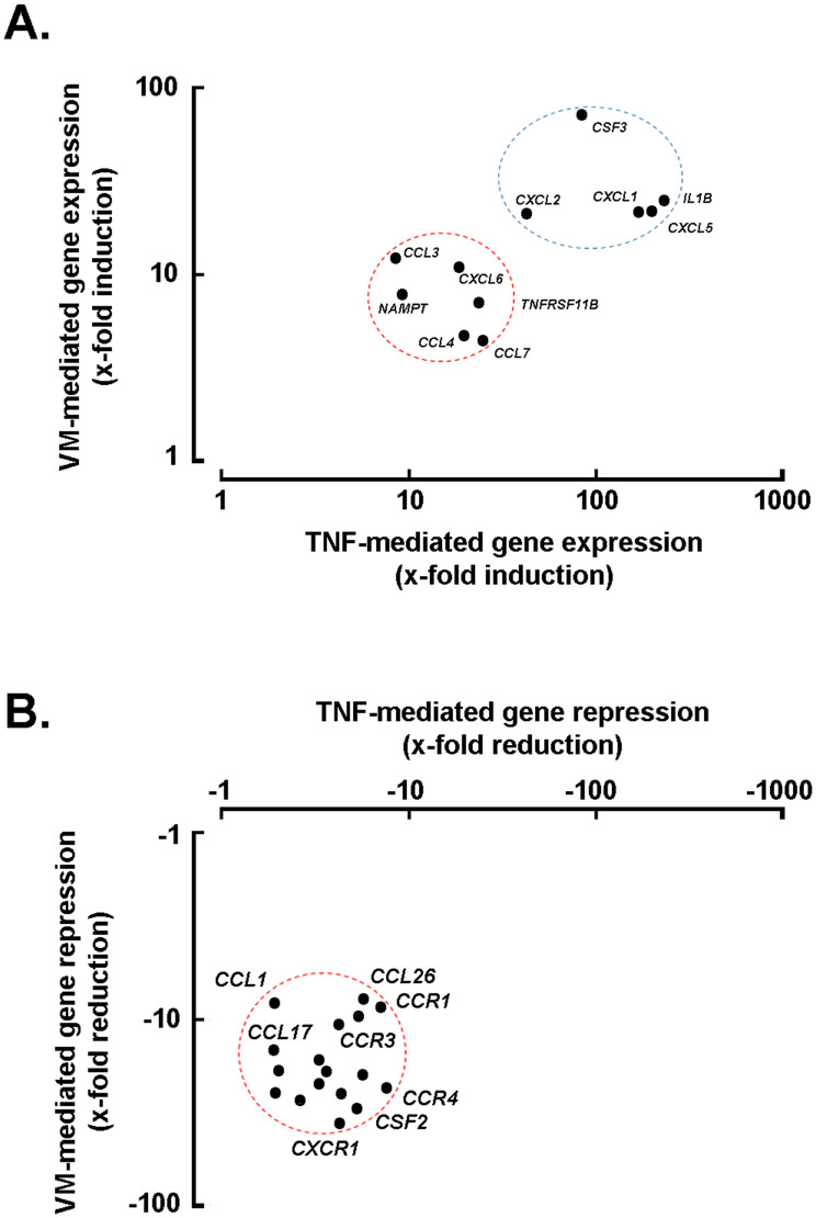
Results: Our findings revealed that in vitro priming of ADMSCs with Cultrex led to the formation of 3D capillary-like structures and the acquisition of an inflammatory molecular signature. VM-derived ADMSCs share a common proinflammatory molecular signature similar to that induced in 2D ADMSC monolayers by tumor necrosis factor (TNF)-alpha and are characterized by upregulated expression of COX2, CCL2, CCL5, CXCL5, CXCL8, IL-6, SNAI1, and MMP9. Interestingly, pharmacological inhibition or gene silencing of the JAK2/STAT3 signaling pathway reduced chemotactic cell migration, in vitro VM and the expression of proinflammatory and invasive biomarkers.

Conclusions: Overall, we provide novel evidence that inhibiting JAK2/STAT3-regulated VM can also alter the acquisition of a proinflammatory signature and prevent the contribution of ADMSCs to alternative tumor neovascularization processes.

Keywords: ADMSC; AG490; Inflammation; JAK2/STAT3; TNFα; Vasculogenic mimicry.


Conflict of interest statement

Declarations. Ethics approval and consent to participate: Not applicable. Consent for publication: Not applicable. Competing interests: The authors declare no competing interests.

Figures

Fig. 1
Fig. 1
Effect of JAK2/STAT3 signaling pathway inhibition on the formation of 3D capillary-like structures in ADMSCs. ADMSCs were trypsinized and seeded on top of a Cultrex layer as described in the Methods section. (A) Representative phase contrast images were taken to monitor the formation of capillary-like structures (upper panels) and were analyzed via Wimasis (lower panels). (B) Quantification from Wimasis analysis of the total loops and branching points over time. (C) Representative phase contrast images were taken to monitor the inhibition of capillary-like structure formation by increasing concentrations of AG490 after 5 h (upper panels). The structures were analyzed via Wimasis (lower panels). (D) Representative phase contrast pictures were taken to monitor the inhibition of capillary-like structure formation by RNA-mediated interference via siSTAT3 in transiently transfected ADMSCs after 5 h (upper panels) compared with that in control cells transiently transfected with a scrambled sequence (siScr); the structures were analyzed via Wimasis (lower panels). (E) Quantification from Wimasis analysis of the total loops and of branching points at 10 µM AG490 (black bars) and upon siSTAT3-transfection (gray bars) are shown as percentages of their respective controls (0.1% DMSO and siScr). (F) Relative gene expression of STAT3 after pharmacological inhibition with 10 µM AG490 (black bars) and upon transient interference with siSTAT3 (gray bars). The VM data are representative of three independent experiments, and the transfection data are representative of two independent experiments
Fig. 2
Fig. 2
Effect of pharmacological inhibition of the JAK2/STAT3 signaling pathway on the chemotactic migration capacity of ADMSCs. Chemotactic real-time cell migration was performed as described in the Methods section. (A) Relative cell migration in serum-supplemented or serum-free media was monitored over a 3-hour period. (B) A representative profile of the effects of the indicated increasing concentrations of AG490 on relative cell migration was compared to that of the vehicle (0.1% DMSO) over 3 h. (C) Quantification of the inhibitory effect of AG490 concentration on the chemotactic response. All the data are representative of two independent experiments, each performed in triplicate
Fig. 3
Fig. 3
Transcriptomic analysis of the inflammatory molecular signature induced upon capillary-like structure formation. ADMSCs were seeded on top of Cultrex dishes and left to form 3D capillary-like structures for 5 h in the presence or absence of 10 µM AG490 (the control consisted of vehicle-treated 2D monolayers). Total RNA was extracted, and gene expression was assessed via an RT2 Profiler PCR array for human inflammatory cytokines and receptors as described in the Methods section. (A) Gene expression levels of upregulated genes (green arrow) and downregulated genes (red arrow) are represented as the fold regulation between the control 2D monolayers and VM-forming ADMSCs, with a cutoff value greater than or equal to two. (B) Number of VM-induced genes and extent of their AG490-mediated inhibition of expression. (C) Protein‒protein interaction network of the 10 most VM-induced genes whose expression was inhibited by AG490, as retrieved with STRING. (D) Number of VM-downregulated genes and extent of their AG490-mediated inhibition of expression. (E) Protein‒protein interaction network of the 10 most VM-downregulated genes whose expression was also inhibited by AG490, as determined with STRING
Fig. 4
Fig. 4
Effects of AG490 on inflammation and invasion markers related to capillary-like structure formation. ADMSC response to priming on Cultrex for 5 h for capillary-like structure formation in the presence or absence of 10 µM AG490 (control, 2D monolayers). Total RNA was extracted, and gene expression was assessed via RT‒qPCR, as described in the Methods section. Relative gene expression of inflammation and invasion markers was compared between the control (vehicle-treated 2D monolayers, white bars) and VM-forming ADMSCs in the presence (3D + AG490; gray bars) or absence (vehicle-treated 3D; black bars) of 10 µM AG490. The results are presented as percentages of VM-related gene expression levels. The data are representative of two to three independent experiments performed in triplicate, except for the AG490 data, which are representative of one to two independent experiments performed in triplicate
Fig. 5
Fig. 5
Involvement of the JAK2/STAT3 signaling pathway in the TNFα-induced inflammatory signature in ADMSCs. (A) Monolayer ADMSC culture response to increasing TNFα concentrations for 24 h. COX-2, phospho-STAT3 (pSTAT3) and total STAT3 protein lysates were immunoblotted, with GAPDH serving as a loading control. (B) Densitometry analysis of COX-2 and pSTAT3 expression, represented as the percentage of the respective maximal effect of TNFα. (C) ADMSC 2D monolayer cultures treated with 30 ng/mL TNFα in the presence of the indicated increasing concentrations of AG490 for 24 h (vehicle, 0.1% DMSO). COX-2, pSTAT3 and STAT3 protein lysates were immunoblotted, with GAPDH serving as a loading control. (D) Densitometry analysis of TNFα-induced COX-2/GAPDH inhibition, represented as the percentage of the maximal effect of TNFα (labeled COX-2; left panel), and of TNFα-induced pSTAT3/STAT3 (labeled pSTAT3; right panel). (E) Monolayer-ADMSC cultures subjected to 30 ng/mL TNFα in the presence or absence of 10 µM AG490 for 24 h (the control was 0.1% DMSO). Modulation profiles of inflammation and invasion gene markers by TNFα (black bars) and by AG490 (gray bars) were measured via RT‒qPCR. All the data are representative of three independent experiments
Fig. 6
Fig. 6
Implication of JAK2/STAT3 in the inflammatory molecular signature of ADMSCs stimulated with TNFα. ADMSCs were treated with 30 ng/mL TNFα in the presence or absence of 10 µM AG490 for 24 h (the control was treated with 0.1% DMSO) to perform a RT2 PCR array for human inflammatory cytokines and receptors, as described in the Methods section. (A) Gene expression of upregulated genes (green arrow) and downregulated genes (red arrow) is represented as the fold regulation between the control and TNFα-treated cells, with a cutoff value greater than or equal to two. (B) Number of TNFα-induced genes and extent to which AG490 inhibited their expression. (C) Protein‒protein interaction network of the 10 TNFα-induced genes whose expression was inhibited the most by AG490, as determined via the Search Tool for the Retrieval of Interacting Genes/Proteins (STRING). (D) Number of TNFα-downregulated genes and extent to which AG490 inhibited their expression. (E) Protein‒protein interaction network of the 10 genes most downregulated by TNFα that were inhibited by AG490, as determined with STRING. The data are representative of one of two independent experiments
Fig. 7
Fig. 7
Toward a shared inflammatory molecular signature between VM- and TNFα-treated ADMSC phenotypes. VM- and TNFα-mediated gene expression was compared via the RT2 PCR gene array shown in Figs. 3 and 6 and is expressed as x-fold induction (A) or x-fold reduction (B)

Similar articles

References

    1. World Health Organization. Cancer. 2025. https://www.who.int/news-room/fact-sheets/detail/cancer
    1. Lauby-Secretan B, Scoccianti C, Loomis D, Grosse Y, Bianchini F, Straif K, International Agency for Research on Cancer Handbook Working Group. Body fatness and Cancer–Viewpoint of the IARC working group. N Engl J Med. 2016;375(8):794–8. - PMC - PubMed
    1. Mukherjee A, Bilecz AJ, Lengyel E. The adipocyte microenvironment and cancer. Cancer Metastasis Rev. 2022;41(3):575–87. - PubMed
    1. Brown KA, Scherer PE. Update on adipose tissue and cancer. Endocr Rev. 2023;44(6):961–74. - PMC - PubMed
    1. Park Y, Colditz GA. Obesity elevates cancer survivors’ risk of second cancer: identifying modifiable risk factors for second cancer. J Natl Cancer Inst. 2021;113(9):1113–4. - PMC - PubMed

MeSH terms

LinkOut - more resources