. 2025 Jun 19;27(1):111.
doi: 10.1186/s13058-025-02014-9.

Hope for OTHERS (Our Tissue Helping Enhance Research & Science): research results from the University of Pittsburgh rapid autopsy program for breast cancer

Affiliations

Hope for OTHERS (Our Tissue Helping Enhance Research & Science): research results from the University of Pittsburgh rapid autopsy program for breast cancer

Alexander Chih-Chieh Chang et al. Breast Cancer Res. .

Abstract

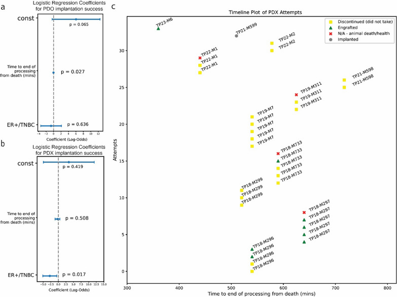
Breast cancer affects 1/8 of women throughout their lifetimes, with over 90% of cancer deaths being caused by metastasis. However, metastasis poses unique challenges to research, as complex changes in the microenvironment in different metastatic sites and difficulty obtaining tissue for study hinder the ability to examine in depth the changes that occur during metastasis. Rapid autopsy programs thus fill a unique need in advancing metastasis research. Here, we describe our protocol and processes for establishing and improving the US-based Hope for OTHERS (Our Tissue Helping Enhance Research and Science) program for organ donation in metastatic breast cancer. As of August 2024, we consented 114 patients and performed 37 autopsies, from which we collected 551 unique metastatic frozen tumor samples, 1244 FFPE blocks, 90 longitudinal liquid biopsy samples and developed 14 patient-derived organoid and 8 patient-derived xenograft models. We report in-depth clinical and histopathological information and discuss extensive new research and novel findings in patient outcomes, metastatic phylogeny, and factors in successful living model development. Our results reveal key logistical and protocol improvements that are uniquely beneficial to certain programs based on identifiable features, such as working closely with patient advocates, methods to rescue RNA quality in cases where tissue quality may degrade due to time delays, as well as guidelines and future expansions of our program.


Conflict of interest statement

Declarations. Ethics approval and consent to participate: All research involving human participants was conducted with informed consent obtained according to the ethical guidelines of the University of Pittsburgh Institutional Review Board under STUDY19060376. Consent for publication: All images of individuals on Fig. 3 are from a public website and are of authors who consent to this publication. Competing interests: The authors declare no competing interests.

Figures

Fig. 1
Fig. 1
Study design and workflow of the HfO program. Diagram illustrating the study design and workflow of the HfO Tissue Donation Program
Fig. 2
Fig. 2
Summary of patients’ treatment timelines and clinical details. Mixture line and normalized timeline chart showing a summary of HfO program, including primary tumor molecular subtype, race, gender, histological subtype, stage at time of diagnosis (pathological if available, clinical if not), treatment markers and treatment categories up to August 2024
Fig. 3
Fig. 3
Examples of patient advocate media and figures demonstrating impact of key support staff. a Promotion of tissue donation programs for advancing metastatic breast cancer (MBC) research via new media approaches such as podcasts, published media, and logo of the HfO program. Photos are of co-authors in this manuscript. b Graph showing number of patients engaged at conferences/events. c. Bar plot shows the considerable standard deviation in mean transport time due to unique complexities within institutional and geographic contexts. d Line graph shows the exponential increase in consent after an additional review of our operational protocols and rebranding (n = 114 consents, n = 34 autopsies [pre-2018 are not counted]). P value from segmented regression 6.38E−31 for a change in consent rate slope post protocol review
Fig. 4
Fig. 4
Summary of collected samples, and statistics on tissue diversity and counts in autopsy and non-autopsy settings. a Histogram that summarizes statistics for FFPE. Total of 1244, median of 41, range of 7 to 69. b Histogram that summarizes statistics for frozen tumors. Total of 511, median of 12, range of 3–27. c. Histogram that summarizes statistics for cryovials from autopsy. Total of 1952, median of 46 per patient, range from 0 to 142. d Histogram that summarizes our longitudinal blood collections. e Segment pie charts using data from the US Aurora, EU Aurora, MET500, and HfO reports showing the distinctly different range of tissues collected in autopsy and non-autopsy settings
Fig. 5
Fig. 5
Diverse causes of death and subclinical metastases seen in autopsy settings, necessitating consistent collection. a Bar plot showing the frequency of different causes of death in our program based on clinical note and autopsy report review (n = 37). b Bar chart showing the mean increase in organs identified with metastases after careful pathological review on autopsy that were not identified in regular clinical monitoring, error bars are standard deviation. P value 0.15 with paired t-test. Images of c ultrasound and d. MRI in a patient with ILC showing the CT-undetectable liver metastases. e CT image and f autopsy image at time of death for the patient with ILC illustrating the discrepancy between a ‘normal’ CT and the organ status. g Scatter plot showing the difference in metastases seen clinically and in autopsy for patients with ILC. Patients with ILC have much more spread in peritoneal tissues that are undetectable clinically. h Scatter bubble plot showing our FFPE collection, red box highlights our improved protocol to increase consistency in grossly normal tissues. Color corresponds to size of bubble. i Figure showing top 7 organ sites, collected whenever available, even if grossly normal under our revised protocol
Fig. 6
Fig. 6
Single nuclei sequencing of paired frozen and fixed tissue samples. a Side by side comparison of frozen versus fixed single nuclei sequencing data metrics shows significant improvement in fixed technologies with increase in genes detected, molecules detected, and less mitochondrial contamination. (n = 3) Run using 10 × Genomics kit targeting 8000 cells. b UMAP plot showing that frozen single nuclei sequencing lead to loss of signal causing failure of integration and removal of batch effects (n = 3), with diminished ESR1, and CD4 signal. c Fixed single nuclei sequencing has better integration due to better signal recovery, with visible improvements in ESR1 and CD4 signal. PGR signal consistency across both sets shows that fixed technologies are not artificially introducing signal that isn’t there. d Bar plot showing the number of Hallmark pathways that have adjusted p value less than 0.05 after pathway analysis between fixed and frozen cells, showing that fixed tissue almost always has better pathway signal. P value calculated using chi-square test
Fig. 7
Fig. 7
Logistic regression results for PDO and PDX generation success. a Logistic regressions for PDO attempts (n = 27 attempts) show that time to end of processing is predictive of success for PDO success. b Logistic regressions for PDX attempts (n = 34 attempts) show that TNBC lesions are more likely to be successful. c There are also significantly different probabilities of success on an intra-patient basis, implying that tissue of origin or tumor cellularity may play a role (n = 34)
Fig. 8
Fig. 8
Validation of expression of the ESR1-ARNT2 fusion detected in HfO samples. a Growth of PDX model CTG-3533. b ER-immunoblot of TP18-M733 metastatic lesion protein samples. Red asterisks denote the expected size for the ESR1-ARNT2 fusion protein. β-actin serves as loading control in both blots, n = 1 (Samples are in order 7 = thoracic lymph node, 8/10 = diaphragm, 12-18 = liver, 19 = peri-pancreatic lymph node, 3 = mesentary, 8 = diaphragm, 11 = gallbladder, 22 = lung). c PDXO from CTG-3533 PDX can be cultivated from the PDX to extend back to in vitro experimentation. d PDXO shows low levels of expression of the fusion construct and heterogenous signal. Sequencing also allows design of the fusion construct to be transfected into cells lines like T47D, as seen on ER-western blot. Red asterisks denote the expected size for the ESR1-ARNT2 fusion protein. β-actin serves as loading control in both blots, n = 1

Update of

References

    1. Breast Cancer Risk in American Women - NCI. December 17, 2020. Accessed 2 Aug 2024. https://www.cancer.gov/types/breast/risk-fact-sheet
    1. Survival Rates for Breast Cancer. Accessed 1 Feb 2024. https://www.cancer.org/cancer/types/breast-cancer/understanding-a-breast...
    1. Najjar MK, Manore SG, Regua AT, Lo HW. Antibody-drug conjugates for the treatment of HER2-positive breast cancer. Genes. 2022;13(11):2065. 10.3390/genes13112065. - PMC - PubMed
    1. A Perspective on Cancer Cell Metastasis | Science. Accessed 5 Aug 2023. https://www.science.org/doi/10.1126/science.1203543
    1. Dillekås H, Rogers MS, Straume O. Are 90% of deaths from cancer caused by metastases? Cancer Med. 2019;8(12):5574–6. 10.1002/cam4.2474. - PMC - PubMed