. 2025 Jun 19;17(1):136.
doi: 10.1186/s13195-025-01784-w.

A longitudinal study of the 5xFAD mouse retina delineates Amyloid beta (Aβ)-mediated retinal pathology from age-related changes

Affiliations

A longitudinal study of the 5xFAD mouse retina delineates Amyloid beta (Aβ)-mediated retinal pathology from age-related changes

Savannah A Lynn et al. Alzheimers Res Ther. .

Abstract

Background: Age-related macular degeneration (AMD) is the commonest cause of irreversible blindness in developed societies. AMD coincides with advanced age to which genetic and lifestyle factors contribute additional risks. High levels of the Alzheimer's-linked Amyloid beta (Aβ) proteins are correlated with aged/AMD retinas. To delineate the role of Aβ in retinopathy from age-related changes, we used transgenic 5xFAD mice in a longitudinal study to recapitulate the aged/AMD Aβ-burden of the human retina.

Methods: Mice were genotyped to exclude the retinal degeneration alleles Pde6brd1, Pde6brd8, Agouti, Tyr and Oca2. Retinas of 5xFAD and wildtype littermates (97 males/females in total) were longitudinally assessed until 15 months using non-invasive retinal scans: multi-focal electroretinography, optokinetic tracking, optical coherence tomography (OCT), colour fundus photography and fluorescein angiography. Mice were killed at 4, 8 and 15 months, and eyes enucleated for analyses by light, confocal and electron microscopy.

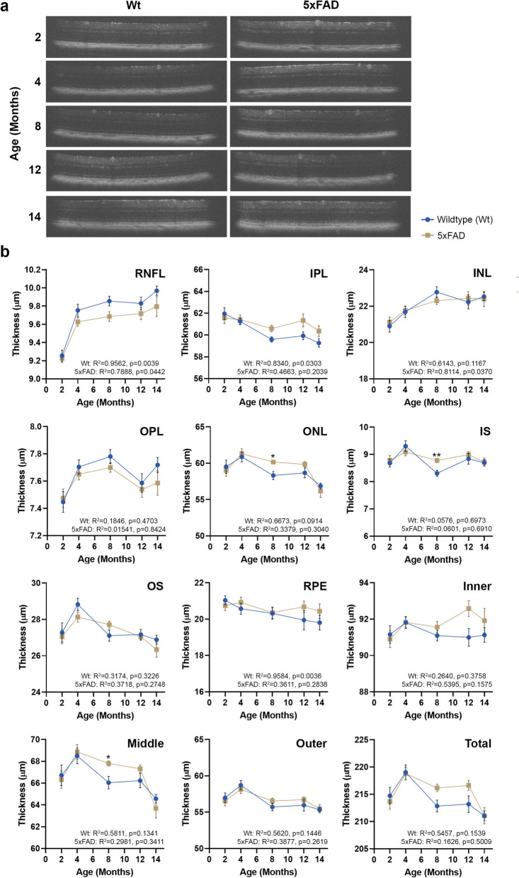
Results: Age-related changes included a gradual decline of retinal activity in all mice. Subretinal/drusen-like deposits increased with age, but, like retinal vessel morphology and vessel integrity, showed no differences between cohorts. Diminished PSD95 levels indicated impaired photoreceptor-bipolar connectivity which correlated with age. Ultrastructural imaging showed increased electron-dense granules and undigested outer segments within retinal pigment epithelial cells with age. 5xFAD pathology included significant weight reduction vs. wildtype/littermates, which were pronounced in females. 8 month old 5xFAD mice had diminished A and B waves, though the age-related decline in wildtype mice abolished these subsequently. Visual acuity/function was also reduced in 14 month 5xFAD eyes. OCT revealed thickened photoreceptor nuclei and inner segments in 8 month 5xFAD retinae. Scrutiny of chorioretinal tissues revealed diminished photoreceptor nuclei in 4 month 5xFAD eyes, though differences were abolished as both cohorts aged. From 8 months onwards, 5xFAD mice possessed fewer bipolar cell nuclei.

Conclusions: Chronic Aβ exposure led to the earlier development of retinopathy-linked features, the identification of which advances our understanding of how Aβ contributes to multifaceted retinopathies. These were distinguishable from wider age-related changes and non-specific influences of retinal degeneration alleles in 5xFAD mice. Longitudinal analyses revealed sex and age-related limitations and important 3Rs considerations for future studies using 5xFAD mice.

Keywords: 5xFAD; Age; Age-related macular degeneration (AMD); Amyloid beta (Aβ); Mouse model; Retina; Retinopathy.


Conflict of interest statement

Declarations. Ethics approval and consent to participate : Studies were overseen by the institutions’ Animal Welfare and Ethical Review Board (AWERB) and were consistent with the Association for Research in Vision and Ophthalmology statement (ARVO) for the Use of Animals in Ophthalmic procedures and the UK Animal (Scientific Procedures) Act of 1986. The study complied with the ARRIVE guidelines with work undertaken under a UK Home Office project licence. Consent for publication: Not applicable. Competing interests: The authors declare no competing interests.

Figures

Fig. 1
Fig. 1
Experimental plan and muti-focal electroretinography (ERG) in 5xFAD and wildtype littermate mice. a Experimental set-up showing key milestones and workflow. b An overall decrease in the amplitude of A wave and B waves were observed in 5xFAD retinae from 4 months onwards compared to control littermates, suggesting a potential impairment of photoreceptor and inner retinal function, respectively. However, these differences only reached significance at 8 months, after which retinal function was indistinguishable from those in wildtype control mice. Averaged A and B wave implicit times T(A) and T(B) showed no significant differences between the two groups. Statistical comparisons were made using two-way ANOVA (mixed model) with Bonferroni post-hoc test with significance shown as * p ≤ 0.05. Data is expressed as means ± SEM (standard error of the mean). c Grouped average ERG traces for all timepoints with shaded regions representing SEM. Experimental replicates were as follows. 2 months: Wt (n = 22), 5xFAD (n = 25); 4 months: Wt (n = 22), 5xFAD (n = 30); 8 Months: Wt (n = 18), 5xFAD (n = 23); 12 Months: Wt (n = 13), 5xFAD (n = 17) and 14 Months: Wt (n = 20), 5xFAD (n = 18). Assessed by Pearson’s correlation coefficient
Fig. 2
Fig. 2
Optokinetic tracking (OKT) responses in 5xFAD and wildtype mice. The optokinetic tracking responses were assessed longitudinally as a measure of visual acuity and function. No significant differences in detectable spatial frequencies were recorded until 14 months, when 5xFAD mice exhibited a decline in clockwise (CW), counter-clockwise (CCW), up and down responses. Data is expressed as means ± SEM (standard error of the mean). Statistical comparisons were made using two-way ANOVA with Bonferroni post-hoc test with significance shown as * p ≤ 0.05, ** p ≤ 0.01, *** p ≤ 0.001, **** p ≤ 0.0001. Experimental replicates were as follows. 2 Months: Wt (n = 11), 5xFAD (n = 14); 4 Months: Wt (n = 11), 5xFAD (n = 14); 6 Months: Wt (n = 11), 5xFAD (n = 14); 8 Months: Wt (n = 10), 5xFAD (n = 14); 10 months: Wt (n = 24), 5xFAD (n = 28); 12 months: Wt (n = 24), 5xFAD (n = 27) and 14 months: Wt (n = 14), 5xFAD (n = 13)
Fig. 3
Fig. 3
Optical coherence tomography (OCT) measurements of 5xFAD and wildtype mice. a OCT scans obtained at 2, 4, 6, 8, 10 12 and 14 months were subject to automatic segmentation using InVivoVue Diver analysis software to assess differences in component retinal layer thicknesses. b No differences were recorded in the RNFL, IPL, INL, OPL, OS or RPE layers. However, differences between 5xFAD and wildtype littermate retinae were recorded in the ONL and IS layers (photoreceptors) at 8 months. Measurement of the inner (RNFL-INL), middle (OPL-ONL) and outer (IS-RPE) retinal thicknesses showed changes in the middle retina at 8 months between the two cohorts, which was not evident when summed as the total retinal thickness. Data is expressed as means ± SEM (standard error of the mean). Statistical comparisons were made using a two-way ANOVA with Bonferroni’s post-hoc test with statistical significance shown as * p ≤ 0.05 and ** p ≤ 0.01. Experimental replicates were as follows. 2 months: Wt (n = 22), 5xFAD (n = 25); 4 months: Wt (n = 22), 5xFAD (n = 28); 8 Months: Wt (n = 22), 5xFAD (n = 25); 12 Months: Wt (n = 13), 5xFAD (n = 17) and 14 Months: Wt (n = 19), 5xFAD (n = 18). Correlation was assessed using Pearson’s coefficient. Inner plexiform layer (IPL), Inner nuclear layer (INL), Inner segments (IS), Outer plexiform layer (OPL), Outer nuclear layer (ONL), Outer segments (OS), Retinal ganglion cells (RGC), Retinal nerve fibre layer (RNFL), Retinal pigment epithelium (RPE)
Fig. 4
Fig. 4
Funduscopy and fluorescein angiography images of 5xFAD and wildtype mouse retinae. a Anonymised colour fundus photography (CFP) taken at 4, 8 and 14 months were scrutinised for any evidence of pathology, which showed evidence of punctate/white subretinal deposits in both cohorts that gradually increased with age. b Fluorescein angiography (FFA) images were quantified in MATLAB using Quant BV software to assess retinal vessel area, the avascular region, vein width and vessel tortuosity. No differences were observed between 5xFAD and wildtype littermate mice. Similarly, longitudinal studies revealed no evidence of dye leakage, indicative of compromised retinal vessels in either group. Data is expressed as means ± SEM (standard error of the mean). Statistical comparisons were made using a two-way ANOVA with Bonferroni’s post-hoc test. Experimental replicates were as follows. 4 Months: Wt (n = 7), 5xFAD (n = 5); 4 months: Wt (n = 7) 5xFAD (n = 5); 8 months: Wt (n = 7), 5xFAD (n = 5) and 14 months: Wt (n = 5), 5xFAD (n = 5)
Fig. 5
Fig. 5
Light microscopy analyses of chorioretinal sections of 5xFAD and wildtype mice. a Representative images of wildtype mice and 5xFAD retinae at 4, 8 and 14 months, which were stained with toluidine blue showing cross-sections through the retina and associated tissues. Scale bars correspond to 50µm. Sections highlighted in white boxes are shown as magnified inserts which elicited closer scrutiny: RNFL—IPL (top panel), INL—ONL (middle panel) and IS—Choroid (lower panel). Occasionally, evidence of detached or multinuclear clusters were observed subretinally in 5xFAD eyes (white arrows). Scale bars in inserts correspond to 20µm. b Quantification of nuclei in the RGC layer in anonymised sections showed no significant differences between 5xFAD and wildtype retinae at any timepoint, though 5xFAD retinae contained fewer nuclei with increasing age. By contrast, analyses of the INL revealed significantly fewer bipolar cell nuclei in 5xFAD retinae compared to controls at 8 and 14 months. Scrutiny of the adjacent ONL revealed significantly fewer photoreceptor nuclei in 5xFAD retinae compared to wildtype mice at an earlier timepoint of 4 months. However, these differences became less evident afterwards, though 5xFAD mice contained fewer ONL nuclei compared to controls at 8 and 14 months. Data is expressed as means ± SEM (standard error of the mean). Statistical comparisons were made using a two-way ANOVA with Bonferroni’s post-hoc test with significance shown as * p ≤ 0.05 and ** p ≤ 0.01. Experimental replicates were as follows. 4 Months: Wt (n = 3), 5xFAD (n = 3); 8 Months Wt (n = 3), 5xFAD (n = 3) and 14 Months: Wt (n = 5), 5xFAD (n = 4). Bruch’s membrane (BrM), Inner plexiform layer (IPL), Inner nuclear layer (INL), Inner segments (IS), Outer plexiform layer (OPL), Outer nuclear layer (ONL), Outer segments (OS), Retinal ganglion cells (RGC), Retinal nerve fibre layer (RNFL), Retinal pigment epithelium (RPE)
Fig. 6
Fig. 6
Ultrastructural comparison of 5xFAD and wildtype mouse retinae. Mice culled at 4, 8 and 14 months provided details of Aβ and/or age-related changes in each retinal layer. a A representative low-powered electron micrograph (EM) (× 3000) showing the cross-section of a 14 month old wildtype mouse retina. Wildtype littermates at this experimental end-point showed no evidence of any retinal abnormalities. b Scrutiny of the 5xFAD outer retina at 4 months as illustrated by this representative EM micrograph also showed no evidence of pathology, though intracellular vacuoles (red arrows) were prevalent. c EM micrograph of 8 month old 5xFAD outer retina, further magnified (× 8000) and focused on the RPE layer, showing undigested POS (white arrowhead) as well as electron-dense structures such as melanosomes (white arrows) in RPE cells. d Evidence of charcoal-like granules (red arrowhead) alongside electron-dense bodies including lipofuscin (white arrows) in RPE cells of a 14 month old 5xFAD eye. e Representative EM micrograph showing undigested POS (white arrowheads) amongst other electron-dense granules in 14 month old 5xFAD RPE cells. However, the apical RPE microvilli and their basolateral infolds appear normal in these transgenic mice. f High-powered representative EM micrograph (× 12,000) of 8 month old 5xFAD retina showing abundant electron-dense granules including intracellular vacuoles (red arrowhead) in RPE cells with no further evidence of any morphological abnormalities. Histograms showing the quantification of aforementioned features in anonymised sections revealed no significant differences between 5xFAD and wildtype littermates. However, the frequencies of most such features appear to increase with age in both groups. Quantification of BrM thicknesses revealed no differences between cohorts or across different age groups. Scale bars are indicated in each micrograph which corresponds to 5µm (a, b), 2µm (c, e) and 1µm (d, f). Bruch’s membrane (BrM); Outer segments (OS); Retinal pigment epithelium (RPE)
Fig. 7
Fig. 7
Study concept and summary of changes in 5xFAD mice retinae driven by Amyloid beta (Aβ) pathology and age. Venn diagram: Age-related macular degeneration (AMD) is caused by the confluence of advanced age combined with genetic as well as lifestyle risk factors. Although AMD is not associated with any known mutations in the amyloid precursor protein (APP) or presenilin-1 (PSEN1) genes which are over-expressed in 5xFAD mice, the resulting high levels of retinal Aβ effectively recapitulates the age/AMD-associated Aβ-burden in the human retina. Schematic of summary findings: Lifecourse of 5xFAD mice showing key experimental timepoints correlated with notable features of Aβ-driven retinopathy over any age-related changes. Statistically significant differences are indicated by a red asterisk. Age-related changes are also noted in both cohorts. Measurements which produced no obvious changes/trends in either cohort are listed within the box. Light microscopy (LM) and transmission electron microscopy (TEM), indicate where data were obtained using these approaches. Amyloid beta (Aβ); Bruch’s membrane (BrM); electroretinography (ERG); inner segments (IS); optical coherence tomography (OCT); outer nuclear layer (ONL); photoreceptor outer segments (POS) and retinal pigment epithelium (RPE)

References

    1. Owen CG, Jarrar Z, Wormald R, Cook DG, Fletcher AE, Rudnicka AR. The estimated prevalence and incidence of late stage age related macular degeneration in the UK. Br J Ophthalmol. 2012;96(5):752–6. - PMC - PubMed
    1. Wong WL, Su X, Li X, Cheung CM, Klein R, Cheng CY, et al. Global prevalence of age-related macular degeneration and disease burden projection for 2020 and 2040: a systematic review and meta-analysis. Lancet Glob Health. 2014;2(2):e106–16. - PubMed
    1. Hykin P, Chakravarthy U, Lotery A, McKibbin M, Napier J, Sivaprasad S. A retrospective study of the real-life utilization and effectiveness of ranibizumab therapy for neovascular age-related macular degeneration in the UK. Clin Ophthalmol. 2016;10:87–96. - PMC - PubMed
    1. Fritsche LG, Igl W, Bailey JN, Grassmann F, Sengupta S, Bragg-Gresham JL, et al. A large genome-wide association study of age-related macular degeneration highlights contributions of rare and common variants. Nat Genet. 2015. - PMC - PubMed
    1. Merle BM, Silver RE, Rosner B, Seddon JM. Adherence to a mediterranean diet, genetic susceptibility, and progression to advanced macular degeneration: a prospective cohort study. Am J Clin Nutr. 2015;102(5):1196–1206. - PMC - PubMed

Substances

LinkOut - more resources