. 2025 Jun 17:35:e2505015.
doi: 10.4014/jmb.2505.05015.

Skin ECM Provides a Bio-Derived Platform for Supporting Dermal Renewal and Matrix Synthesis

Affiliations

Skin ECM Provides a Bio-Derived Platform for Supporting Dermal Renewal and Matrix Synthesis

Sewon Park et al. J Microbiol Biotechnol. .

Abstract

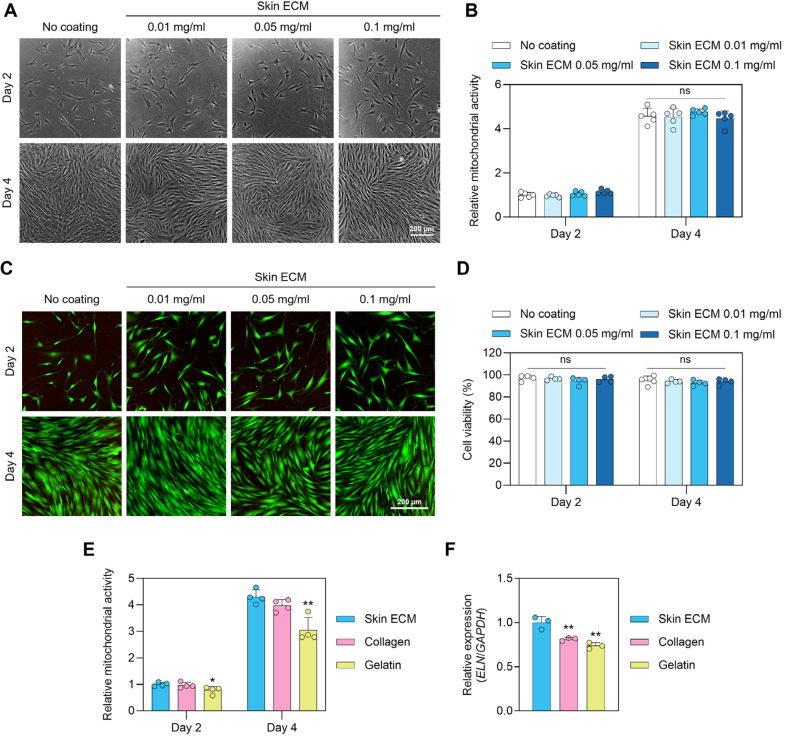
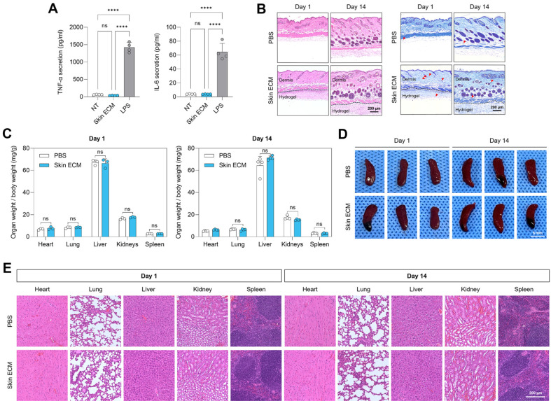
The extracellular matrix (ECM) plays a central role in directing dermal fibroblast behavior and coordinating tissue regeneration through its structural organization and biochemical signaling. In this study, we investigate the composition and regenerative bioactivity of Skin ECM in the context of dermal remodeling as a soluble supplement and surface coating for fibroblast culture. Proteomic profiling demonstrates that Skin ECM preserves the molecular complexity and skin-specific composition of native dermal ECM, including key structural and signaling proteins essential for tissue repair, with over 95% overlap with the human skin proteome, highlighting its strong tissue specificity and biological relevance. Functionally, Skin ECM enhances fibroblast migration during wound healing, upregulates elastin expression, and suppresses transforming growth factor beta 1 (TGF-β1)-induced expression of profibrotic and inflammatory markers, indicating inhibition of fibroblast activation. In vivo subcutaneous implantation confirms high local and systemic biocompatibility without signs of inflammation or toxicity. Collectively, these findings establish Skin ECM as a bioactive, tissue-specific, and immunologically compatible ECM material, offering broad utility in regenerative medicine, three-dimensional (3D) skin model systems, and dermal therapeutics including cosmetic interventions.

Keywords: Skin extracellular matrix; dermal fibroblast; skin regeneration; wound healing.


Conflict of interest statement

Conflict of Interest

Seung-Woo Cho (Chief Executive Officer) and Yoonhee Jin (Chief Technology Officer) are affiliated with Cellartgen. The authors have no financial conflicts of interest to declare.

Figures

Fig. 1
Fig. 1. Proteomic characterization of Skin ECM reveals tissue-specific composition and enrichment of dermal matrisome proteins.
(A) Pie chart showing the proportion of ECM proteins (based on the MatrisomeDB classification) in Skin ECM. (B) Distribution of matrisome subcategories within Skin ECM, human skin tissue, and recombinant human collagen III (rhCollagen), including core matrisome components (collagens, glycoproteins, proteoglycans) and matrisome-associated proteins (ECM regulators, ECM-affiliated proteins, secreted factors). (C) Correlation matrix with Pearson correlation coefficients between matrisome protein expression in human skin tissue, Skin ECM, and rhCollagen. (D) Top 10 most abundant proteins in the core matrisome categories of Skin ECM, including collagens, glycoproteins, and proteoglycans. Proteins shown in red indicate to be expressed in the native human skin tissue. (E) Pie chart showing the proportion of Skin ECM proteins that are annotated as skin-expressed proteins (95.12% by total abundance), based on the Human Protein Atlas. (F) Venn diagrams showing protein overlap between Skin ECM and human skin proteomes at the level of total proteins (left) and matrisome proteins (right). Scatterplots of (G) gene ontology molecular function (GOMF) and (H) gene ontology biological process (GOBP) terms significantly enriched in matrisome proteins of Skin ECM. The terms were positioned based on semantic similarity using REVIGO. The bubble size represents the log-transformed ‘Size’, which corresponds to the number of annotations for each term in the European Bioinformatics Institute Gene Ontology Annotation (EBI GOA) database. (I) A Voronoi diagram illustrating the Reactome pathway analysis results of matrisome proteins in Skin ECM. Color intensity indicates the p-value from the statistical test. Pathways shown in dark grey are not significantly overrepresented, while those with no associated proteins are depicted in light grey. (J) Protein–protein interaction (PPI) network of skin-elevated proteins in Skin ECM. Node color represents relative abundance. (K) A chord plot showing relationships between GOBP terms (skin development, epidermis development, structural constituent of skin epidermis, and tissue regeneration) and associated proteins in Skin ECM.
Fig. 2
Fig. 2. Skin ECM coating supports fibroblast viability and matrix gene expression without inducing hyperproliferation.
(A) Phase-contrast images of human dermal fibroblasts cultured on tissue culture surfaces coated with increasing concentrations of Skin ECM (0, 0.01, 0.05, and 0.1 mg/ml) at day 2 and day 4. Scale bar = 200 μm. (B) MTT assay showing mitochondrial activity at day 2 and day 4 (n = 5; ns: not significant). (C) Representative Live/Dead fluorescence staining images of fibroblasts at each coating concentration. Green: live cells; red: dead cells. Scale bar = 200 μm. (D) Quantification of cell viability based on Live/Dead staining (n = 4; ns: not significant). (E) MTT assay comparing fibroblast viability on Skin ECM (0.05 mg/ml), type I collagen from rat tail (0.05 mg/ml), or gelatin (1 mg/ml) (n = 4, *p < 0.05 and **p < 0.01 versus Skin ECM). (F) qRT-PCR analysis of elastin (ELN) expression in fibroblasts cultured on Skin ECM (0.1 mg/ml), type I collagen from rat tail (0.05 mg/ml), or gelatin (1 mg/ml) (n = 3, **p < 0.01 versus Skin ECM). Data are presented as mean ± SD. Statistical significance was assessed by one-way ANOVA followed by Tukey’s multiple comparisons test.
Fig. 3
Fig. 3. Skin ECM supplement enhances fibroblast migration and suppresses profibrotic and inflammatory gene expression.
(A) Representative scratch wound healing images of human dermal fibroblasts treated with vehicle control (serum-free), Skin ECM (1 mg/ml), or rhCollagen (1 mg/ml) at 0 to 32 h. Dotted lines delineate wound boundaries. Scale bar = 500 μm. (B) Quantification of wound closure at 32 h demonstrates significantly increased fibroblast migration in the Skin ECM group compared to control and rhCollagen-treated groups (n = 3, *p < 0.05 and **p < 0.01 versus indicated group). (C) ELISA results showing levels of secreted pro-collagen type I in fibroblast cultures treated with vehicle (low serum), Skin ECM (1 mg/ ml), or rhCollagen (1 mg/ml). Skin ECM increased collagen output compared to control (n = 5; *p < 0.05 and **p < 0.01; ns: not significant). (D) Gene expression analysis of ACTA2, COL1A1, and IL6 in fibroblasts stimulated with TGF-β1 (10 ng/ml), with or without co-treatment with Skin ECM (0.01 or 0.05 mg/ml) (n = 3, *p < 0.05, **p < 0.01, ***p < 0.001, and ****p < 0.0001 versus indicated group). Data are presented as mean ± SD. Statistical analysis was performed using one-way ANOVA followed by Tukey’s multiple comparisons test.
Fig. 4
Fig. 4. Skin ECM exhibits excellent biocompatibility in vitro and in vivo.
(A) ELISA quantification of TNF-α and IL-6 secretion from RAW 264.7 macrophages treated with Skin ECM (5 mg/ml) or LPS (1 μg/ml) stimulation. Skin ECM did not induce pro-inflammatory cytokine release (n = 4; ****p < 0.0001, ns: not significant; one-way ANOVA with Tukey’s post hoc test). (B) Representative histological images (H&E and TB staining) of skin tissue at the subcutaneous injection site 1 and 14 days after implantation of Skin ECM (5 mg/ml). Dashed lines denote the boundary between the mouse skin layers and hydrogels. Mast cells with purple granules are indicated with red arrowheads in the TB-stained images. Scale bars = 200 μm. (C) Quantification of organ-to-body weight ratios (mg/g) for heart, lung, liver, kidney, and spleen at days 1 and 14 following subcutaneous administration of Skin ECM or PBS (n = 3–4 per group; ns: not significant; unpaired two-tailed t-test). (D) Macroscopic spleen images showing no morphological abnormalities or splenomegaly in Skin ECM-treated mice compared to PBS controls. Scale bar = 5 mm. (E) H&E staining of major organs (heart, lung, liver, kidney, and spleen) 1 and 14 days postinjection. Scale bar = 200 μm. Data are presented as mean ± SD.

Similar articles

References

    1. Yue B. Biology of the extracellular matrix: an overview. J. Glaucoma. 2014;23:S20–23. doi: 10.1097/IJG.0000000000000108. - DOI - PMC - PubMed
    1. Noro J, Vilaca-Faria H, Reis RL, Pirraco RP. Extracellular matrix-derived materials for tissue engineering and regenerative medicine: A journey from isolation to characterization and application. Bioact. Mater. 2024;34:494–519. doi: 10.1016/j.bioactmat.2024.01.004. - DOI - PMC - PubMed
    1. Heo TH, Gu BK, Ohk K, Yoon JK, Son YH, Chun HJ, et al. Polynucleotide and hyaluronic acid mixture for skin wound dressing for accelerated wound healing. Tissue Eng. Regen. Med. 2025;22:55–256. doi: 10.1007/s13770-025-00712-1. - DOI - PMC - PubMed
    1. Tracy LE, Minasian RA, Caterson EJ. Extracellular matrix and dermal fibroblast function in the healing wound. Adv. Wound Care (New Rochelle) 2016;5:119–136. doi: 10.1089/wound.2014.0561. - DOI - PMC - PubMed
    1. Boraldi F, Lofaro FD, Bonacorsi S, Mazzilli A, Garcia-Fernandez M, Quaglino D. The role of fibroblasts in skin homeostasis and repair. Biomedicines. 2024;12:1586. doi: 10.3390/biomedicines12071586. - DOI - PMC - PubMed