

doi: 10.1002/joa3.70118.
eCollection 2025 Jun.
Successful pacemaker implantation using left bundle branch area pacing in a patient with dextrocardia: A case report
Affiliations
- PMID: 40538501
- PMCID: PMC12177230
- DOI: 10.1002/joa3.70118
Item in Clipboard
Successful pacemaker implantation using left bundle branch area pacing in a patient with dextrocardia: A case report
J Arrhythm.
.
Abstract
The left image shows an intraoperative fluoroscopic view with left-right inversion, and the right image is a postoperative noncontrast CT. Both demonstrate the right ventricular lead positioned in the interventricular septum.
Keywords: complete atrioventricular block; dextrocardia; left bundle branch area pacing; pacemaker; stylet‐driven leads.
© 2025 The Author(s). Journal of Arrhythmia published by John Wiley & Sons Australia, Ltd on behalf of Japanese Heart Rhythm Society.
Conflict of interest statement
Authors declare no conflict of interests for this article.
Figures

A 12‐lead electrocardiogram at presentation showing complete atrioventricular block with a ventricular rate of 37 beats/min, negative QRS waves in leads I and aVL, and inverted P waves in leads I and aVL.

Both the chest radiograph (posteroanterior view) (A) and computed tomography images (B) confirmed that the patient had dextrocardia. Ao, aorta; LA, left atrium; LV, left ventricle; RA, right atrium; RV, right ventricle.

(A) shows an intraoperative image taken at right anterior oblique 20° and mirrored to simulate a left anterior oblique 20° view. During the procedure, imaging was performed with a setup in which the left–right orientations were reversed. Contrast material injection reveals the lead positioned deep in the interventricular septum in the left–right mirrored image. (B) shows a postoperative image with left–right mirroring.
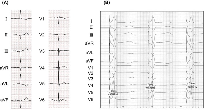
(A) A 12‐lead electrocardiogram after pacemaker implantation, showing reversed arm and leg leads, as well as right‐sided chest leads. (B) The intracardiac electrocardiogram showed a QRS complex duration of 121 ms, a left ventricular activation time of 76 ms in lead V6, and a V6–V1 interpeak interval of 39 ms.
References
LinkOut - more resources
Full Text Sources
