. 2025 Jun 5:16:1516836.
doi: 10.3389/fphar.2025.1516836. eCollection 2025.

Zileuton protects against arachidonic acid/5-lipoxygenase/leukotriene axis-mediated neuroinflammation in experimental traumatic brain injury

Affiliations

Zileuton protects against arachidonic acid/5-lipoxygenase/leukotriene axis-mediated neuroinflammation in experimental traumatic brain injury

Xiaofeng Xu et al. Front Pharmacol. .

Abstract

Introduction: Traumatic brain injury (TBI) is a leading cause of death and disability globally. Several studies have shown that 5-lipoxygenase (5-LOX) inhibition reduces leukotriene (LT) release and the inflammatory response, attenuating the development of respiratory diseases, myocardial infarction, and ischemic cerebral injury. However, its role in the pathophysiology of TBI remains unclear.

Methods: Controlled cortical impact injury was induced to construct a mouse model of TBI. Pericontusional brain tissue samples from sham and TBI mice at 7 days after injury were used for RNA-seq analysis. Altered gene enrichment following TBI, based on Kyoto Encyclopedia of Genes and Genomes (KEGG) pathway analysis, was quantified through real-time polymerase chain reaction (RT-PCR). Immunocytochemistry, Western blotting, and single-cell sequencing experiments were also performed to analyze 5-Lox protein expression. Arachidonic acid (AA) was detected through liquid chromatography mass spectrometry/mass spectrometry. Enzyme-linked immunosorbent assay was used to detect LTB4 release after TBI with or without zileuton treatment. Brain damage, blood-brain barrier disruption, and neuronal apoptosis were detected through histological examination. Neurological outcomes were determined through rotarod and fear conditioning tests.

Results: TBI induced significant upregulation of genes related to the AA metabolic pathway, particularly the AA/5-LOX/LT axis, as verified by RT-PCR. AA and LTB4 production increased significantly after TBI. The expression levels of Pla2g4a, which hydrolyses phospholipids to release AA, and 5-Lox, which in turn act downstream to convert AA to LT, were dramatically upregulated up to 7 days after TBI. 5-LOX accumulated in the cytoplasm of activated ameboid microglial cells. In vivo, 5-LOX inhibition with zileuton blocked LT release and reduced microglial activation and the production of inflammatory cytokines, including Il-1β, Ccl7, Spp1, Ccr1, Ccl2, and Il-10. Zileuton also reduced TBI-induced lipid ROS and neuronal cell apoptosis, ameliorating brain damage compared to the vehicle group and improving neurological outcomes after TBI. Mechanically, TBI-induced LT upregulation may stimulate BV2 microglial activation through the ERK, NF-κB, and Akt pathways.

Conclusion: Our findings demonstrated the role of 5-LOX in TBI and its potential as a therapeutic target in TBI treatment.

Keywords: 5-lipoxygenase; leukotriene; microglial cells; neuroinflammation; traumatic brain injury; zileuton.


Conflict of interest statement

The authors declare that the research was conducted in the absence of any commercial or financial relationships that could be construed as a potential conflict of interest.

Figures
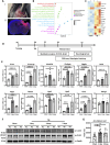
FIGURE 1
FIGURE 1
Arachidonic acid (AA) metabolism was upregulated after experimental traumatic brain injury (TBI). [(A)i] The process of controlled cortical impact (CCI) in mice. [(A)ii] Dashed box illustrates the pericontusional region of interest after TBI. (B) Kyoto Encyclopedia of Genes and Genomes (KEGG) pathway enrichment analysis of the genes upregulated after TBI. Significantly enriched metabolic, immune system, signal transduction, and cellular process pathways are shown. (C) Heat map showing differentially-expressed genes related to AA metabolism in the TBI-induced pericontusional brain tissue at 7 days based on RNA-seq analysis (n = 3 for each group). (D) Schematic overview of the experimental design. Mice were subjected to TBI using the CCI model, and the resulting damage phenotypes were analyzed at indicated time points post-injury. (E) mRNA levels of 5-Lox, 5-Lox-ap, Gpx3, Gpx7, Cbr2, Pla2g4a, Ltc4s, Tbxas1, Hpgds, and Ptges were measured in sham and TBI mice at 1 and 3 days after TBI (n = 6 for each group, *p < 0.05, **p < 0.01, ***p < 0.001, ****p < 0.0001). (F) Time course of 5-LOX and Pla2g4a/cPLA2 protein expression in pericontusional tissue. (G) AA concentrations in brain tissue were measured in sham and TBI mice at 1 and 3 days after TBI (n = 10, = 12, and = 11 for the sham, 1-day, and 3-day groups, respectively, *p < 0.05).
FIGURE 2
FIGURE 2
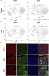
The expression of 5-LOX was upregulated in activated microglia/monocytes after TBI. (A, B) tSNE maps of mouse brain cells showing cell type-specific expression of Pla2g4a and 5-Lox in sham and TBI mice at 3 days after TBI. (C) Representative 5-LOX/CD68 immunofluorescence staining in the brains of sham and TBI mice at 3 and 7 days after TBI. White arrows indicate 5-LOX/CD68 co-localized inflammatory cells (scale bar = 10 μm).
FIGURE 3
FIGURE 3
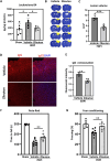
Zileuton reduced TBI-induced brain damage and neurological deficits. (A) Leukotriene B4 levels in brain tissue were measured in sham, TBI, and zileuton-treated mice at 3 days after TBI (n = 6 for each group, *p < 0.05, **p < 0.01). (B) Representative cresyl violet-stained brain sections from the vehicle- and zileuton-treated groups at 7 days after TBI. Lesions are outlined in yellow (scale bar = 10 μm). (C) Lesion volume was quantified and reported as mean ± SEM. (D) Representative IgG immunostaining of brain sections from vehicle- and zileuton-treated groups at 3 days after TBI (scale bar = 10 μm). (E) Quantification of fluorescence intensity for IgG extravasation in vehicle- and zileuton-treated groups 3 days after TBI (n = 6 for each group, *p < 0.05). (F) Rotarod test in vehicle- and zileuton-treated groups at 7 days after TBI (n = 6–8 mice/group) (G) Fear conditioning test; percentage of freezing time during the 5-minute test at 7 days after TBI (n = 6–8 mice/group, *p < 0.05).
FIGURE 4
FIGURE 4
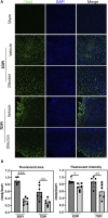
Zileuton reduced TBI induced microglia/macrophage activation. (A) Immunofluorescence staining for CD68 in the pericontusional regions of TBI and zileuton-treated mice at 3 and 7 days after TBI (Scale bar = 10 μm). (B) Quantification of CD68 fluorescence area and intensity relative to 4′6-diamidino-2-phenylindole (DAPI, n = 6 for each group, *p < 0.05).
FIGURE 5
FIGURE 5
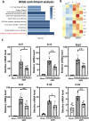
(A) Kyoto Encyclopedia of Genes and Genomes (KEGG) pathway enrichment analysis of the genes downregulated by zileuton treatment. The top most 10 significantly enriched pathways (p < 0.05 by Fisher’s exact test) are shown. (B) Heat map showing differentially expressed genes in the pericontusional brain tissue between the vehicle- and zileuton-treated groups at 7 days after TBI based on RNA-seq analysis (n = 3 for each group). (C) mRNA levels of ccl7, ccr1, ssp1, ccl2, il1β, and il10 were measured in the vehicle- and zileuton-treated mice at 3 days after TBI (n = 6 for each group, *p < 0.05, **p < 0.01).
FIGURE 6
FIGURE 6
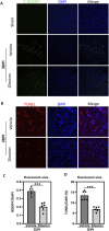
5-LOX inhibition reduced TBI-induced lipid ROS generation and neuronal cell apoptosis. (A) Representative confocal images of brain sections labeled with C11-BODIPY and DAPI from vehicle- and zileuton-treated mice at 3 days after TBI. Green and blue colors indicate lipid ROS (peroxidized lipids) and the nucleus, respectively. (B) Apoptotic cells in brain tissue sections were detected through TUNEL staining. (C) Quantification of the area of C11-BODIPY/DAPI fluorescence in (A). (D) Quantification of the area of TUNEL/DAPI fluorescence in (B). (n = 6 for each group, *p < 0.05, **p < 0.01).
FIGURE 7
FIGURE 7
Leukotrienes (LTs) induced inflammatory pathway activation in BV2 microglial cells. (A) Representative dose-dependent and (B) time-dependent Western blot results for NF-κB p65, phospho-NF-κB p65, phospho-Erk1/2, and Erk1/2 expression in LTB4-treated BV2. (C) Representative Western blot results for NF-κB p65, phospho-NF-κB p65, phospho-Erk1/2, Erk1/2 Akt, phospho-Akt at T308, and phospho-Akt at S473 in LTE4-treated BV2. (D) Proposed model of the suppression of TBI-induced microglial activation, cytokine release, brain damage, and neurobehavioral deficits by zileuton through the inhibition of LT release.

Similar articles

References

    1. Abueid L., Uslu U., Cumbul A., Velioglu Ogunc A., Ercan F., Alican I. (2017). Inhibition of 5-lipoxygenase by zileuton in a rat model of myocardial infarction. Anatol. J. Cardiol. 17, 269–275. 10.14744/AnatolJCardiol.2016.7248 - DOI - PMC - PubMed
    1. Alam A., Thelin E. P., Tajsic T., Khan D. Z., Khellaf A., Patani R., et al. (2020). Cellular infiltration in traumatic brain injury. J. Neuroinflammation 17, 328. 10.1186/s12974-020-02005-x - DOI - PMC - PubMed
    1. Attaluri S., Upadhya R., Kodali M., Madhu L. N., Upadhya D., Shuai B., et al. (2022). Brain-specific increase in leukotriene signaling accompanies chronic neuroinflammation and cognitive impairment in a model of Gulf war illness. Front. Immunol. 13, 853000. 10.3389/fimmu.2022.853000 - DOI - PMC - PubMed
    1. Beura L. K., Hamilton S. E., Bi K., Schenkel J. M., Odumade O. A., Casey K. A., et al. (2016). Normalizing the environment recapitulates adult human immune traits in laboratory mice. Nature 532, 512–516. 10.1038/nature17655 - DOI - PMC - PubMed
    1. Bolte A. C., Lukens J. R. (2021). Neuroimmune cleanup crews in brain injury. Trends Immunol. 42, 480–494. 10.1016/j.it.2021.04.003 - DOI - PMC - PubMed

LinkOut - more resources