. 2025 Jun 5:16:1591394.
doi: 10.3389/fphar.2025.1591394. eCollection 2025.

Aqueous extracts of Portulaca oleracea L. alleviate atopic dermatitis by restoring skin barrier function

Affiliations

Aqueous extracts of Portulaca oleracea L. alleviate atopic dermatitis by restoring skin barrier function

Haobin Wei et al. Front Pharmacol. .

Abstract

Background: Portulaca oleracea L. (PO) is an edible plant with a long medicinal history in traditional Chinese medicine for various inflammatory diseases, including skin disorders such as atopic dermatitis (AD). However, the anti-inflammatory effects and AD-alleviating mechanisms of PO remain unclear.

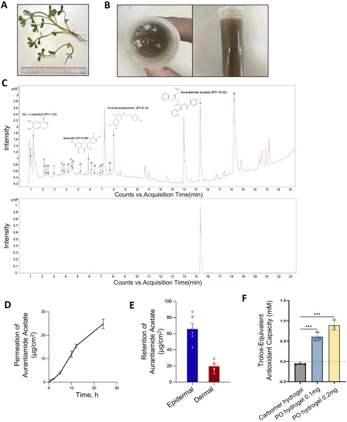
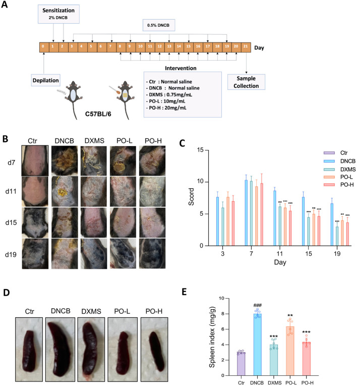
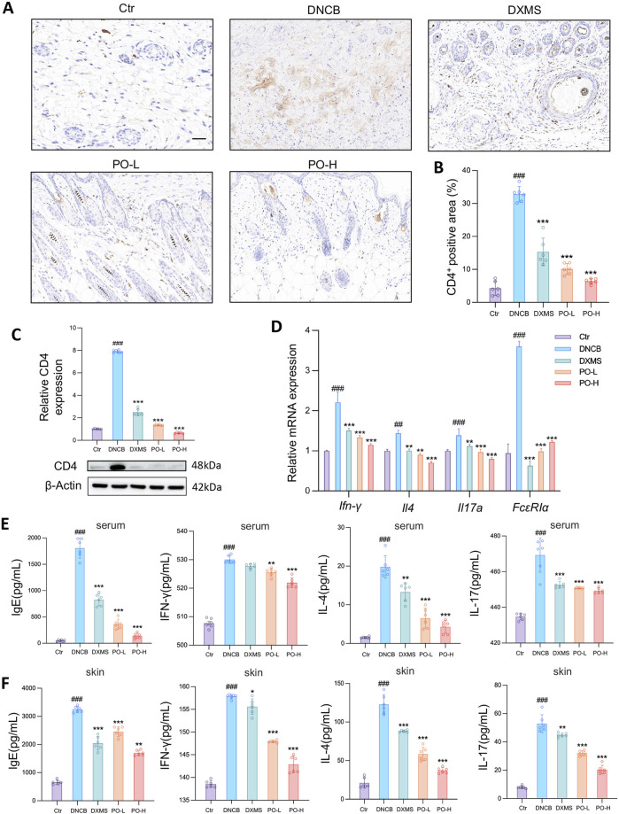
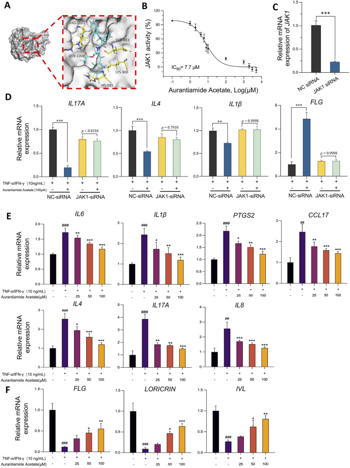
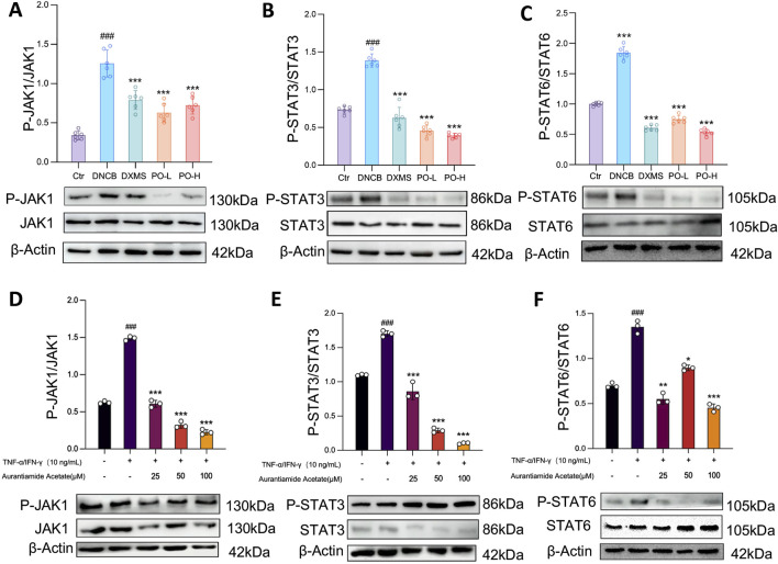
Methods: PO aqueous extract was prepared from a water-soluble portion and then mixed with carbomer to obtain a hydrogel, which provided stable drug permeation and absorption in mouse skin. Amantadine acetate was identified as an abundant ingredient and further predicted to be a Janus kinase 1 (JAK1) inhibitor via molecular binding simulation. Mice with AD, established by repeated sensitization with 2,4-dinitrochlorobenzene (DNCB), were topically treated with PO hydrogel. Aurantiamide acetate was applied to HaCaT keratinocytes prior to inflammatory challenge in the presence or absence of JAK1-siRNA. Transcriptional and translational gene expressions associated with cutaneous inflammation or skin barriers were assessed by qPCR and Western blotting, respectively. Enzyme-linked immunosorbent assays were performed to detect immunoglobulin E and proinflammatory factors in skin tissue or serum. The phosphorylation of JAK1, signal transducer and activator of transcription (STAT)3, and STAT6 in keratinocytes and skin were analyzed by Western blotting.

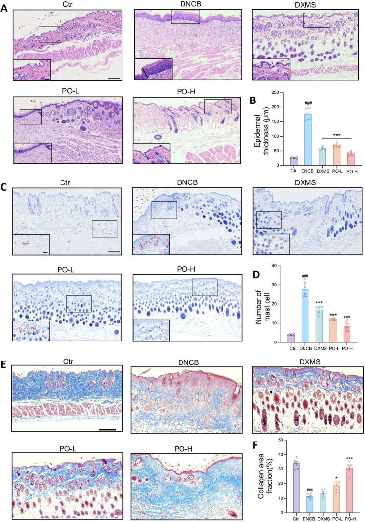
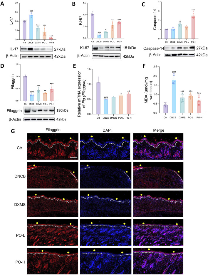
Results: In DNCB-sensitized mice, the PO hydrogel ameliorated skin lesions, lowered symptom scores, and reduced epidermal thickness by suppressing proinflammatory factor generation, oxidative stress, and the expression of CD4+. The PO hydrogel promoted the expression of caspase-14 and filaggrin, thereby helping restore skin barrier function in AD. The PO hydrogel and/or aurantiamide acetate inhibited the enzymatic activity of JAK1 and downstream (STAT)3/STAT6 signaling pathways in vitro and in vivo.

Conclusion: PO significantly ameliorated skin lesions and restored epidermal barrier function in AD mice. This was achieved by suppressing JAK1 enzymatic activity and JAK1-mediated STAT signaling pathways.

Keywords: JAK1 inhibition; aurantiamide acetate; cutaneous inflammation; epidermal barrier; filaggrin; purslane.


Conflict of interest statement

The authors declare that the research was conducted in the absence of any commercial or financial relationships that could be construed as a potential conflict of interest.

Figures

FIGURE 1
FIGURE 1
Preparation and characterization of the carbomer hydrogel of PO aqueous extract. (A) Fresh Portulaca oleracea L. (B) Prepared carbomer hydrogel of PO aqueous extract. (C) Chromatography of PO aqueous extracts by UHPLC-Q-TOF-MS/MS analysis in positive ion mode. The identified compounds are numbered in order on the basis of retention time. Upper panel, PO aqueous extracts; lower panel, reference compound aurantiamide acetate, with a retention peak at 15.32 min. Rt, retention time. (D) The cumulative permeation of aurantiamide acetate, one of the active ingredients in PO aqueous extract, was determined by UHPLC-Q-TOF-MS/MS in dissected rat skin at various time points after topical administration. n = 6. (E) Epidermal and dermal retention of aurantiamide acetate was determined by UHPLC-Q-TOF-MS/MS in dissected rat skin 24 h after administration. n = 6. (F) In vitro antioxidant ability of PO hydrogel. n = 3. The data are expressed as means ± SD.
FIGURE 2
FIGURE 2
PO carbomer hydrogel attenuates DNCB-induced AD clinical symptoms in mice. (A) Schematic diagram of the animal study protocol. (B) Representative photographs of the dorsal skin lesions on the 7th, 11th, 15th, and 19th days of the experiment. (C) SCORAD scores were assessed every 4 days. (D,E) Representative photographs of the spleen and the spleen indices of the mice. The data are expressed as means ± SD, n = 6. ### p < 0.001 vs. the vehicle control (Ctr) group; * p < 0.05, ** p < 0.01, and *** p < 0.001 vs. the DNCB-induced AD group.
FIGURE 3
FIGURE 3
PO carbomer hydrogel mitigated histopathological defects in mice with DNCB-induced AD. (A) Representative HE-stained dorsal skin on the 21st day of the experiment. The epidermal thickness was determined as indicated by the solid black lines. (B) Measurement of epidermal thickness as indicated in (A). (C) Representative toluidine blue-stained mast cells, as indicated by the red circles in the insets. (D) The number of mast cells per random microscopic field of the dorsal skin (×400 magnification). (E) Representative Masson’s trichrome-stained dorsal skin on the 21st day of the experiment. (F) Calculated collagen area fraction (blue). The data are expressed as means ± SD. ### p < 0.001 vs. the vehicle control (Ctr) group; * p < 0.05, ** p < 0.01, and ***p < 0.001 vs. the DNCB-induced AD group. Scale bars indicate 200 μm and 60 μm (insets of higher magnification), respectively.
FIGURE 4
FIGURE 4
PO carbomer hydrogel modulates cutaneous immune responses and suppresses the generation of proinflammatory factors in DNCB-induced AD mice. (A) Immunohistochemical staining of CD4+ cell infiltration (brown) in the skin. The scale bar represents 100 μm. (B) Quantification of the CD4+ area in the skin. (C) Relative CD4 protein expression in dorsal skin. (D) Relative mRNA expression of Ifn-γ, Il4, FcεRIα, and Il17a in dorsal skin, as detected by quantitative PCR. n = 6. (E,F) Determination of IgE, INF-γ, IL-4, and IL-17 concentrations in mouse serum (E) and affected skin (F) via ELISA. The data are expressed as means ± SD. # p < 0.05, ## p < 0.01, ### p < 0.001 vs. the vehicle control (Ctr) group; * p < 0.05, ** p < 0.01, and *** p < 0.001 vs. the DNCB-induced AD group.
FIGURE 5
FIGURE 5
PO carbomer hydrogel modulates immune responses and partially restores the damaged skin barrier. (A–D) Relative protein expression of IL-17, Ki-67, caspase-14, and filaggrin was detected by Western blotting. Blots are representative of 6 independent experiments. (E) Relative mRNA expression of Flg (Fillagrin) detected by quantitative PCR in the dorsal skin. n = 6. (F) Content of MDA in the affected skin. n = 6. MDA, malondialdehyde. (G) Immunostaining of dorsal sections for filaggrin (red) and nuclei (blue). The epidermis is shown along the white dotted lines. The yellow arrows indicate the filaggrin-derived skin barrier in the epidermis. Scale bar = 100 μm. The data are expressed as means ± SD. # p < 0.05, ## p < 0.01, ### p < 0.001 vs. the vehicle control (Ctr) group; * p < 0.05, ** p < 0.01, and *** p < 0.001 vs. the DNCB-induced AD group.
FIGURE 6
FIGURE 6
Aurantiamide acetate suppresses dermal inflammation and restores skin barrier function through targeting JAK1. (A) Diagram of the molecular docking between aurantiamide acetate, an active ingredient in PO, and JAK1 (PDB: 6BBU). (B) Inhibitory effects of aurantiamide acetate on JAK1 tyrosine kinase activity detected by the ADP-Glo™ reagent. (C) siRNA-JAK1 silencing efficiency 24 h after transfection. (D) Relative mRNA expression levels of IL17A, IL4, IL1β and FLG were detected via quantitative PCR in HaCaT cells transfected with NC or JAK1 siRNA. (E) Aurantiamide acetate inhibits proinflammatory gene transcription in non-siRNA-treated HaCaT keratinocytes stimulated with TNF-α/IFN-γ. (F) Aurantiamide acetate upregulates skin barrier protective gene transcription in non-siRNA-targeted HaCaT keratinocytes stimulated with TNF-α/IFN-γ. (C–F) Cells were pretreated with aurantiamide acetate (25, 50, or 100 μM) or vehicle (0.1% DMSO) for 1 h before TNF-α (10 ng/mL) and IFN-γ (10 ng/mL) challenge for 18 h. Relative mRNA expression levels of IL6, IL1β, PTGS2 (COX-2), CCL17 (TARC), IL8, IL4, IL17A, FLG, LORICRIN and IVL were detected via quantitative PCR. The data were normalized to internal GAPDH or β-Actin gene expression. The data are expressed as means ± SD of 3 independent experiments performed in duplicate. # p < 0.05, ## p < 0.01, ### p < 0.001 vs. the nonstimulated group; * p < 0.05, ** p < 0.01, and *** p < 0.001 vs. the vehicle group.
FIGURE 7
FIGURE 7
Aurantiamide acetate and PO carbomer hydrogel suppress the JAK1-STAT3/6 signaling pathway. (A–C) Western blot analysis of JAK1-STAT3/6 phosphorylation in mouse skin. n = 6. (D–F) Western blot analysis of JAK1-STAT3/6 phosphorylation in keratinocytes. The cells were pretreated with aurantiamide acetate (25, 50, or 100 μM) or vehicle (0.1% DMSO) for 1 h before being challenged with TNF-α (10 ng/mL) or IFN-γ (10 ng/mL) for 18 h. The data are expressed as means ± SD of 3 independent experiments performed in duplicate. # p < 0.05, ## p < 0.01, ### p < 0.001 vs. the Ctr/non-stimulated group; * p < 0.05, ** p < 0.01, and *** p < 0.001 vs. the vehicle/untreated group. The data were normalized to that of β-Actin.

Similar articles

Cited by

References

    1. Bai X., Rao X., Wang Y., Shen H., Jin X. (2023). A homogeneous Lonicera japonica polysaccharide alleviates atopic dermatitis by promoting Nrf2 activation and NLRP3 inflammasome degradation via p62. J. Ethnopharmacol. 309, 116344. 10.1016/j.jep.2023.116344 - DOI - PubMed
    1. Bakker D., de Bruin-Weller M., Drylewicz J., van Wijk F., Thijs J. (2023). Biomarkers in atopic dermatitis. J. Allergy Clin. Immunol. 151 (5), 1163–1168. 10.1016/j.jaci.2023.01.019 - DOI - PubMed
    1. Bieber T. (2022). Atopic dermatitis: an expanding therapeutic pipeline for a complex disease. Nat. Rev. Drug Discov. 21 (1), 21–40. 10.1038/s41573-021-00266-6 - DOI - PMC - PubMed
    1. Bonefeld C. M., Petersen T. H., Bandier J., Agerbeck C., Linneberg A., Ross-Hansen K., et al. (2016). Epidermal filaggrin deficiency mediates increased systemic T-helper 17 immune response. Br. J. Dermatol 175 (4), 706–712. 10.1111/bjd.14570 - DOI - PubMed
    1. Chovatiya R., Paller A. S. (2021). JAK inhibitors in the treatment of atopic dermatitis. J. Allergy Clin. Immunol. 148 (4), 927–940. 10.1016/j.jaci.2021.08.009 - DOI - PMC - PubMed

LinkOut - more resources