. 2025 Jun 1:37:100736.
doi: 10.1016/j.ynstr.2025.100736. eCollection 2025 Jul.

Probiotics restore impaired spatial cognition and synaptic plasticity of prenatally-stressed male rats: focus on hippocampal and intestinal tight junctions

Affiliations

Probiotics restore impaired spatial cognition and synaptic plasticity of prenatally-stressed male rats: focus on hippocampal and intestinal tight junctions

Fatemeh Aghighi Bidgoli et al. Neurobiol Stress. .

Abstract

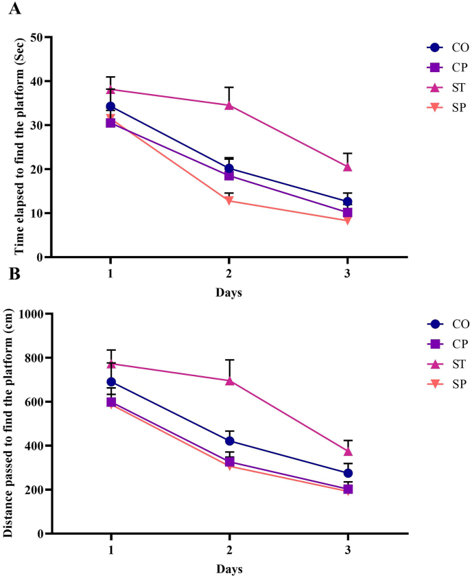
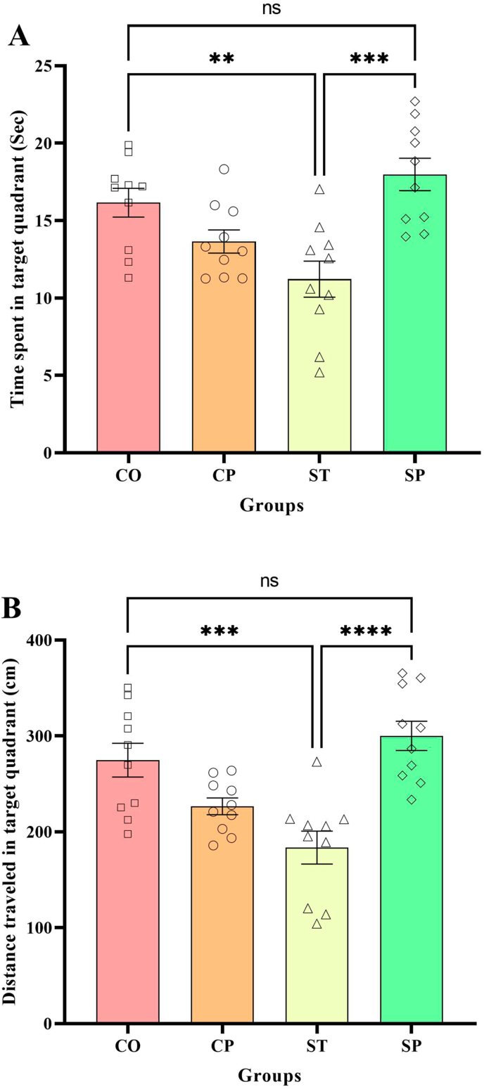
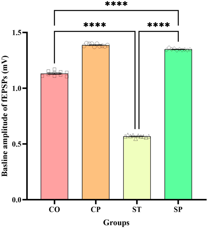
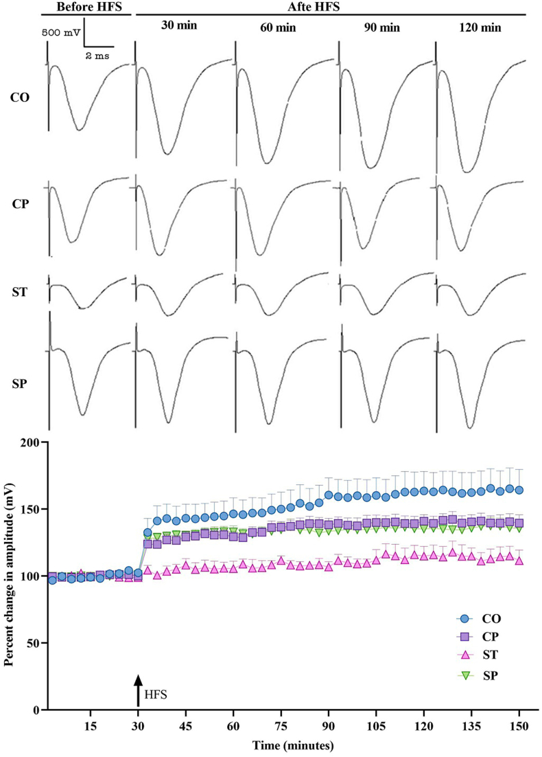
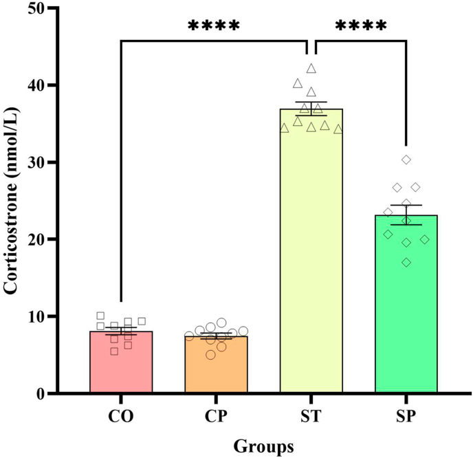
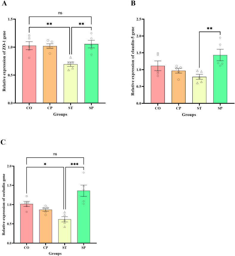
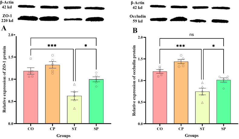
Bidirectional communication between the gut microbiota and the nervous system founded the gut-microbiota-brain axis, substantially affects numerous vital functions of the body. Stress, as the body's natural reaction to stressful situations, in turn, affects the functioning of various organs. Through evaluating long-term potentiation (LTP) and spatial memory assessment using the Morris water maze, we aimed to examine the effect of prenatal stress on the electrophysiological and behavioral aspects of hippocampus-dependent spatial memory. The relationship of the synaptic plasticity and learning and memory with the hypothalamus-pituitary-adrenal (HPA) axis and the integrity of blood-brain and intestinal barriers were also examined. The experimental subjects were introduced to probiotic treatment to assess how the supplementation influences stress-related alterations. The prenatal stress effectively impaired both LTP occurrence and behavioral function. It also led to disruption of blood-brain and gut barriers and increased serum level of corticosterone. The probiotic supplementation positively affected the synaptic plasticity and learning and memory. It also improved the integrity of both barriers and reduced the stress hormone corticosterone. Whereas there is a reverse relationship between stress and the hippocampus-dependent spatial memory, normal stress hormone, and the integrity of intestinal and brain barriers, the probiotic supplements improve all impairments. We conclude that the HPA axis plus the blood-brain and intestinal barriers play a role in hippocampus-dependent spatial memory that are substantially affected by the beneficial gut and probiotic bacteria.

Keywords: Blood-brain barrier; Hypothalamus-pituitary-adrenal axis; Intestinal barrier; Learning and memory; Long-term potentiation; Probiotics; Stress.


Conflict of interest statement

The authors declare the following financial interests/personal relationships which may be considered as potential competing interests: Mahmoud Salami reports financial support was provided by 10.13039/501100004048Kashan University of Medical Sciences. If there are other authors, they declare that they have no known competing financial interests or personal relationships that could have appeared to influence the work reported in this paper.

Figures

Fig. 1
Fig. 1
The CUS protocol was applied for 21 days. The control mother group was not submitted to the CUS protocol. Treatments occurred from day 22 to day 52 (saline or probiotic). On days 53–57, the animals were subjected to behavioral and electrophysiological tests. After the electrophysiological test, the animals were euthanized, and the hippocampus, duodenum, and blood were sampled for further analysis (n = 10).
Fig. 2
Fig. 2
Morris water maze performance of different groups. The time elapsed (A) and the distance traveled (B) to reach the hidden platform during the learning phase. Prenatally stressed (ST) animals exhibited significantly impaired performance compared to CO animals, as indicated by increased latency (P < 0.01) and distance traveled (P < 0.05). In contrast, prenatally stressed animals supplemented with probiotics (SP) demonstrated significantly reduced latency and distance compared to the ST group (P < 0.0001). Data are shown as mean ± SEM, and each point indicate the average of four daily trials (n = 10). CO: Control, CP: Control + Probiotic, ST: Stress, SP: Stress + Probiotic.
Fig. 3
Fig. 3
Spatial memory evaluated in the probe trial test. The data are shown as spent time (A) and traveled path (B) in the target quadrant. The comparison of the spent time (P < 0.01) and traveled path (P < 0.001) in the target quadrant between the CO and ST rats indicates that the CUS exposure disturbs spatial memory. Probiotic administration improved spatial memory, so SP rats spent more time (P < 0.001) and traveled more distance (P < 0.0001) in the target quadrant compared to their ST counterparts. Data are shown as mean ± SEM (n = 10). ∗∗P < 0.01; CO group vs ST. ∗∗∗P < 0.001; CO and SP groups vs ST group. ∗∗∗∗P < 0.0001; ST group vs SP group. CO: Control, CP: Control + Probiotic, ST: Stress, SP: Stress + Probiotic.
Fig. 4
Fig. 4
The amplitude of the baseline fEPSPs measured in the CA1 region of the hippocampus. The comparison of ST and CO rats indicates that the CUS exposure markedly affects the basic hippocampal fEPSPs (P < 0.0001). The probiotic administration resulted in a statistically significant increase in baseline fEPSP amplitudes in stressed rats receiving probiotics (SP) compared to ST rats (P < 0.0001). Data are presented as mean ± SEM (n = 10). ∗∗∗∗P < 0.0001; CO and SP groups vs ST group. CO: Control, CP: Control + Probiotic, ST: Stress, SP: Stress + Probiotic.
Fig. 5
Fig. 5
Induction of LTP in the EPSPs recorded in the CA1 region following CA3–CA1 pathway tetanization. While the tetanization augmented the fEPSPs in the CO rats (P < 0.0001), it failed to potentiate the responses in the ST animals. The postnatal probiotic treatment robustly increased the amplitude of the fEPSPs in both the CP and SP groups (P < 0.0001). There was a significant difference between the ST and SP groups (P < 0.0001). The arrow indicates the application of high frequency stimulation (HFS). Each point shows average data obtained during 3 min (n = 10). CO: Control, CP: Control + Probiotic, ST: Stress, SP: Stress + Probiotic.
Fig. 6
Fig. 6
The Serum corticosterone levels were assessed in all experimental groups. Animals subjected to prenatal stress (ST group) exhibited significantly elevated corticosterone levels compared to CO animals (P < 0.0001). The corticosterone level was decreased in the SP rats (P < 0.0001). All data are shown as mean ± SEM (n = 10). ∗∗∗∗P < 0.0001; CO and SP groups vs ST group. CO: Control, CP: Control + Probiotic, ST: Stress, SP: Stress + Probiotic.
Fig. 7
Fig. 7
Hippocampal tight junction gene expression levels in experimental groups. Gene expression levels of ZO-1 (A), claudin-5 (B), and occludin (C) were determined using real-time PCR. (A) CUS significantly decreased the expression of ZO-1 (P < 0.01), and treatment with probiotics significantly increased it (P < 0.01). (B) Probiotic administration significantly increased expression of claudin-5 in SP rats (P < 0.01). (C) The occludin gene expression in ST rats was lower than in CO rats (P < 0.05). The occludin expression was increased in the SP rats (P < 0.001). Data were analyzed using a two-way ANOVA followed by Tukey's post-hoc test. Values are presented as the mean ± SEM (n = 5). ∗P < 0.05; CO group vs ST. ∗∗P < 0.01; CO and SP groups vs ST group. ∗∗∗P < 0.0001; ST group vs SP group. CO: Control, CP: Control + Probiotic, ST: Stress, SP: Stress + Probiotic.
Fig. 8
Fig. 8
ZO-1 (A), occludin (B) tight junction protein expression in duodenal cells of four experimental groups. The densitometric quantification of ZO-1 and occludin was normalized to GAPDH. CUS significantly decreased the expression of ZO-1 and occludin (P < 0.001), and treatment with probiotics significantly increased its (P < 0.05). Data were analyzed using two-way ANOVA followed by Tukey's post-hoc test. The values are represented as mean ± SEM (n = 5). ∗P < 0.05; ST group vs SP group. ∗∗∗P < 0.0001; CO group vs ST. CO: Control, CP: Control + Probiotic, ST: Stress, SP: Stress + Probiotic.

Similar articles

References

    1. Aghighi Bidgoli F., Salami M., Talaei S.A. Environmental enrichment restores impaired spatial memory and synaptic plasticity in prenatally stress exposed rats: the role of GABAergic neurotransmission. Int. J. Dev. Neurosci. 2020;80(7):573–585. - PubMed
    1. Algamal M., Pearson A.J., Hahn-Townsend C., Burca I., Mullan M., Crawford F., Ojo J.O. Repeated unpredictable stress and social isolation induce chronic HPA axis dysfunction and persistent abnormal fear memory. Prog. Neuro Psychopharmacol. Biol. Psychiatr. 2021;104 - PubMed
    1. Aoudia N., Rieu A., Briandet R., Deschamps J., Chluba J., Jego G., Garrido C., Guzzo J. Biofilms of Lactobacillus plantarum and Lactobacillus fermentum: effect on stress responses, antagonistic effects on pathogen growth and immunomodulatory properties. Food Microbiol. 2016;53:51–59. - PubMed
    1. Barzegar M., Sajjadi F.S., Talaei S.A., Hamidi G., Salami M. Prenatal exposure to noise stress: anxiety, impaired spatial memory, and deteriorated hippocampal plasticity in postnatal life. Hippocampus (New York, N. Y.) 2015;25(2):187–196. - PubMed
    1. Benoit J.D., Rakic P., Frick K.M. Prenatal stress induces spatial memory deficits and epigenetic changes in the hippocampus indicative of heterochromatin formation and reduced gene expression. Behav. Brain Res. 2015;281:1–8. - PMC - PubMed

LinkOut - more resources