. 2025 Jun 7:52:200-212.
doi: 10.1016/j.bioactmat.2025.05.034. eCollection 2025 Oct.

Near-field electrospun 3D anisotropic fiber-hydrogel scaffold integrated with photothermal effect for skin wound healing

Affiliations

Near-field electrospun 3D anisotropic fiber-hydrogel scaffold integrated with photothermal effect for skin wound healing

Ruinan Hao et al. Bioact Mater. .

Abstract

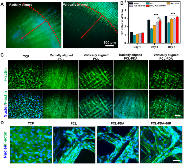
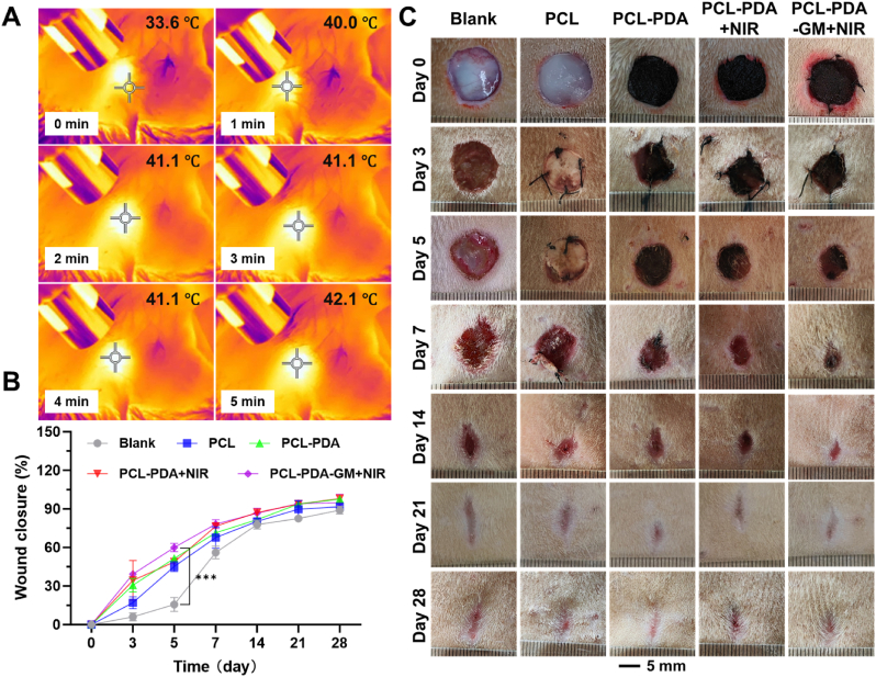
Wound healing remains a critical clinical challenge due to inflammatory responses, oxidative stress in the wound microenvironment, and impaired tissue remodeling. In this study, an anisotropic scaffold was developed by integrating photothermal stimulation with topographical cues to modulate wound healing. The scaffold consisted of gelatin methacryloyl (GM) hydrogel and radially aligned poly (ε-caprolactone) (PCL) fibers integrated with polydopamine (PDA). The anisotropic scaffold not only exhibited anti-inflammatory effects but also enabled localized thermal stimulation under near-infrared (NIR) light to promote wound healing. It guided cell migration and proliferation from the wound edge toward the center, while the GM hydrogel maintained a moist environment and mitigated uncontrolled thermal damage. In a full-thickness skin wound model in rats, the anisotropic scaffold accelerated wound healing, epidermal regeneration, angiogenesis, and collagen deposition. This approach offers a safe, efficient, and bioactive-factor-free therapeutic strategy for wound repair, showing great potential for clinical translation.

Keywords: Cell migration; Mild heat stimulation; Near-field electrospinning; Radially aligned fibers; Wound healing.


Conflict of interest statement

The authors declare that they have no known competing financial interests or personal relationships that could have appeared to influence the work reported in this paper.

Figures

Image 1
Graphical abstract
Fig. 1
Fig. 1
(A) Schematic of the PCL scaffold composed of radially aligned fiber and vertically aligned fibers prepared using near-field electrospinning device. (B) Photographs of PCL scaffolds with the different counts of the radially aligned fibers. (C) Schematic and photographs of a single-layer PCL fiber scaffold with 200 radially aligned fibers. The scaffold was divided into five areas, with a diameter of 15 mm. (D) The gap distance between the radially aligned fibers. (E) Photographs of the radially aligned layer and (F) the thickness of the PCL scaffold. Scar bar = 3 mm. (G) SEM images of the PCL scaffold and (H) magnification for the scaffold. (I) SEM image of the PCL-PDA scaffold. (J) Energy dispersive spectrometer mapping for representative elements (C, O, N) of the PCL-PDA scaffold. (K) Representative tensile stress–strain curves of the PCL, PCL-PDA, and PCL-PDA-GM scaffolds. (L) Photograph of the PCL-PDA-GM scaffold. (M) SEM image of the PCL-PDA-GM scaffold. (N) SEM image of the cross-section of the PCL-PDA-GM scaffold.
Fig. 2
Fig. 2
(A) Temperature rise curves of the PCL-PDA scaffold under NIR laser irradiation at different power densities (1, 1.25, 1.5 and 1.75 W/cm2). (B) Temperature rise curves of the PCL-PDA-GM scaffold under NIR laser irradiation at different power densities (1.25, 1.5, 1.75, 2.0 and 2.5 W/cm2). (C) Temperature rise curves of the PCL, PCL-PDA and PCL-PDA-GM scaffolds under NIR laser irradiation at a power density of 1.5 W/cm2. (D) Temperature-time curve of the PCL-PDA-GM scaffold after five cycles of irradiation and cooling. (E) Temperatures beneath the PCL-PDA and PCL-PDA-GM scaffolds measured by a temperature probe under NIR irradiation. (F) DSC of the GM hydrogel.
Fig. 3
Fig. 3
(A) DPPH scavenging capabilities of PCL, PCL-PDA, PCL-PDA-GM, and PCL-PDA-GM + NIR scaffolds. (B) Fluorescence intensity and (C) representative fluorescence images of Rosup-stimulated L929 cells after co-incubation with different scaffolds, stained with DCFH-DA. Scale bar = 50 μm.
Fig. 4
Fig. 4
(A) Representative fluorescence images of L929 cells on vertically aligned and radially aligned PCL-PDA scaffolds after 3 days of migration. Cell nuclei were stained with DAPI (blue fluorescence), and F-actin cytoskeletons were stained with phalloidin (green fluorescence). (B) OD values of L929 cells co-cultured with different scaffolds for 1, 3, and 5 days. (C) Representative fluorescence images of L929 cells on TCP, vertically aligned PCL-PDA scaffold, and radially aligned PCL-PDA scaffold. Cell nuclei were stained with DAPI (blue fluorescence) and F-actin cytoskeletons were stained with phalloidin (green fluorescence). Scale bar = 500 μm. (D) Representative fluorescent images of NIH-3T3 cells cultured on TCP, PCL, PCL-PDA, and PCL-PDA + NIR scaffolds. Scale bar = 50 μm.
Fig. 5
Fig. 5
(A) Representative thermal images of rats treated with the PCL-PDA-GM scaffold under laser irradiation (1.5 W/cm2, 5 min). (B) Proportion of wound closure in different groups at various time points. (C) Representative photographs of the wounds after treatment with the different scaffolds at 0, 3, 5, 7, 14, 21, and 28 days.
Fig. 6
Fig. 6
(A) Immunofluorescence staining of TNF-α, IL-6, TGF-β, and IL-10 in wound tissues on day 7. Scale bar = 50 μm. Quantitative analysis of (B) TNF-α, (C) IL-6, (D) TGF-β, and (E) IL-10 expression levels in wound tissues on day 7, based on immunofluorescence staining results.
Fig. 7
Fig. 7
(A) Representative H&E staining and Masson's trichrome staining images from different treatment groups on days 14 and 28. Quantitative analysis of (B) the epidermis thickness and (C) collagen deposition on days 14 and 28. (D) Representative immunofluorescence images of CD31 expression in wound tissues on day 28. Scale bar = 500 μm.

References

    1. Liang Y., He J., Guo B. Functional hydrogels as wound dressing to enhance wound healing. ACS Nano. 2021;15:12687–12722. doi: 10.1021/acsnano.1c04206. - DOI - PubMed
    1. Jeschke M.G., van Baar M.E., Choudhry M.A., Chung K.K., Gibran N.S., Logsetty S. Burn injury. Nat. Rev. Dis. Primers. 2020;6:11. doi: 10.1038/s41572-020-0145-5. - DOI - PMC - PubMed
    1. Eming S.A., Martin P., Tomic-Canic M. Wound repair and regeneration: mechanisms, signaling, and translation. Sci. Transl. Med. 2014;6 doi: 10.1126/scitranslmed.3009337. - DOI - PMC - PubMed
    1. Peña O.A., Martin P. Cellular and molecular mechanisms of skin wound healing. Nat. Rev. Mol. Cell Biol. 2024;25:599–616. doi: 10.1038/s41580-024-00715-1. - DOI - PubMed
    1. Hao R., Cui Z., Zhang X., Tian M., Zhang L., Rao F., Xue J. Rational design and preparation of functional hydrogels for skin wound healing. Front. Chem. 2021;9 doi: 10.3389/fchem.2021.839055. - DOI - PMC - PubMed

LinkOut - more resources