Review
. 2025 Jun 5:16:1573499.
doi: 10.3389/fendo.2025.1573499. eCollection 2025.

Endoplasmic reticulum-mitochondria crosstalk: new mechanisms in the development of atherosclerosis

Affiliations
Review

Endoplasmic reticulum-mitochondria crosstalk: new mechanisms in the development of atherosclerosis

Mingxiao Li et al. Front Endocrinol (Lausanne). .

Abstract

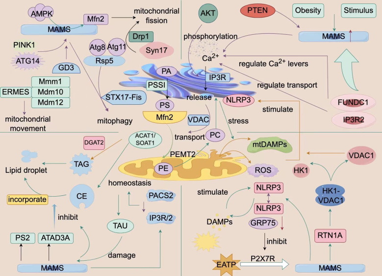
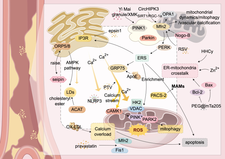
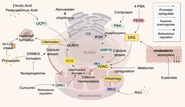
Atherosclerosis (AS) is a global public health concern and involves a complex pathogenesis characterized by lipid abnormalities, oxidative stress, and inflammatory responses at the cellular and molecular levels. The crosstalk between the endoplasmic reticulum (ER) and mitochondria, mediated by mitochondria-associated membranes (MAMs), plays a critical role in the pathogenesis of atherosclerosis. As two key cellular organelles, the ER and mitochondria interact physically and functionally through MAMs, which serve as bridges between their close contact and interdependence. MAMs maintain lipid homeostasis, promote calcium ion transport, the oxidative stress response, apoptosis, and autophagy. Recent studies have highlighted the significance of ER-mitochondria crosstalk in the progression of AS, as indicated by mitochondrial and ER structural and functional integrity, redox homeostasis, and calcium homeostasis. This review comprehensively explores the novel mechanisms of ER-mitochondria crosstalk in AS and emphasizes the potential of MAMs as therapeutic targets, aiming to provide new perspectives and strategies for the treatment of cardiovascular diseases.

Keywords: atherosclerosis; endoplasmic reticulum; endoplasmic reticulum contact complex; endoplasmic reticulum-mitochondrial crosstalk; mitochondria; mitochondria-associated membranes (MAMs).


Conflict of interest statement

The authors declare that the research was conducted in the absence of any commercial or financial relationships that could be construed as a potential conflict of interest.

Figures

Figure 1
Figure 1
Biological Processes of ER-Mitochondria Crosstalk. This figure is a schematic diagram of the biological processes of interaction between the endoplasmic reticulum and mitochondria within a cell. It shows a variety of molecules and signaling pathways, including mitochondrial fission and fusion-related proteins (e.g., Mfn2, Drp1), autophagy-related proteins (e.g., PINK1, PARKIN), calcium ion signaling, and ER-related proteins (e.g., ERMES, VDAC1). These molecules and signaling pathways interact to regulate cellular metabolism, energy balance, and autophagy processes. Additionally, the figure illustrates the impact of external factors such as obesity and stimulation on these processes. The structures and molecules in the figure represent the biological processes of ER-mitochondria crosstalk (MAMs). The main structural molecules are described as follows: AMPK: AMP-activated protein kinase; MAMs: Mitochondria-associated membranes; PINK1: PTEN-induced putative kinase 1; ATG14: Autophagy-related protein 14; GD3: Disialyl ganglioside; ERMES: Endoplasmic reticulum-mitochondria contact site complex; STX17-Fis: Synaptophysin 17-mitochondrial fission protein 1 complex; PACS2: Phosphorylated protein sorting and transport protein 2; TAU: Microtubule-associated protein Tau; CE: Cholesterol ester; PS2: Presenilin 2; ATAD3A: ATPase family AAA domain-containing protein 3A; NLRP3: NOD-like receptor pyrin domain-containing protein 3; RTN1A: Reticulon 1A; FUNDC1: FUN14 domain-containing protein 1; VDAC1: Voltage-dependent anion channel 1; Mfn2: Mitofusin 2. The image is created with Figdraw.
Figure 2
Figure 2
ER-Mitochondria Crosstalk in Atherosclerosis. This figure provides a detailed illustration of the complex mechanisms by which the endoplasmic reticulum (ER) and mitochondria interact through mitochondria-associated membranes (MAMs) and their associated proteins during atherosclerosis. It highlights key molecules and signaling pathways, including calcium ion flux and lipid transfer. Mfn2 serves as a central hub for communication between the ER and mitochondria. The figure also shows the roles of various proteins (such as ORP5/8, IP3R, GRP75, PERK, PAC5-2, VDAC, and Mfn2) in these interactions. Additionally, it illustrates abnormal biological processes such as ER stress (ERS), mitochondrial autophagy (mitophagy), and apoptosis, which collectively impact the progression of atherosclerosis. Through the interplay of these molecules and signaling pathways, ER-mitochondria crosstalk plays a critical role in the development of atherosclerosis. Structures and Molecules in the Figure: White rectangular areas: Mitochondria-associated membranes (MAMs); Yellow structure (upper left): Endoplasmic reticulum (ER); Yellow structures (upper right and lower right): Mitochondria; Yellow arrow (center): Calcium ion flow between the ER and mitochondria. The main structural molecules are described as follows: PINK1: PTEN-induced putative kinase 1; Parkin: Parkin protein; Mfn2: Mitofusin 2; Nogo-B: Reticulon 4B; Bax: Bcl-2-associated X protein; Bcl-2: B-cell lymphoma-2; Fis1: Mitochondrial fission protein 1; OX-LDL: Oxidized low-density lipoprotein; Seipin: Fat differentiation-related protein; ACAT: Acyl-CoA:cholesterol acyltransferase; ORP5/8: Oxysterol-binding protein-related proteins 5/8; GRP75: Glucose-regulated protein 75; CAMK1: Calcium/calmodulin-dependent protein kinase 1; NLRP3: NOD-like receptor pyrin domain-containing protein 3; LDs: Lipid droplets; PERK: RNA-dependent protein kinase-like ER kinase. The image is created with Figdraw.
Figure 3
Figure 3
Drugs Targeting ER-Mitochondria Crosstalk in AS. This figure illustrates the signaling exchange between the ER and mitochondria via MAMs contact sites in an atherosclerotic environment. The ER transfers Ca2+ to mitochondria to maintain their function, but Ca2+ overload can inhibit mitochondrial function, reducing ATP and increasing ROS, which triggers apoptosis. Statins stabilize endothelial function by inhibiting a key enzyme in cholesterol synthesis. Various drugs (e.g., pravastatin, atorvastatin, 4-PBA) target ER-mitochondria crosstalk to improve calcium ion flow, maintain mitochondrial function, reduce inflammation, and decrease foam cell formation, thereby improving AS. Traditional Chinese medicine components, such as curcumin and resveratrol, modulate key proteins (e.g., OPA1, MFN2) to influence mitochondrial function and show therapeutic potential. The structures and molecules in the figure represent the targets of AS drugs that act on ER-mitochondria crosstalk. The gray structure at the top represents the ER, the pink structure at the bottom represents the mitochondrion, and the gray-pink area between them represents the MAMs. The meanings of the various arrows are labeled in the legend in the top right corner of the figure. The main structural molecules are described as follows: UCP1: Uncoupling protein 1; ROS: Reactive oxygen species; OPA1: Optic atrophy protein 1; NLRP3: NOD-like receptor pyrin domain-containing protein 3; VDAC: Voltage-dependent anion channel; HK1: Hexokinase 1; PERK: Protein kinase R-like ER kinase; PKA: Protein kinase A; RSV: Resveratrol; ERS: Endoplasmic reticulum stress; GRP75: Glucose-regulated protein 75; IP3R: Inositol 1,4,5-trisphosphate receptor; 4-PBA: 4-phenylbutyric acid. The image is created with Figdraw.

Similar articles

References

    1. Perrotta I. Atherosclerosis: from molecular biology to therapeutic perspective 2.0. Int J Mol Sci. (2022) 23:15158. doi: 10.3390/ijms232315158 - DOI - PMC - PubMed
    1. Copeland DE, Dalton AJ. An association between mitochondria and the endoplasmic reticulum in cells of the pseudobranch gland of a teleost. J Biophys Biochem Cytol. (1959) 5:393–6. doi: 10.1083/jcb.5.3.393 - DOI - PMC - PubMed
    1. Luan Y, Luan Y, Yuan RX, Feng Q, Chen X, Yang Y, et al. Structure and function of mitochondria-associated endoplasmic reticulum membranes (MAMs) and their role in cardiovascular diseases. Oxid Med Cell Longev. (2021) 2021:4578809. doi: 10.1155/2021/4578809 - DOI - PMC - PubMed
    1. Janikiewicz J, Szymański J, Malinska D, Patalas-Krawczyk P, Michalska B, Duszyński J, et al. Mitochondria-associated membranes in aging and senescence: structure, function, and dynamics. Cell Death Dis. (2018) 9:332. doi: 10.1038/s41419-017-0105-5 - DOI - PMC - PubMed
    1. Wang N, Wang C, Zhao H, He Y, Lan B, Sun L, et al. The MAMs structure and its role in cell death. Cells. (2021) 10(3):657. doi: 10.3390/cells10030657 - DOI - PMC - PubMed