

GPD1L downregulation in colorectal cancer: a novel obesity-related biomarker linking metabolic dysregulation to tumor progression
- PMID: 40538846
- PMCID: PMC12177464
- DOI: 10.3389/fonc.2025.1582728
GPD1L downregulation in colorectal cancer: a novel obesity-related biomarker linking metabolic dysregulation to tumor progression
Abstract
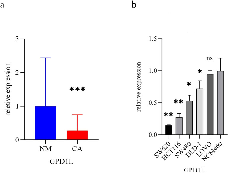
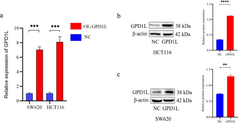
Objective: To delineate the expression profile and tumor-suppressive function of the metabolism-associated gene GPD1L in colorectal carcinogenesis. Methods: Transcriptomic datasets from TCGA and GEO repositories (GSE74602, GSE113513, GSE164191) were computationally analyzed. Paired tumor/adjacent mucosal specimens (n=58) from CRC patients at Jincheng People's Hospital were analyzed alongside the NCM460 colon epithelial line and five CRC lines (SW620, HCT116, SW480, DLD-1, LOVO). Following GPD1L quantification via qPCR, selected cell models underwent pcDNA3.1-GPD1L transfection for functional characterization. Then Western blot analysis was used to explore its possible mechanism.
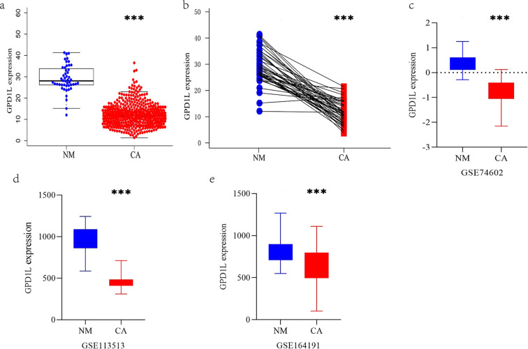
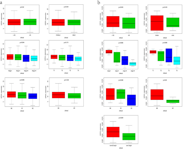
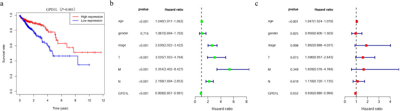
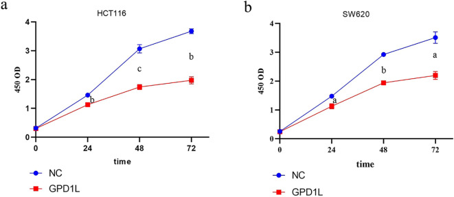
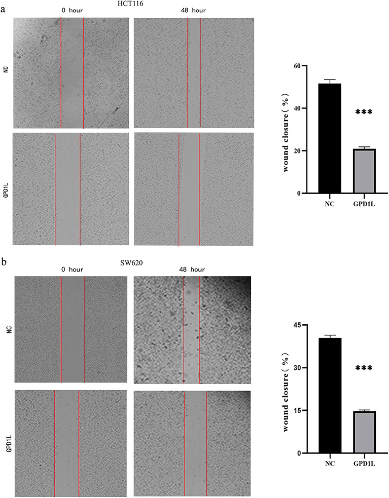
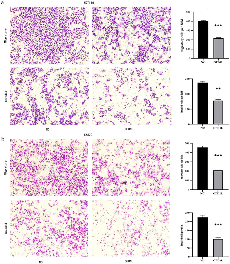
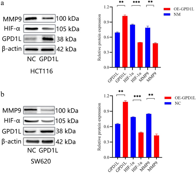
Results: Comparative analysis revealed a marked elevation of GPD1L expression in non-neoplastic tissues relative to tumor specimens (P<0.001). Transcriptional profiling further identified significant depletion of GPD1L mRNA levels across malignant cell lines versus the NCM460 epithelial reference (P<0.05), with HCT116/SW620 showing maximal downregulation. Ectopic GPD1L expression attenuated oncogenic phenotypes: proliferation decreased (P<0.001), while Transwell quantification revealed 46.0% (HCT116: 605.0 ± 9.2 vs 326.7 ± 8.50 cells/field) and 54.3% (SW620: 455.3 ± 17.2 vs 208.0 ± 14.0 cells/field) reductions in migratory capacity (both P<0.001). Invasion assays showed parallel inhibition (HCT116: 43.3% decrease, P<0.01; SW620: 54.8% decrease, P<0.001). After overexpression of GPD1L, the expression levels of HIF-1α and MMP9 were reduced (P<0.05).
Conclusion: GPD1L downregulation represents a hallmark of CRC progression, with affecting the expression of HIF-1α and MMP9 significantly impeding malignant behaviors, nominating it as a candidate tumor suppressor in colorectal neoplasia.
Keywords: GPD1L; bioinformatics; colorectal cancer; metabolism-associated gene; tumor suppressor genes.
Copyright © 2025 Zhu, Li, Liu and Wang.
Conflict of interest statement
The authors declare that the research was conducted in the absence of any commercial or financial relationships that could be construed as a potential conflict of interest.
Figures
Similar articles
-
RSU1 Mediates Caco-2 Colorectal Cancer Cells Proliferation and Migration via PI3K/AKT Signaling Pathway.Cell Biochem Biophys. 2025 Jun 21. doi: 10.1007/s12013-025-01809-z. Online ahead of print. Cell Biochem Biophys. 2025. PMID: 40544194
-
Strategies for detecting colon cancer in patients with inflammatory bowel disease.Cochrane Database Syst Rev. 2017 Sep 18;9(9):CD000279. doi: 10.1002/14651858.CD000279.pub4. Cochrane Database Syst Rev. 2017. PMID: 28922695 Free PMC article.
-
PIK3CA mutations-mediated downregulation of circLHFPL2 inhibits colorectal cancer progression via upregulating PTEN.Mol Cancer. 2022 May 26;21(1):118. doi: 10.1186/s12943-022-01531-x. Mol Cancer. 2022. PMID: 35619132 Free PMC article.
-
Strategies for detecting colon cancer and/or dysplasia in patients with inflammatory bowel disease.Cochrane Database Syst Rev. 2006 Apr 19;(2):CD000279. doi: 10.1002/14651858.CD000279.pub3. Cochrane Database Syst Rev. 2006. Update in: Cochrane Database Syst Rev. 2017 Sep 18;9:CD000279. doi: 10.1002/14651858.CD000279.pub4. PMID: 16625534 Updated.
-
Topotecan, pegylated liposomal doxorubicin hydrochloride and paclitaxel for second-line or subsequent treatment of advanced ovarian cancer: a systematic review and economic evaluation.Health Technol Assess. 2006 Mar;10(9):1-132. iii-iv. doi: 10.3310/hta10090. Health Technol Assess. 2006. PMID: 16545208
References
-
- Guercio BJ, Zhang S, Ou FS, Venook AP, Niedzwiecki D, Lenz HJ, et al. Polite BN et al: Associations of Physical Activity With Survival and Progression in Metastatic Colorectal Cancer: Results From Cancer and Leukemia Group B (Alliance)/SWOG 80405. J Clin Oncol. (2019) 37:2620–31. doi: 10.1200/JCO.19.01019 - DOI - PMC - PubMed
LinkOut - more resources
Full Text Sources
Miscellaneous