. 2025 Sep;32(3):231.
doi: 10.3892/mmr.2025.13596. Epub 2025 Jun 20.

Recombinant chromosome 6 open reading frame 120 protein promotes angiogenesis and endothelial‑to‑mesenchymal transition in human umbilical vein endothelial cells via the PI3K/Akt signaling pathway

Affiliations

Recombinant chromosome 6 open reading frame 120 protein promotes angiogenesis and endothelial‑to‑mesenchymal transition in human umbilical vein endothelial cells via the PI3K/Akt signaling pathway

Yingying Lin et al. Mol Med Rep. 2025 Sep.

Abstract

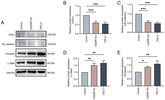
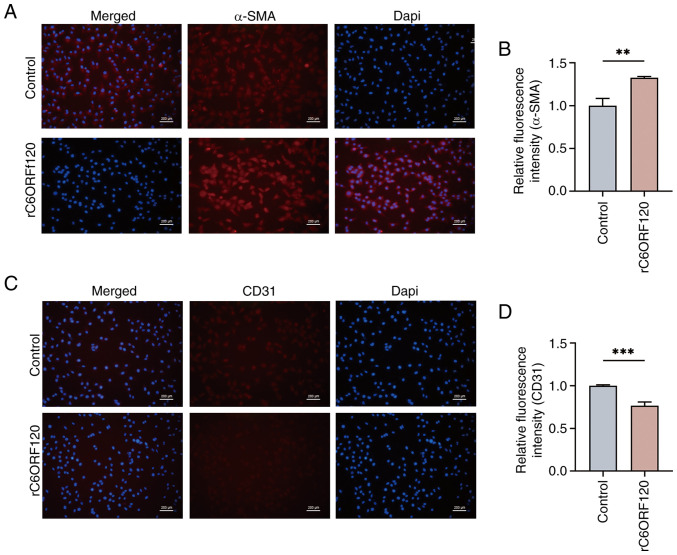
The vascular endothelium plays a pivotal role in modulating various physiological processes and its dysfunction is fundamental to the development of numerous vascular and non‑vascular diseases. Chromosome 6 open reading frame 120 (C6ORF120) has been implicated in cellular processes such as apoptosis, inflammation, immunomodulation and fibrosis. However, the specific effects of C6ORF120 on endothelial cell function remain unclear. The present study aimed to explore the potential role of C6ORF120 in endothelial dysfunction and its underlying molecular mechanisms. It synthesized recombinant C6ORF120 protein (rC6ORF120) and assessed its effects on human umbilical vein endothelial cells (HUVECs) through various functional assays, including the CCK‑8 assay for proliferation, scratch assay for migration and tube formation assay for angiogenesis. Additionally, immunofluorescence (IF) and western blotting (WB) were employed to evaluate endothelial‑mesenchymal transition (EndMT). The present study also quantified the expression of key proteins within the PI3K/Akt signaling pathway to elucidate its role in mediating the effects of rC6ORF120 on HUVECs. Treatment with rC6ORF120 significantly enhanced HUVEC proliferation (200 ng/ml vs. control at 72 h, 1.14±0.01 vs. 1.05±0.02; t=8.15; P<0.001) and induced phenotypic changes. In migration and angiogenesis assays, rC6ORF120‑treated HUVECs exhibited increased wound closure (37.69±2.74% vs. 66.16±6.13%; t=7.35; P=0.002) and angiogenesis assays showed significant improvements in tube formation parameters such as total tubule length (77,199.67±4,684.88 µm vs. 96,203.00±3,354.89 µm; t=5.71; P=0.002). WB and IF analyses both indicated that rC6ORF120 promotes EndMT in HUVECs. Furthermore, rC6ORF120 treatment increased PI3K/Akt phosphorylation significantly compared with controls (p‑PI3K; 1.57±0.18 vs. 1.00±0.00; t=5.64; P=0.005). LY294002 significantly reversed these effects on EndMT and angiogenesis (P<0.05), while the effect on cell migration was less pronounced (P=0.565). Our study highlights the critical role of C6ORF120 in HUVECs, promoting proliferation, migration, angiogenesis and EndMT, which are mediated, at least in part, by the PI3K/Akt pathway.

Keywords: PI3K/Akt; chromosome 6 open reading frame 120; endothelial dysfunction; endothelial‑mesenchymal transition; human umbilical vein endothelial cells.


Conflict of interest statement

The authors declare that they have no competing interests.

Figures

Figure 1. Effect of rC6ORF120 on HUVECs viability and morphology. (A) CCK8 assay was used to determine the viability of HUVECs at 24, 48 and 72 h after stimulation with rC6ORF120 (0, 100, 200 and 400 ...
Figure 1.
Effect of rC6ORF120 on HUVECs viability and morphology. (A) CCK8 assay was used to determine the viability of HUVECs at 24, 48 and 72 h after stimulation with rC6ORF120 (0, 100, 200 and 400 ng/ml). (B) Morphological alterations in HUVECs following 48-h exposure or five passages of rC6ORF120 treatment. ns indicates no statistically significant difference; *P<0.05; ***P<0.001. rC6ORF120, recombinant C6ORF120 protein; HUVECs, human umbilical vein endothelial cells.
Figure 2. rC6ORF120 induced the EndMT in HUVECs. (A) Representative western blot images of endothelial (CD31 and VE–cadherin) and mesenchymal (Vimentin and α –SMA) marker proteins in HUVECs following ...
Figure 2.
rC6ORF120 induced the EndMT in HUVECs. (A) Representative western blot images of endothelial (CD31 and VE-cadherin) and mesenchymal (Vimentin and α-SMA) marker proteins in HUVECs following treatment with rC6ORF120 and TGF-β1. (B-E) Corresponding bar graph representing relative expression of protein expression levels, normalized to GAPDH. *P<0.05; **P<0.01; ***P<0.001. rC6ORF120, recombinant C6ORF120 protein; EndMT, endothelial-mesenchymal transition; HUVECs, human umbilical vein endothelial cells; α-SMA, α-smooth muscle actin.
Figure 3. rC6ORF120 alters the fluorescence intensity of EndMT markers. Immunocytofluorescence staining of (A) α –SMA and (C) CD31 in HUVECs following rC6ORF120 treatment, with (B and D) correspondin...
Figure 3.
rC6ORF120 alters the fluorescence intensity of EndMT markers. Immunocytofluorescence staining of (A) α-SMA and (C) CD31 in HUVECs following rC6ORF120 treatment, with (B and D) corresponding statistical analysis. **P<0.01; ***P<0.001. Scale bar, 200 µm. rC6ORF120, recombinant C6ORF120 protein; EndMT, endothelial-mesenchymal transition; HUVECs, human umbilical vein endothelial cells.
Figure 4. Effects of rC6ORF120 on the migration and angiogenic capacity of HUVECs. (A) Comparison of wound closure between the rC6ORF120–treated group and the control group after a 48–h incubation per...
Figure 4.
Effects of rC6ORF120 on the migration and angiogenic capacity of HUVECs. (A) Comparison of wound closure between the rC6ORF120-treated group and the control group after a 48-h incubation period. (B) Bar graph representing relative wound closure. (C) The impact of rC6ORF120 on the tube-forming capacity of HUVECs. Bar graph representing (D) number of junctions and (E) total tubule length. **P<0.01. Scale bar, 200 µm. rC6ORF120, recombinant C6ORF120 protein; HUVECs, human umbilical vein endothelial cells.
Figure 5. rC6ORF120 modulates the PI3K / Akt signaling pathway in HUVECs (A) Western blot analysis of PISK, p–PI3K, Akt and p–Akt in the HUVECs treated with rC6ORF120. Bar graph representing relative ...
Figure 5.
rC6ORF120 modulates the PI3K/Akt signaling pathway in HUVECs (A) Western blot analysis of PISK, p-PI3K, Akt and p-Akt in the HUVECs treated with rC6ORF120. Bar graph representing relative expression of (B) p-PI3K/PI3K and (C) p-Akt/Akt. **P<0.01; ***P<0.001. rC6ORF120, recombinant C6ORF120 protein; HUVECs, human umbilical vein endothelial cells; p-, phosphorylated.
Figure 6. Involvement of the PI3K / Akt pathway in rC6ORF120–induced EndMT in HUVECs. Cells were treated with rC6ORF120, the PI3K inhibitor LY294002, or a combination of both. (A) Representative blots...
Figure 6.
Involvement of the PI3K/Akt pathway in rC6ORF120-induced EndMT in HUVECs. Cells were treated with rC6ORF120, the PI3K inhibitor LY294002, or a combination of both. (A) Representative blots depicting the PI3K/Akt signaling pathway and proteins indicative of EndMT. (B-G) Quantitative assessment of protein expression levels. ns indicates no statistically significant difference; *P<0.05; **P<0.01; ***P<0.001. rC6ORF120, recombinant C6ORF120 protein; EndMT, endothelial-mesenchymal transition; HUVECs, human umbilical vein endothelial cells; p-, phosphorylated.
Figure 7. Inhibition of the PI3K / Akt pathway reverses the induction of EndMT by rC6ORF120. Immunofluorescence analysis of (A) α –SMA and (C) CD31. The bar graphs depict the relative fluorescence in...
Figure 7.
Inhibition of the PI3K/Akt pathway reverses the induction of EndMT by rC6ORF120. Immunofluorescence analysis of (A) α-SMA and (C) CD31. The bar graphs depict the relative fluorescence intensity of (B) α-SMA and (D) CD31. ns indicates no statistically significant difference; ***P<0.001. Scale bar, 200 µm. EndMT, endothelial-mesenchymal transition; rC6ORF120, recombinant C6ORF120 protein; α-SMA, α-smooth muscle actin.
Figure 8. Effect of PI3K / Akt inhibition on rC6ORF120–mediated effects on HUVEC migration and angiogenesis. (A) Effect of rC6ORF120 on the migration of HUVECs after addition of LY294002. (B) Bar grap...
Figure 8.
Effect of PI3K/Akt inhibition on rC6ORF120-mediated effects on HUVEC migration and angiogenesis. (A) Effect of rC6ORF120 on the migration of HUVECs after addition of LY294002. (B) Bar graph representing relative wound closure. (C) The reversing effect of LY294002 on rC6ORF120-induced angiogenic activity in HUVECs. Bar graph representing (D) number of junctions and (E) total tubule length. ns indicates no statistically significant difference; **P<0.01; ***P<0.001. Scale bar, 200 µm. rC6ORF120, recombinant C6ORF120 protein; HUVECs, human umbilical vein endothelial cells.
Figure 9. C6ORF120 enhances HUVEC migration, tube formation and angiogenesis, which are hallmark features of endothelial dysfunction. These processes are mediated, at least in part, by the PI3K / Akt ...
Figure 9.
C6ORF120 enhances HUVEC migration, tube formation and angiogenesis, which are hallmark features of endothelial dysfunction. These processes are mediated, at least in part, by the PI3K/Akt signaling pathway. rC6ORF120, recombinant C6ORF120 protein; HUVECs, human umbilical vein endothelial cells; EndMT, endothelial-mesenchymal transition.

Similar articles

References

    1. Xu Y, Kovacic JC. Endothelial to mesenchymal transition in health and disease. Annu Rev Physiol. 2023;85:245–267. doi: 10.1146/annurev-physiol-032222-080806. - DOI - PubMed
    1. Zhang L, Wu X, Hong L. Endothelial reprogramming in atherosclerosis. Bioengineering (Basel) 2024;11:325. doi: 10.3390/bioengineering11040325. - DOI - PMC - PubMed
    1. Liu Y, Wu Z, Li Y, Chen Y, Zhao X, Wu M, Xia Y. Metabolic reprogramming and interventions in angiogenesis. J Adv Res. 2024;70:323–338. doi: 10.1016/j.jare.2024.05.001. - DOI - PMC - PubMed
    1. Piera-Velazquez S, Jimenez SA. Endothelial to mesenchymal transition: Role in physiology and in the pathogenesis of human diseases. Physiol Rev. 2019;99:1281–1324. doi: 10.1152/physrev.00021.2018. - DOI - PMC - PubMed
    1. Naderi-Meshkin H, Setyaningsih WAW. Endothelial cell dysfunction: Onset, progression, and consequences. Front Biosci (Landmark Ed) 2024;29:223. doi: 10.31083/j.fbl2906223. - DOI - PubMed

MeSH terms

Substances