. 2025 Jul 18;20(7):1527-1543.
doi: 10.1021/acschembio.5c00112. Epub 2025 Jun 20.

Discovery and Validation of a Novel Class of Necroptosis Inhibitors Targeting RIPK1

Affiliations

Discovery and Validation of a Novel Class of Necroptosis Inhibitors Targeting RIPK1

Lior Soday et al. ACS Chem Biol. .

Abstract

Necroptosis is a form of programmed cell death that, when dysregulated, is associated with cancer and inflammatory and neurodegenerative diseases. Here, starting from hits identified from a phenotypic high-throughput screen for inhibitors of necroptosis, we synthesized a library of compounds containing a 7-phenylquinoline motif and validated their anti-necroptotic activity in a novel live-cell assay. Based on these data, we designed an optimized photoaffinity probe for target engagement studies and through biochemical and cell-based assays established receptor-interacting kinase 1 (RIPK1) as the cellular target, with inhibition of necroptosis arising from the prevention of RIPK1 autophosphorylation and activation. X-ray crystallography and mass spectrometry revealed that these compounds bind at the hinge region of the active conformation of RIPK1, establishing them as type I kinase inhibitors. In addition, we demonstrated in vitro synergy with type III kinase inhibitors, such as necrostatin-1 and found that lead compounds protected mice against acute inflammation in necroptosis models in vivo. Overall, we present a novel pharmacophore for inhibition of human RIPK1, a key protein involved in necroptosis, and provide a photoaffinity probe to explore RIPK1 target engagement in cells.


Figures
1
1
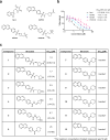
Structure–activity relationship (SAR) studies of a novel series of necroptosis inhibitors. (a) Structures of necroptosis inhibitors Nec-1, AZ’902, and AZ’320 and the photoaffinity-based probe 7PQYnD from the 7PQ compound series. (b) Dose–response curves for Nec-1, AZ’902, AZ’320, and 7PQYnD in I.21 FADD–/– human Jurkat cells. Results are representative of at least three independent repeats; error bars represent SD (n ≥ 3). (c) Structures and EC50 values (95% confidence interval (CI)) for compounds within the 7PQ SAR series. EC50 values in (b,c) are expressed as the geometric mean with a 95% CI of at least three biological replicates.
2
2
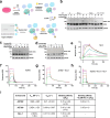
Validation of the 7PQ series as inhibitors of RIPK1. (a,b) Western blot analysis of RIPK1 Ser166 phosphorylation in I2.1 cells with treatment of necroptosis inhibitors and the 7PQ series. Results are representative of two experiments. GAPDH was used as a loading control. Uncropped immunoblots associated with all figures can be found in Figure S5. (c–f) ADP-Glo kinase assay to determine AZ’902 and Nec-1 inhibition of RIPK1 kinase activity with increasing concentrations of ATP. (c–d) Dose response curves for AZ’902 (c) and Nec-1 (d) with ATP concentrations of 10 μM and 500 μM. (e,f) IC50 values of AZ’902 (e) and Nec-1 (f) as a function of ATP concentration divided by the Km for RIPK1. Experiments were carried out with AZ’902 or Nec-1 at ten concentrations (0–100 μM), varied ATP concentrations (10–500 μM), and constant GST-RIPK1 concentration (75 nM). Data are shown as means ± SD (for c,d) and as IC50 ± 95% CI (for e,f). Experiments were conducted in triplicate.
3
3
7PQ series binds to the kinase domain of RIPK1. (a) Schematic of the streptavidin mass shift assay workflow used for target engagement studies in I2.1 cells. (b–d) Western blot analysis of the streptavidin mass shift assay in I2.1 cells. Streptavidin is abbreviated as STA. GAPDH was used as a loading control. (b) 7PQYnD probe used alone. (c) Competition assay where AZ’902 was preincubated before 7PQYnD was added to cells. (d) Preincubation of Nec-1 does not impact 7PQYnD binding to RIPK1. (e–h) SPR representative sensorgrams of increasing concentrations of AZ’902 and Nec-1 (green, pink, teal, gray, orange, purple) and 1:1 fitting (black) of the interactions with the kinase domain of RIPK1. (i) SPR fitted values of AZ’320 and AZ’902 in the presence and absence of Nec-1.
4
4
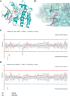
Structural determination of 7PQ binding mode to RIPK1. (a) Co-crystal structure of AZ’320 (light pink sticks) and Nec-1s (dark pink sticks) bound to RIPK1 (teal cartoon) (PDB = 9GTY). AZ’902 (purple sticks) overlaid for comparison. The normally disordered glycine-rich loop, which was able to be modeled, is highlighted in orange. (b) Surface representation of the 7PQ binding site, with hydrogen bonds to key residue Met95 shown as yellow dashes. The 7PQ pharmacophore fits into a defined pocket, while the sulfonamide motifs are exposed to solvent. (c,d) HDX-MS experiment representing the differences observed with 7PQ compounds bound to RIPK1 as opposed to RIPK1 alone. The different colored lines represent different incubation times in D2O buffer, yellow = 3 s, red = 30 s, blue = 300 s, and black = 3000 s. The experiments were performed in triplicate. (c) HDX-MS difference plot for AZ’902 bound to RIPK1. The glycine-rich loop (21–35) and hinge region (94–100) are protected from deuterium exchange by the compound. (d) HDX-MS difference plot for AZ’320 bound to RIPK1. The glycine-rich loop (21–35) is protected from deuterium exchange by the compound.
5
5
7PQ series synergizes with necrostatins in vitro and exhibits comparable protection to Nec1 against necroptosis-related death in an in vivo mouse model for systemic inflammatory response syndrome (SIRS). (a) Synergy/antagonism scores of necroptosis inhibition by Nec-1s and AZ’902 using the HSA model generated from Combenefit software in I2.1 cells. Blue represents synergism and red represents antagonism. The synergy scores were calculated from three independent biological replicates. (b) Quantification of cell death from the synergy assay. The combination of AZ’902 (0.78 μM) and Nec-1s (0.25 μM) was more protective than either compound alone or compared to vehicle (DMSO). (c–e) Inhibitory effects of TNF-induced SIRS murine model. 6–8 weeks old C57BL/6J mice were treated intravenously with Nec-1 (5 μg/g) or AZ’320 (5 μg/g) for 15 min prior to being injected intravenously with TNF (0.5 μg/g). Control mice were injected with TNF and vehicle (44.5% phosphate-buffered saline (PBS) 1×, 17% DMSO, 10% ethanol, 2.5% cyclodextrin, 18% PEG 400, and 8% cremophor). During the first 12 h, mice were frequently monitored for temperature and weight. (c) Survival curve of mice, (d) temperature change of mice, and (e) % body weight change of mice after TNF injection. 24 h after TNF administration, a single injection was lethal in 100% of control mice, accompanied by a reduction in body temperature and weight. Nec-1 partially protected mice from TNF-induced mortality and hypothermia, while AZ’320 administration completely reverted the toxic effects induced by TNF. Data is expressed as mean ± SEM arbitrary units or percentage of 3 individual mice. *p < 0.5, **p < 0.01.

References

    1. Weinlich R., Oberst A., Beere H. M., Green D. R.. Necroptosis in Development, Inflammation and Disease. Nat. Rev. Mol. Cell Biol. 2017;18(2):127–136. doi: 10.1038/nrm.2016.149. - DOI - PubMed
    1. Nailwal H., Chan F. K.. Necroptosis in Anti-Viral Inflammation. Cell Death Differ. 2019;26(1):4–13. doi: 10.1038/s41418-018-0172-x. - DOI - PMC - PubMed
    1. Fritsch M., Günther S. D., Schwarzer R., Albert M. C., Schorn F., Werthenbach J. P., Schiffmann L. M., Stair N., Stocks H., Seeger J. M., Lamkanfi M., Krönke M., Pasparakis M., Kashkar H.. Caspase-8 Is the Molecular Switch for Apoptosis, Necroptosis and Pyroptosis. Nature. 2019;575(7784):683–687. doi: 10.1038/s41586-019-1770-6. - DOI - PubMed
    1. Chen J., Kos R., Garssen J., Redegeld F.. Molecular Insights into the Mechanism of Necroptosis: The Necrosome as a Potential Therapeutic Target. Cells. 2019;8(12):1486. doi: 10.3390/cells8121486. - DOI - PMC - PubMed
    1. Zhang Y., Su S. S., Zhao S., Yang Z., Zhong C.-Q., Chen X., Cai Q., Yang Z.-H., Huang D., Wu R., Han J.. RIP1 Autophosphorylation Is Promoted by Mitochondrial ROS and Is Essential for RIP3 Recruitment into Necrosome. Nat. Commun. 2017;8:14329. doi: 10.1038/ncomms14329. - DOI - PMC - PubMed

MeSH terms

Substances

LinkOut - more resources