. 2025 Jul 9;16(7):e0139325.
doi: 10.1128/mbio.01393-25. Epub 2025 Jun 20.

Legionella pneumophila type II secretome reveals a polysaccharide deacetylase that impacts intracellular infection, biofilm formation, and resistance to polymyxin- and serum-mediated killing

Affiliations

Legionella pneumophila type II secretome reveals a polysaccharide deacetylase that impacts intracellular infection, biofilm formation, and resistance to polymyxin- and serum-mediated killing

Carlton O Adams et al. mBio. .

Erratum in

Abstract

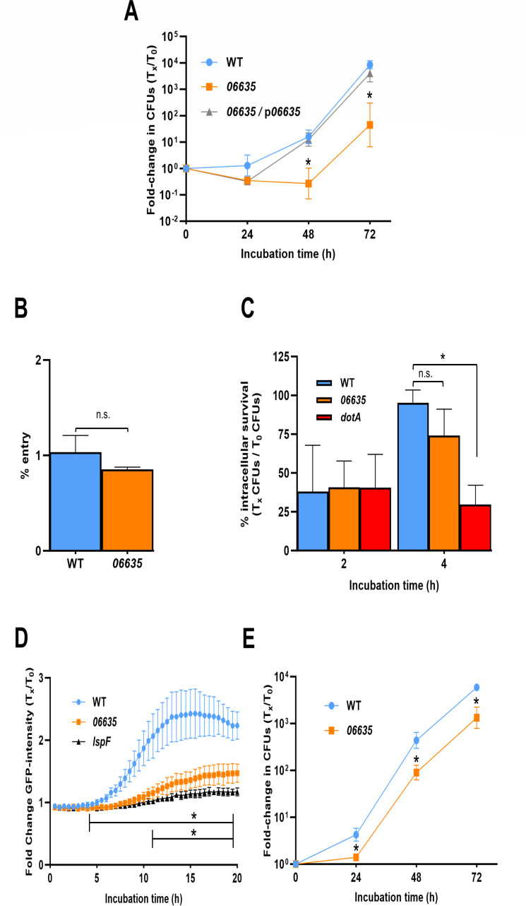
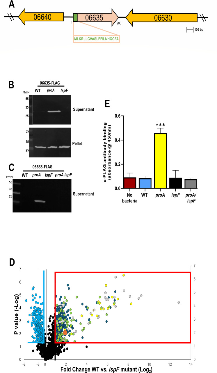
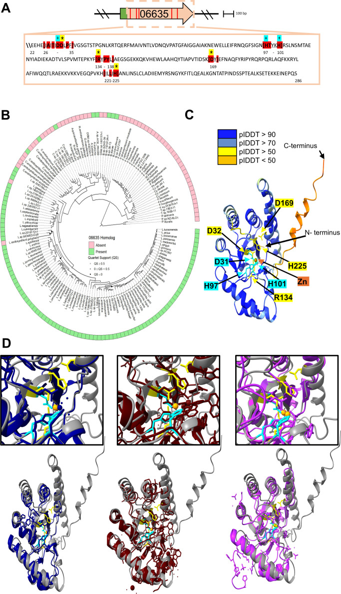
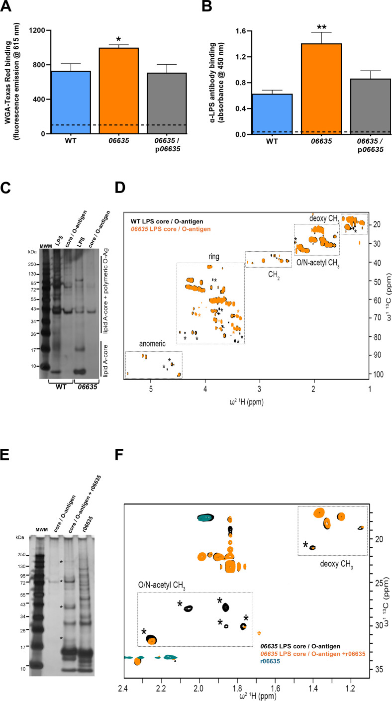
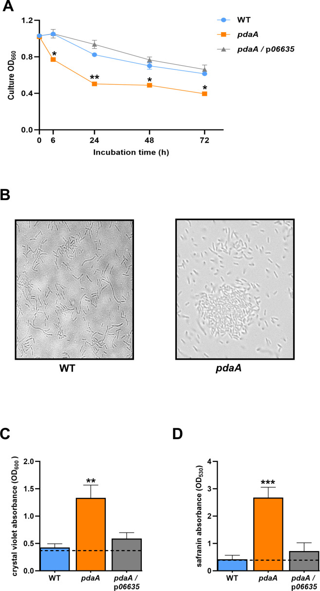
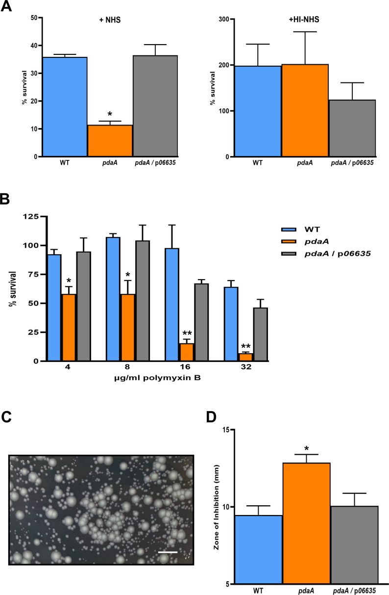
Prior analyses suggested that the type II secretion system (T2SS) of Legionella pneumophila secretes ≥47 proteins beyond its 26 known substrates. Upon examination of mutants of wild-type strain 130b that lack those exoproteins most conserved across the Legionella genus, we discovered that protein "06635" majorly promotes L. pneumophila replication within amoebae. Immunoblotting, proteomics, and whole-cell enzyme-linked immunosorbent assay (ELISA) confirmed that 06635 exists in culture supernatants and on the bacterial outer surface and does so in a T2SS-dependent manner. Bioinformatic analyses identified 06635 as a novel member of the carbohydrate esterase family 4, whose members deacetylate bacterial surface polysaccharides, peptidoglycan, chitins, and/or xylans. Given 06635's T2SS-dependent secretion, low-level amino acid similarity to known peptidoglycan deacetylases, and the unaltered lysozyme resistance of a 06635 mutant, we pursued the hypothesis that 06635 deacetylates a polysaccharide on L. pneumophila's surface. Supporting this, the 06635 mutant exhibited increased binding to both wheat germ agglutinin (i.e., more surface N-acetylglucosamine) and antibodies that recognize acetylated lipopolysaccharide (LPS). Nuclear magnetic resonance (NMR) analysis of isolated mutant vs wild-type LPS confirmed that 06635 promotes LPS deacetylation. Thus, we designated 06635 as PdaA, for polysaccharide deacetylase A. Compatible with its altered surface, the pdaA mutant showed greater autoaggregation, increased biofilm formation, and heightened sensitivity to both polymyxin and human serum. Thus, we hypothesize that, following its secretion via the T2SS, PdaA deacetylates LPS, and perhaps other moieties, impacting many significant processes. While defining PdaA, we identified many more putative substrates of the L. pneumophila T2SS, bringing the size of the T2SS output to approximately 120.IMPORTANCELegionella pneumophila is the principal cause of Legionnaires' disease, an increasingly common form of pneumonia. Although prior work demonstrated that the bacterium utilizes its type II protein secretion system (T2SS) to survive in aquatic environments and to cause lung infection, the full scope and impact of this Legionella secretion system is still relatively underappreciated. By utilizing an expanded proteomic approach and testing newly made mutants in a wide range of assays, we have determined that the L. pneumophila type II secretome encompasses approximately 120 proteins, and among these proteins is a novel polysaccharide deacetylase (PdaA) that modulates the L. pneumophila surface and lipopolysaccharide, impacting intracellular infection, biofilm formation, and resistance to both antibiotics and human serum. Moreover, since T2SSs and homologs of PdaA were found in many other bacteria, our findings should also have implications for understanding other infectious diseases and environmental processes.

Keywords: L. pneumophila; PdaA; T2SS; autoaggregation; biofilm formation; intracellular infection; lipopolysaccharide; polysaccharide deacetylase; resistance to polymyxin B; serum-resistance.


Conflict of interest statement

The authors declare no conflict of interest.

Figures

Fig 1
Fig 1
Effect of 06635 on L. pneumophila intracellular infection of amoebae. (A) Monolayers of A. castellanii were infected with WT strain 130b (WT), the 06635 mutant NU486 (06635), or NU486 containing plasmid-carried 06635 (06635/p06635) at an multiplicity of infection (MOI) = 0.1, and then immediately (i.e., t = 0) and at 24, 48, and 72 h post-inoculation, aliquots taken from the culture supernatants were assessed for bacterial numbers by plating for CFU on buffered charcoal yeast extract (BCYE) agar. Because L. pneumophila does not replicate in the medium, increases in CFU are due to bacterial growth in the amoebae. The values presented are the means and standard deviations from three technical replicates, and asterisks denote significant differences between the 06635 mutant and both WT and the complemented mutant; P < 0.05. (B and C) A. castellanii monolayers were infected with WT, 06635 mutant NU486, or dotA mutant NU428 at an MOI = 20. After centrifugation and a 1 h incubation to permit bacterial uptake by the amoebae, gentamicin was added to kill any remaining extracellular legionellae. After a final wash step, the amoebal cells were lysed either immediately (i.e., t = 0) (B) or at t = 0 and then 2 and 4 h later (C), and the numbers of released CFU were ascertained by plating. In panel B, entry is presented as the percentage of the inoculum that was protected from gentamicin treatment, and in panel C, early intracellular survival is presented as the ratio of recovered CFU at 2 and 4 h over the CFU recovered at time 0 × 100. The values presented are the means and standard deviations from three technical replicates, and the asterisk in panel C denotes a significant reduction for the dotA mutant; P < 0.05. (D) Monolayers of A. castellanii were infected were infected with green fluorescent protein (GFP)-expressing WT, 06635 mutant NU486, or lspF mutant NU275 at an MOI = 20. After centrifugation and a 1 h incubation to permit L. pneumophila uptake, gentamicin was added to kill any remaining extracellular bacteria. GFP fluorescence originating from the intracellular bacteria was monitored kinetically every 30 min for the next 20 h, and the fluorescence values obtained were normalized to the GFP signal at t = 0 following gentamicin treatment. The values presented are the means and standard deviations from six technical replicates. The asterisk above the upper horizontal line denotes the significant difference emerging between the two mutants and WT, whereas the asterisk above the lower horizontal line signifies when the 06635 mutant is different from the lspF mutant; P < 0.05. (E) Monolayers of Vermamoeba vermiformis were infected with WT strain 130b (WT) and the 06635 mutant NU486 (06635), and bacterial growth was monitored, analogously to what was described in panel A. Asterisks denote significant differences between the 06635 mutant and WT; P < 0.05. In panels B and C, n.s. = not significant. (A–E) All data presented are representative of the results obtained from at least three independent experiments.
Fig 2
Fig 2
The 06635 gene locus and the T2SS-dependent secretion of the 06635 protein. (A) Depiction of the chromosomal locus of L. pneumophila strain 130b that encodes 06635. In the map, the arrows indicate the relative length of each gene and the direction of their transcription. The 06630 gene (i.e., ABXK18_RS06630) downstream of 06635 is transcribed in the opposite direction and is annotated as a putative transglycosylase, whereas the 06640 gene (i.e., ABXK18_RS06640) upstream of 06635 is also divergently transcribed and is annotated as encoding a novel hypothetical protein. The box below the 06635 open reading frame (ORF) contains the amino acid sequence of the 06635 protein’s predicted signal sequence. A locus, like the one depicted here, occurs in the other L. pneumophila strains examined, including Philadelphia-1 and the Philadelphia-1 derivatives JR32 and Lp02. (B) WT strain 130b (WT), proA mutant AA200 (proA), and lspF mutant NU275 (lspF), each containing an isopropyl-beta-d-thiogalactopyranoside (IPTG)-inducible 06635 gene that encodes 06635 with a C-terminal FLAG tag (on p06635-FLAG1), were grown in BYE broth. Concentrated, cell-free culture supernatants were subjected to SDS-PAGE and then immunoblotted using anti-FLAG antibodies (top panel). Cell pellets obtained from the cultures were subjected to SDS-PAGE and the same immunoblot analysis (bottom panel). The top and bottom panels represent portions of the same gel, whose left-most lane contained pre-stained molecular weight markers (MWMs), with sizes in kDa. For the top panel, the image of the MWM lane was duplicated and (as the white space denotes) assembled next to the supernatant-containing lanes. (C) Supernatants obtained from wild-type strain 130b (WT), proA mutant AA200 (proA), lspF mutant NU275 (lspF), and proA lspF mutant NU493 (proA lspF), each carrying p06635-FLAG2, were subjected to immunoblot analysis, similarly to what was indicated in panel B. In both panels B and C, the images presented are the (only) portions of the blots showing proteins recognized by the antibodies, i.e., the 06635-FLAG protein. (D) Volcano plot depicting the differences in secreted proteins of L. pneumophila WT strain 130b (WT) vs lspF mutant NU275 (lspF) grown in BYE broth, as determined by proteomic analysis. The box outlined in red encompasses those proteins that were more abundant by at least twofold (P < 0.05) in the WT supernatant. The yellow dots denote proteins that had been previously confirmed to be T2SS substrates, the gray dots indicate proteins that had been previously detected in culture supernatants and predicted by in silico analysis to be T2SS substrates, the orange dot marks the protein 06635, and the green dots signify proteins that, based on in silico analysis, have a signal sequence, which is compatible with them being T2SS substrates. Light blue dots to the left of the vertical blue line denote proteins that were more abundant by at least twofold (P < 0.05) in the lspF mutant supernatant. Black dots indicate those proteins whose abundances were not different between WT and the lspF mutant. (E) WT strain 130b (WT), proA mutant AA200 (proA), lspF mutant NU275 (lspF), and proA lspF mutant NU493 (proA lspF), each expressing 06635-FLAG, were grown in BYE broth and then subjected to whole-cell enzyme-linked immunosorbent assay (ELISA) (five technical replicates/strain), where the presence of 06635-FLAG on the bacterial surface was measured by anti-FLAG-specific antibody binding, as detected by secondary antibody-associated absorbance at 450 nm. A phosphate buffered saline (PBS) buffer alone (no bacteria) was included as a control. The asterisks indicate greater surface expression of 06635-FLAG for the proA mutant relative to the other strains; ***, P < 0.0005. For panels B, C, and E, the results presented are representative of the outcome of at least three independent experiments. For panel D, the data are the results pooled from three biological replicates.
Fig 3
Fig 3
Signature residues, phylogenetic distribution, and predicted structure of the 06635 protein. (A) The box below the gene map contains the amino acid sequence of 06635, minus its N-terminal signal sequence. Residues highlighted in red are the conserved sequence motifs of the “NodB domain” of the CE-4 superfamily. A “+” symbol denotes those residues that typically contribute to the Zn-binding domain. The asterisks denote the residues that typically constitute the enzyme active site. (B) Distribution of genes encoding 06635 homologs within species of Legionella and related genera. (Center) A maximum-likelihood phylogenetic tree for unique species in the orders Berkiellales, Coxiellales, Diplorickettsiales, DSM-16500, Francisellales, Legionellales, and Piscirickettsiales. Scale, one amino acid substitution per site. (Outer ring) The presence of a gene corresponding to 06635 is marked by green squares, whereas the absence of a gene is denoted by pink squares, as determined using BLASTP. (C) Predicted 3-D structure of 06635 (without its N-terminal signal sequence) bound with Zn, as determined by AlphaFold 3. The predicted structure is color-coded in accordance with the levels of confidence (i.e., the predicted local distance difference test [plDDT] values) determined by the program (upper left). The protein’s N-terminus and C-terminus are denoted, as are the putative Zn-binding and active-site residues (in blue and yellow) and the location of the bound Zn (in red). (D) Alignments of the predicted 06635 structure with known structures, as analyzed using the DALI server. Presented here are the alignments of 06635 (dark gray) with the acetylxylan esterase of C. subterraneus (Protein Data Bank [PDB] ID: 7Y51) (dark blue, on left), giving a root mean square deviation (RMSD) = 2.1, Z = 21.3, and % identity = 27, the PG GlcNAc deacetylase of B. cereus (PDB 5O6Y) (dark brown, in center), exhibiting an RMSD = 2.2, Z = 20.6, and % identity = 25, and the chitin deacetylase of Colletotrichum lindemuthianum (PDB 2IW0) (dark purple, on right), giving an RMSD = 2.3, Z = 19.7, and % identity = 26. The boxes present an enlarged image of the D-H-H Zn-binding signature and active-site residues of the CE-4 family mapping to a region of alignment.
Fig 4
Fig 4
Effect of protein 06635 on L. pneumophila WGA binding, reactivity with MAb 3-1, and LPS. (A) Following growth in buffered charcoal yeast extract (BYE) broth at 37°C to early stationary phase, WT strain 130b (WT), 06635 mutant NU486 (06635), or NU486 containing plasmid-carried 06635 (06635/p06635) were suspended in phosphate buffered saline (PBS) with or without WGA-Texas Red. Following static incubation for 1 h at 37°C, the bacteria were washed and resuspended in fresh PBS, and the amount of bound WGA-Texas Red was quantified using a fluorimeter where excitation/emission were measured at 595/615 nm. Readings obtained from control wells containing no added bacteria are indicated by the horizontal dashed line. The data presented are the means and standard deviations from four technical replicates, and the asterisk indicates that the mutant behaved differently from the other two strains, P < 0.05. (B) Following 3 days of growth on BCYE agar at 37°C, WT 130b, the 06635 mutant, and the complemented 06635 mutant were subjected to whole-cell ELISA (four technical replicates) utilizing anti-LPS MAb 3-1. Anti-LPS-specific antibody binding was detected by secondary antibody-associated absorbance at 450 nm. Readings obtained from control wells containing no added bacteria are indicated by the horizontal dashed line. The asterisk indicates greater antibody binding for the pdaA mutant relative to the other strains, P < 0.005. (C) Following the growth of WT 130b (WT) and the 06635 mutant (06635) on BCYE agar for 3 days at 37°C, bacterial LPS was purified, and 10 µg of intact LPS or 2 µg core/O-antigen samples were examined by silver staining after SDS-PAGE. Lane 1 contains molecular weight markers (MWMs), as indicated. The bands corresponding to lipid A/core vs core/O-antigen are highlighted. (D) 1H13C HSQC nuclear magnetic resonance (NMR) spectra of purified core/O-antigen fragments (2 mg/mL) from WT (black) and 06635 mutant (orange) strains. Characteristic spectral range for core/O-antigen polysaccharides is highlighted. Chemical shift changes are shown as black arrows. Peak intensities that have increased (orange asterisk) or decreased (black asterisk) >50% in the mutant spectrum compared to the WT spectrum are also highlighted. (E) Silver-stained SDS-PA gels showing 06635 mutant core/O-antigen (0.7 mg/mL) alone, 06635 mutant core/O-antigen (0.7 mg/mL) with r06635 (r06635; 20 µM), or r06635 (20 µM) alone, after incubation at 37°C for 24 h. Lane 1 contains MWMs, as indicated. New bands that appear in the core/O-antigen sample lane when incubated with r06635, which are not present when r06635 is incubated alone, are highlighted with a black asterisk. (F) 1H13C HSQC NMR spectra of samples from panel E, highlighting the O-/N-acetyl methyl and deoxy methyl region. Peak intensities that have decreased (black asterisk) >50% in the 06635 mutant sample after treatment with r06635 are highlighted. In panels A through F, the data presented are representative of the results obtained from at least three independent experiments.
Fig 5
Fig 5
Effect of PdaA on L. pneumophila autoaggregation and biofilm formation. (A) Following 3 days of growth on buffered charcoal yeast extract (BCYE) agar at 37°C, WT strain 130b (WT), pdaA mutant NU486 (pdaA), or NU486 containing plasmid-carried pdaA (pdaA/ppdaA) were suspended in 10% BYE broth to an OD660 of ~1.0. Five-milliliter aliquots were added to glass tubes, and bacterial sedimentation at 37°C was assessed by measuring drops in the OD660 of the statically incubated suspensions. The data presented are the means and standard deviations from three technical replicates, and asterisks indicate that the mutant behaved differently from the other two strains at all time points after t = 0, *, P < 0.05, **, P < 0.005. (B) Following static incubation, as noted in panel A, strain aggregation was assessed at t = 48 h by microscopically examining an aliquot taken from the midpoint in the tube. (C and D) Following 3 days of growth on BCYE agar at 37°C, WT 130b, the pdaA mutant, and the complemented pdaA mutant were resuspended to an OD660 of ~0.2 in BYE broth, and then the suspensions were added into the wells of a 96-well polystyrene microtiter plate. After 2 days at 37°C, the amount of biofilm formed was determined by staining with crystal violet (C) or safranin (D) as read at either 600 nm or 530 nm. The readings obtained from control wells containing only medium (no added bacteria) are indicated by the horizontal dashed lines. Data presented are the means and standard deviations from six technical replicates, and asterisks indicate differences in the levels of biofilm formation between the pdaA mutant and the other two strains, **, P < 0.005; ***, P < 0.0005. The data in panels A–D are representative of the results obtained from at least three independent experiments.
Fig 6
Fig 6
Effect of PdaA on L. pneumophila resistance to NHS and polymyxin B. (A) Following 3 days of growth on buffered charcoal yeast extract (BCYE) agar at 37°C, WT strain 130b (WT), pdaA mutant NU486 (pdaA), or NU486 containing plasmid-carried pdaA (pdaA/ppdaA) were suspended in 90% NHS (left) or heat-inactivated NHS (HI-NHS) (right) and then statically incubated at 37°C. After 1 day of incubation, the percentages of surviving CFU were determined by plating on BCYE agar. The data presented are the means and standard deviations from three technical replicates, and the asterisk indicates that the mutant behaved differently from the other two strains, P < 0.05. (B) Following 3 days of growth on BCYE agar at 37°C, WT 130b, the pdaA mutant, and the complemented pdaA mutant were suspended in BYE broth to an OD660 of 0.3 and then plated for CFU on BCYE agar vs BCYE agar containing 4, 8, 16, or 32 µg/mL of polymyxin B. After 3 days of incubation at 37°C, the percent survival of CFU on the drug-containing media vs the no-drug medium was determined. The data presented are the means and standard deviations from three technical replicates, and the asterisks indicate that the mutant behaved differently from the other two strains, with *, P < 0.05, and **, P < 0.005. (C) Image of pdaA mutant colonies on BCYE containing 32 µg/mL of polymyxin B. The scale bar corresponds to 5 mm. (D) Following 3 days of growth on BCYE agar at 37°C, WT 130b, the pdaA mutant, and the complemented pdaA mutant were spread onto the surface of a BCYE agar plate, and then sterile paper disks containing 5 mg/mL polymyxin B were placed onto the center of the plate. After 3 days of incubation at 37°C, the diameters of the clearing zones around the disks were measured. The data presented are the means and standard deviations from three technical replicates, and the asterisk indicates that the mutant behaved differently from the other two strains, with P < 0.05. The data in panels A–D are representative of the results obtained from at least three independent experiments.

Similar articles

References

    1. Barskey AE, Derado G, Edens C. 2022. Rising incidence of legionnaires’ disease and associated epidemiologic patterns, United States, 1992-2018. Emerg Infect Dis 28:527–538. doi: 10.3201/eid2803.211435 - DOI - PMC - PubMed
    1. Viasus D, Gaia V, Manzur-Barbur C, Carratalà J. 2022. Legionnaires’ disease: update on diagnosis and treatment. Infect Dis Ther 11:973–986. doi: 10.1007/s40121-022-00635-7 - DOI - PMC - PubMed
    1. Nisar MA, Ross KE, Brown MH, Bentham R, Whiley H. 2020. Legionella pneumophila and protozoan hosts: implications for the control of hospital and potable water systems. Pathogens 9:286. doi: 10.3390/pathogens9040286 - DOI - PMC - PubMed
    1. Hochstrasser R, Michaelis S, Brülisauer S, Sura T, Fan M, Maaß S, Becher D, Hilbi H. 2022. Migration of Acanthamoeba through Legionella biofilms is regulated by the bacterial Lqs-LvbR network, effector proteins and the flagellum. Environ Microbiol 24:3672–3692. doi: 10.1111/1462-2920.16008 - DOI - PMC - PubMed
    1. Graham CI, MacMartin TL, Kievit TR, Brassinga AKC. 2023. Molecular regulation of virulence in Legionella pneumophila. Mol Microbiol. doi: 10.1111/mmi.15172 - DOI - PubMed

MeSH terms

LinkOut - more resources