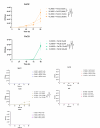
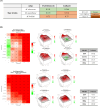
. 2025 Aug 5;13(8):e0071925.
doi: 10.1128/spectrum.00719-25. Epub 2025 Jun 20.

VLX600, an anticancer iron chelator, exerts antimicrobial effects on Mycobacterium abscessus infections

Affiliations

VLX600, an anticancer iron chelator, exerts antimicrobial effects on Mycobacterium abscessus infections

Jaehun Oh et al. Microbiol Spectr. .

Abstract

Mycobacterium abscessus presents significant clinical challenges due to its intrinsic and acquired resistance to antibiotics, resulting in prolonged treatments and poor patient outcomes. Addressing the urgent need for novel therapeutics, this study explores the antimicrobial potential of VLX600, originally developed as an anticancer agent, against M. abscessus. Screening a library of 3,200 clinically evaluated compounds identified VLX600 as a potent antimicrobial with minimal cytotoxicity. VLX600 demonstrated inhibitory effects against various strains of M. abscessus with minimum inhibitory concentrations of 4 µg/mL-16 µg/mL. It also remained effective in intracellular M. abscessus in host cells and exhibited broad-spectrum activity against other bacterial species, including Escherichia coli, Staphylococcus aureus, and Pseudomonas aeruginosa. The antimicrobial activity of VLX600 was abrogated by supplemental iron, indicating a mechanism dependent on iron chelation. VLX600 significantly reduced bacterial burdens and inflammation in a murine model of pulmonary M. abscessus infection. Additionally, synergistic effects were observed when VLX600 was combined with conventional antibiotics such as amikacin and clarithromycin in vitro. These findings highlight VLX600 as a promising candidate for repurposing as an antimicrobial agent against M. abscessus, warranting further clinical investigations.IMPORTANCEMycobacterium abscessus is an opportunistic pathogen that commonly causes pulmonary infections in cystic fibrosis patients. These infections are notoriously difficult to treat due to high levels of antibiotic resistance of M. abscessus, resulting in low cure rates. In this study, we identified a novel antibiotic candidate, VLX600, through high-throughput screening of 3,200 clinical compounds and demonstrated that VLX600 inhibits the growth of M. abscessus by depriving it of ferric and ferrous ions. This study highlights the potential of iron chelators as antimicrobial agents against M. abscessus infections. Since iron is an essential nutrient for the growth of many bacteria, the use of iron chelators could be extended to other infectious diseases. We hope this research will inspire further studies aimed at developing iron chelators as a novel class of antimicrobial agents.

Keywords: Mycobacterium abscessus; VLX600; antibiotics; drug repositioning; iron chelator.


Conflict of interest statement

J.-J. Yim has served as the overall or institutional principal investigator for clinical trials related to non-tuberculous mycobacterial pulmonary disease sponsored by LigaChem Biosciences, Insmed, and AN2 Therapeutics. Additionally, he has received several drugs free of charge as a principal investigator for previous trials related to tuberculosis from Pfizer, Otsuka, and Yuhan.

Figures
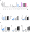
Fig 1
Fig 1
Screening result from the chemical library and discovery of VLX600 as a hit compound. (A) An NRU assay was performed at 48 h after drug treatment to confirm the cytotoxicity of different doses of the 10 selected compounds on J774A.1 cells (n = 3). The samples were treated with 1% Triton X-100 for 10 min before staining and used as a positive control. (B) Antimycobacterial activity of VLX600 in the direct treatment model. VLX600 was applied to the bioluminescent M. abscessus cultures, and the luminescence was measured every 24 h (n = 3). (C) Antimycobacterial activity of VLX600 in the J774A.1 infection model. J774A.1 cells were infected with bioluminescent M. abscessus at an MOI of 10, followed by treatment with VLX600 at different doses (n = 3). Luminescence was measured every 24 h. The error bars represent the standard errors of the means.
Fig 2
Fig 2
Antimycobacterial efficacy of VLX600 against type strains of M. abscessus. (A) The MICs of VLX600 against type strains of M. abscessus were determined with a broth microdilution assay. Each strain of M. abscessus was diluted in CAMHB to McFarland standard 0.5 and further diluted 1:100 in CAMHB. VLX600 was serially diluted in CAMHB and applied to M. abscessus cultures in 96-well plates (n = 3). The plates were then cultured at 37°C for 1 week, and the OD600 was measured to determine the MIC. The MIC was defined as the lowest concentration of VLX600 that inhibited bacterial growth by at least 90%. The MICs are shown as red dots on the graphs. (B) The antimycobacterial efficacy of VLX600 against M. abscessus was evaluated with a CFU assay in the direct treatment model. The type strain of M. abscessus subsp. abscessus (ATCC 19977) culture from the broth microdilution assay was serially diluted in phosphate-buffered saline (PBS) and spread on 7H10 agar plates. The plates were incubated at 37°C until colonies appeared. Asterisks above the bars indicate statistical significance compared to the vehicle. (C). The antimycobacterial efficacy of VLX600 in the J774A.1 infection model was evaluated with a CFU assay. J774A.1 cells were infected with the type strain of M. abscessus subsp. abscessus (ATCC 19977) at an MOI of 10, followed by treatment with different doses of VLX600 (n = 3). The plates were then cultured at 37°C for 48 h. The intracellular bacteria were harvested by lysing the cells with 1% Triton X-100, and then samples were spread on 7H10 agar plates for the CFU assay. The statistical significance was determined by one-way analysis of variance with Tukey’s multiple comparison test, and the results are denoted as follows: *P < 0.05, **P < 0.01, and ***P < 0.001. The error bars represent the standard errors of the means.
Fig 3
Fig 3
Abrogation of the antimycobacterial activity of VLX600 by the addition of iron. The antimycobacterial activity of VLX600 (16 µg/mL) against the type strain of M. abscessus subsp. abscessus (ATCC 19977) was evaluated in the presence of various metal ions. The cultures were incubated at 37°C, and the CFUs were measured daily (n = 3). The antimycobacterial activity of VLX600 was only abrogated by the addition of Fe2+ and Fe3+ in a dose-dependent manner, whereas the addition of other metal ions did not affect its activity except for Cu+. The statistical significance was determined by two-way analysis of variance with Tukey’s multiple comparison test, and the results are denoted as follows: *P < 0.05, **P < 0.01, and ***P < 0.001. The error bars represent the standard errors of the means.
Fig 4
Fig 4
Synergistic effects of VLX600 with amikacin and clarithromycin on M. abscessus. (A) The synergistic effect of VLX600 was evaluated on a direct treatment model using a checkerboard assay. The FICI was calculated, and the drug interaction was considered synergistic for FICI ≤ 0.5, indifferent for 0.5 < FICI ≤ 4, and antagonistic for FICI > 4. (B) The synergistic effect of VLX600 was evaluated on a J774A.1 infection model using a checkerboard assay. The luminescence was measured at 24 h post-infection, and the inhibition rate was calculated from the luminescence value. The drug interaction was evaluated using SynergyFinder with four methods: zero interaction potency (ZIP), Loewe, Bliss, and highest single agent (HSA). The drug interaction was considered synergistic when the score was ≥5, indifferent when the score was −5 < score < 5, and antagonistic when the score was ≤−5.
Fig 5
Fig 5
In vivo efficacy of VLX600 in a mouse model of pulmonary infection (A) Schematic of the schedule of the in vivo experiment. The mice were intraperitoneally injected with 150 mg/kg cyclophosphamide on days 1 and 4 before infection to induce a neutropenic state. On day 0, the mice were challenged with 1 × 10⁶ CFUs of a clinical isolate of M. abscessus via the intranasal route. The drug treatments started on day 1 and were administered daily until sacrifice on day 12 (n = 5). (B) The body weights of the mice were measured daily until sacrifice, and the results revealed a significant improvement in weight loss in all treated groups compared to the PBS group. (C) Lungs were excised and homogenized in PBS on the day of sacrifice. The homogenates were serially diluted and spread on 7H10 agar plates for CFU assays. (D) Spleens were excised and weighed to assess spleen enlargement in mice. The weight of each spleen was normalized to the corresponding body weights. (E) The postcaval lobe of each lung was fixed and stained with hematoxylin and eosin. The paraffin sections were subsequently observed under a light microscope as sectioned paraffin block. The statistical significance was determined by one-way analysis of variance (ANOVA) or two-way ANOVA with Tukey’s multiple comparison test, and the results are denoted as follows: *P < 0.05, **P < 0.01, and ***P < 0.001. The error bars represent the standard errors of the means.

Similar articles

References

    1. To K, Cao R, Yegiazaryan A, Owens J, Venketaraman V. 2020. General overview of nontuberculous mycobacteria opportunistic pathogens: Mycobacterium avium and Mycobacterium abscessus. J Clin Med 9:2541. doi: 10.3390/jcm9082541 - DOI - PMC - PubMed
    1. Dahl VN, Mølhave M, Fløe A, van Ingen J, Schön T, Lillebaek T, Andersen AB, Wejse C. 2022. Global trends of pulmonary infections with nontuberculous mycobacteria: a systematic review. Int J Infect Dis 125:120–131. doi: 10.1016/j.ijid.2022.10.013 - DOI - PubMed
    1. Al-Ghafli H, Al-Hajoj S. 2017. Nontuberculous mycobacteria in Saudi Arabia and gulf countries: a review. Can Respir J 2017:5035932. doi: 10.1155/2017/5035932 - DOI - PMC - PubMed
    1. Recchia D, Stelitano G, Stamilla A, Gutierrez DL, Degiacomi G, Chiarelli LR, Pasca MR. 2023. Mycobacterium abscessus infections in cystic fibrosis individuals: a review on therapeutic options. Int J Mol Sci 24:4635. doi: 10.3390/ijms24054635 - DOI - PMC - PubMed
    1. Griffith DE, Daley CL. 2022. Treatment of Mycobacterium abscessus pulmonary disease. Chest 161:64–75. doi: 10.1016/j.chest.2021.07.035 - DOI - PubMed

MeSH terms

LinkOut - more resources