. 2025 Jun 19;10(14):e190296.
doi: 10.1172/jci.insight.190296. eCollection 2025 Jul 22.

Treatment with efavirenz extends survival in a Creutzfeldt-Jakob disease model by regulating brain cholesterol metabolism

Affiliations

Treatment with efavirenz extends survival in a Creutzfeldt-Jakob disease model by regulating brain cholesterol metabolism

Tahir Ali et al. JCI Insight. .

Abstract

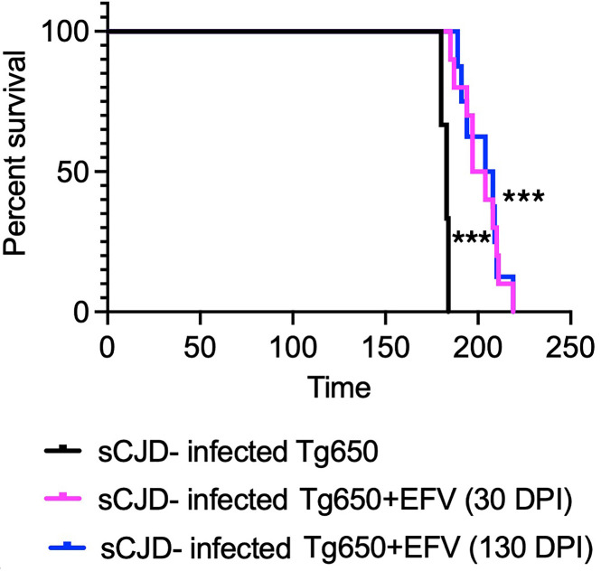
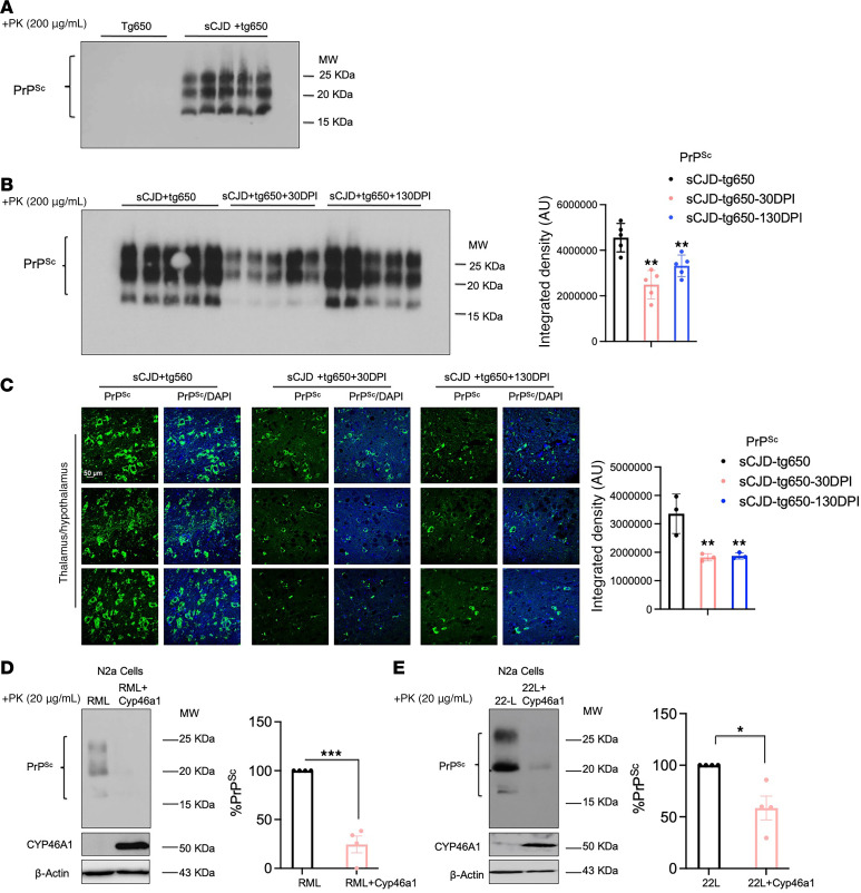
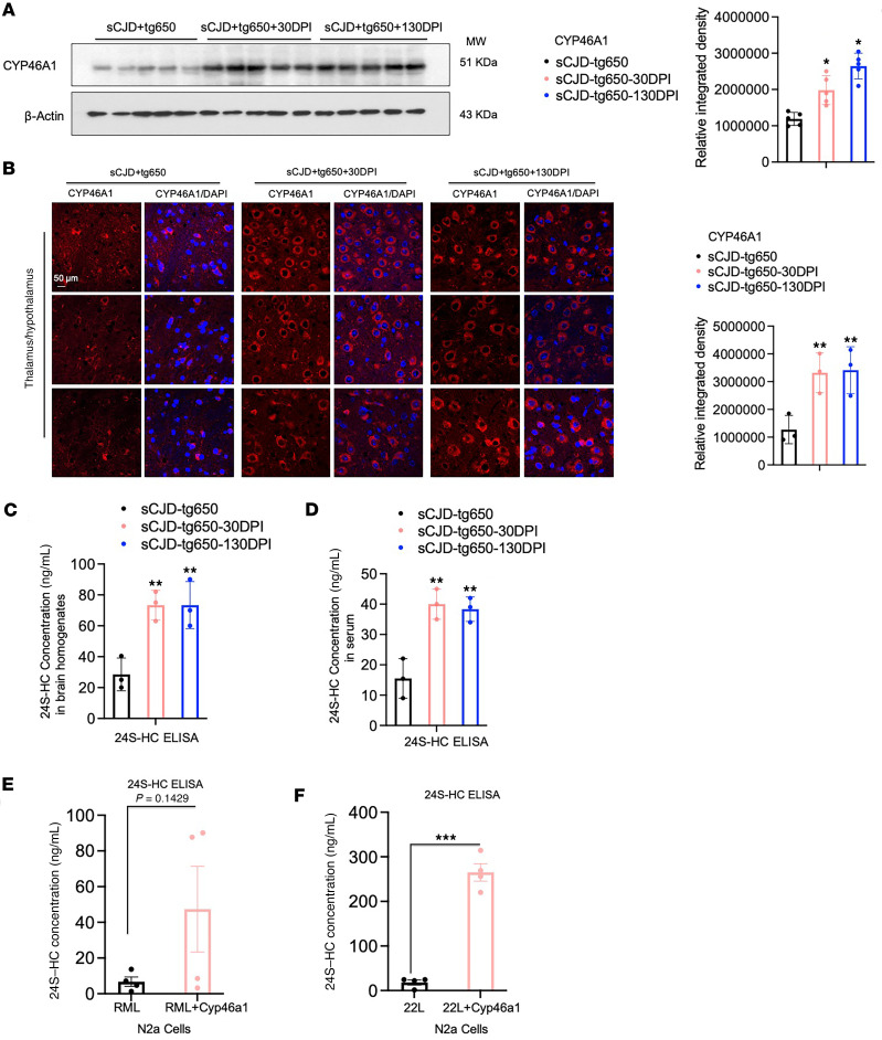
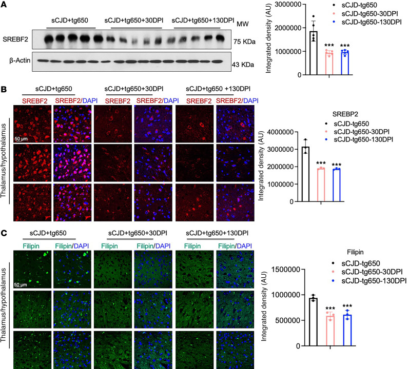
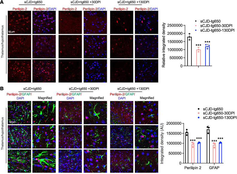
Prion diseases are fatal, infectious, and incurable neurodegenerative conditions affecting humans and animals, caused by the misfolding of the cellular prion protein (PrPC) into its pathogenic isoform, PrPSc. In humans, sporadic Creutzfeldt-Jakob disease (sCJD) is the most prevalent prion disease. Recently, we demonstrated that treatment with the FDA-approved anti-HIV drug efavirenz (EFV) significantly reduced PrPSc and extended survival of scrapie prion-infected mice. Among other effects, EFV activates the brain-specific cholesterol-metabolizing enzyme, CYP46A1, which converts cholesterol into 24S-hydroxycholesterol (24S-HC). However, drugs effective against scrapie prions often fail in human prion diseases, and a relation of the antiprion effects of EFV to CYP46A1 activation is not established. Thus, we evaluated EFV treatment in mice overexpressing human PrPC infected with human sCJD prions. Oral, low-dose EFV treatment starting at 30 or 130 days postinfection significantly slowed disease progression and extended their survival. At early clinical stage, we observed reduced PrPSc accumulation, decreased cholesterol and lipid droplet content, and elevated CYP46A1 and 24S-HC levels in EFV-treated mice. Overexpression of CYP46A1 in prion-infected neuronal cells reduced PrPSc levels and increased 24S-HC, indicating that antiprion effects of EFV correlate with CYP46A1 activation. These findings highlight EFV as a safe and efficacious therapeutic candidate for human prion diseases.

Keywords: Cholesterol; Infectious disease; Neurodegeneration; Prions; Therapeutics.


Conflict of interest statement

Conflict of interest: The authors have declared that no conflict of interest exists.

Figures

Figure 1
Figure 1. EFV oral administration significantly prolongs survival of sCJD-infected tg650 mice.
The Kaplan-Meier plot illustrates the percentage survival of sCJD-infected mice across 3 groups: the untreated group (n = 9), the EFV-treated group starting at 30 DPI (sCJD + EFV 30 DPI-DW) (n = 10), and the EFV-treated group starting at 130 DPI (sCJD + EFV 130 DPI-DW) (n = 10). Log-rank (Mantel-Cox) and Gehan-Breslow-Wilcoxon tests were performed, and statistical significance is ***P < 0.001. DW, drinking water.
Figure 2
Figure 2. EFV reduces PrPres accumulation at the early clinical stage in sCJD-infected tg650 mice.
(A) Analysis of uncropped immunoblot of PrPres using the 3F4 antibody in brain homogenates (BH) from 5 noninfected tg650 mice (mock) and sCJD-tg650 mice at the terminal stage. (B) Analysis of uncropped immunoblotting and quantification of PrPres using 3F4 antibody in BH from 5 different mice/group at the early clinical stage (176 DPI) for all experimental groups including nontreated and treated (EFV treatment started at 30 DPI and 130 DPI) groups. The histograms are represented as the means ± SEM (n = 5 mice/group) of 3 independent experiments. Ordinary 1-way ANOVA was performed, and statistical significance is **P < 0.0092. (C) Immunofluorescence and quantification of PrPres using 3F4 antibody in brain tissue from 3 different mice/group at the early clinical stage (176 DPI) for all experimental groups including nontreated and treated (EFV treatment started at 30 DPI and 130 DPI) groups. Fluorescence intensity was quantified; the histograms represent the means ± SEM of n = 3 mice per group, obtained from 3 independent experiments. Ordinary 1-way ANOVA was performed, and statistical significance is **P < 0.0062. Original magnification: 63×. Scale bar: 50 μm. (D and E) Immunoblot analysis of CYP46A1 and PrPres as well as quantification of PrPres in N2a-RML and N2a-RML-Cyp46a1 overexpression models. The histograms represent the means ± SEM for n = 4 per group, obtained from 3 independent experiments. T test (2-tailed unpaired t test) was performed, and statistical significances are ***P < 0.001 and *P < 0.05, respectively.
Figure 3
Figure 3. EFV increases CYP46A1 and 24S-HC level in brain and serum of sCJD-infected tg650 mice as well as in an in vitro model.
(A) Immunoblotting and quantification of CYP46A1 were performed on BH from 5 sCJD-tg650 mice at 176 days DPI, representing the early clinical stage, from control as well as 30 DPI and 130 DPI treatment groups. Histograms show the means ± SEM (n = 5 mice/group) based on 3 independent experiments. Ordinary 1-way ANOVA was performed, and statistical significance is *P < 0.0224. (B) Immunofluorescence and quantification of CYP46A1 in brain tissue from 3 sCJD-tg650 mice and samples from 30 DPI and 130 DPI treatment groups at 176 DPI (early clinical stage). Fluorescence intensity was quantified, with histograms representing the means ± SEM for n = 3 mice/group, gathered from 3 independent experiments. Ordinary 1-way ANOVA was performed, and statistical significance is **P < 0.0161. Magnification: 63×; scale bar: 50 μm. (C and D) 24S-hydroxycholesterol (24S-HC) quantified by ELISA in BH and serum from sCJD-tg650 control mice and treated groups at the early clinical stage. Histograms represent means ± SEM (n = 3 mice/group) from 3 independent experiments. Ordinary 1-way ANOVA was performed, and statistical significances are **P < 0.0057 (C) and **P < 0.0022 (D). (E and F) 24S-HC levels quantified by ELISA in the media of N2a-RML and N2a-22L cells, with and without CYP46A1 overexpression. The histograms represent the means ± SEM for n = 4 per group, obtained from 3 independent experiments. T test (2-tailed unpaired t test) was performed, and statistical significances are P < 0.1429 (E) and ***P < 0.001 (F).
Figure 4
Figure 4. EFV treatment downregulates SREBF2 in the brains of sCJD-infected tg650 mice.
(A) Immunoblotting and quantification of SREBF2 were conducted on BH from 5 sCJD-tg650 mice at 176 DPI (early clinical stage) as well as in 30 DPI and 130 DPI treatment groups. Histograms show means ± SEM (n = 5 mice/group) from 3 independent experiments. Ordinary 1-way ANOVA was performed, and statistical significance is ***P < 0.0002. (B) Immunofluorescence staining and quantification of SREBF2 (red) and DAPI (blue) in brain tissue from 3 sCJD-tg650 mice, including samples from 30 DPI and 130 DPI treatment groups at the early clinical stage (176 DPI). Fluorescence intensity was measured, with histograms showing means ± SEM (n = 3 mice/group) from 3 independent experiments. Ordinary 1-way ANOVA was performed, and statistical significance is ***P < 0.0007. Magnification: 63×; scale bar: 50 μm. (C) Filipin staining and quantification in brain tissue from 3 sCJD-tg650 mice, as well as 30 DPI and 130 DPI treatment group samples at the early clinical stage (176 DPI). Fluorescence intensity was measured and represented as histograms showing means ± SEM (n = 3 mice/group) from 3 independent experiments. Ordinary 1-way ANOVA was performed, and statistical significance is ***P < 0.0018. Magnification: 63×; scale bar: 50 μm.
Figure 5
Figure 5. EFV treatment downregulates lipid droplet protein in the brains of sCJD-infected tg650 mice.
(A) Immunofluorescence staining and quantification of perilipin-2 (red) and DAPI (blue) in brain tissue from 3 sCJD-tg650 mice, including samples from control (nontreated group), 30 DPI and 130 DPI treatment groups at the early clinical stage (176 DPI). Fluorescence intensity was measured, with histograms showing means ± SEM (n = 3 mice/group) from 3 independent experiments. Ordinary 1-way ANOVA was performed, and statistical significance is ***P < 0.0020. Magnification: 63×; scale bar: 50 μm. (B) Double-immunofluorescence staining for perilipin-2 (red) and GFAP (green) and quantification in brain tissue from 3 sCJD-tg650 mice, as well as 30 DPI and 130 DPI treatment group samples at the early clinical stage (176 DPI). Fluorescence intensity was measured and represented as histograms showing means ± SEM (n = 3 mice/group) from 3 independent experiments. Two-way ANOVA was performed, and statistical significances is ***P < 0.0001 for perilipin-2 and GFAP. Magnification: 63×; digital zoom: 2×; scale bar: 50 μm.

Similar articles

References

    1. Prusiner SB. Novel proteinaceous infectious particles cause scrapie. Science. 1982;216(4542):136–144. doi: 10.1126/science.6801762. - DOI - PubMed
    1. Prusiner SB. Prions. Proc Natl Acad Sci U S A. 1998;95(23):13363–13383. doi: 10.1073/pnas.95.23.13363. - DOI - PMC - PubMed
    1. Mackenzie G, Will R. Creutzfeldt-Jakob disease: recent developments. F1000Res. 2017;6:2053. doi: 10.12688/f1000research.12681.1. - DOI - PMC - PubMed
    1. Colby DW, Prusiner SB. Prions. Cold Spring Harb Perspect Biol. 2011;3(1):a006833. doi: 10.1101/cshperspect.a006833. - DOI - PMC - PubMed
    1. Ladogana A, et al. Mortality from Creutzfeldt-Jakob disease and related disorders in Europe, Australia, and Canada. Neurology. 2005;64(9):1586–1591. doi: 10.1212/01.WNL.0000160117.56690.B2. - DOI - PubMed

MeSH terms