. 2025 Jun 20;20(6):e0325661.
doi: 10.1371/journal.pone.0325661. eCollection 2025.

Active peptides of TSP-1 inhibit retinal angiogenesis through the CD36 pathway in a rat model of choroidal neovascularization

Affiliations

Active peptides of TSP-1 inhibit retinal angiogenesis through the CD36 pathway in a rat model of choroidal neovascularization

Yadi Li et al. PLoS One. .

Abstract

Background: Choroidal neovascularization (CNV) is a key manifestation of intraocular neovascularization, and it is considered one of the main causes of blindness in ophthalmology. Additionally, multiple anti-vascular endothelial growth factor (VEGF) drugs have been used as first-line treatment for CNV. However, several issues posed challenges to the anti-VEGF drugs, which were mainly composed of short duration of action, requirement for repeated injections, and complications. Thrombospondin-1 (TSP-1) is an endogenous protein that was found to regulate multiple biological processes within the body, and it has been proven to exhibit an inhibitory effect on neovascularization. Besides, the function of TSP-1 during the inhibition of neovascularization was currently considered to mainly focus on its type Ⅰ repeats (TSRs), which was attributed to the large molecular weight, complex structure, and possible unknown functions of TSP-1. Therefore, TSRs can be applied as targets and research directions for the further development and exploration of potential therapeutic drugs.

Objectives: Based on the type I repeats (TSRs) of thrombospondin-1 (TSP-1), amino acid sequences of different lengths were designed and synthesized in this study, named as VR-9 VR-10、VR-11、VR-12、VR-13. The objective was to explore the effects of the above five peptides on angiogenesis in Chori-retinal neovascularization, alongside the screening of the best peptides and the deep exploration into the underlying mechanism, aimed to provide a basis for the development and application of peptide drugs in the treatment of CNV.

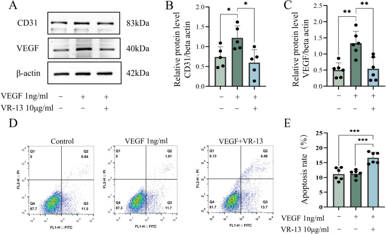
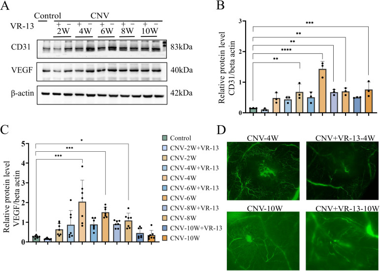
Methods: Wound healing, CCK-8, and 5-ethynyl-2'-deoxyuridine (EdU) assays were employed to evaluate the proliferation and migration ability of cells. CRISPR-Cas9 technology was utilized to establish CD36 knockdown cell lines, alongside the conduction of qPCR to verify the efficiency of gene knockdown. The expression levels of VEGF and CD31 in RF/6A cells and rats were assessed by Western blot. Additionally, Hematoxylin and eosin (HE) staining was performed to examine the structural integrity of the rat retina, while Fluorescein Isothiocyanate-Dextran Cardiac Perfusion (FITC) labeling was used to observe the occurrence and development of choroidal neovascularization (CNV).

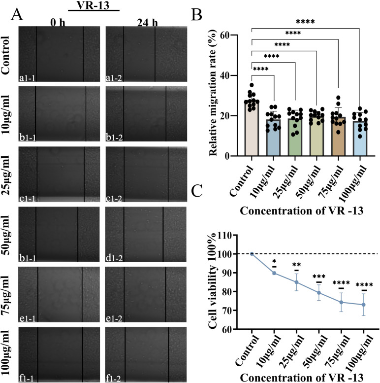
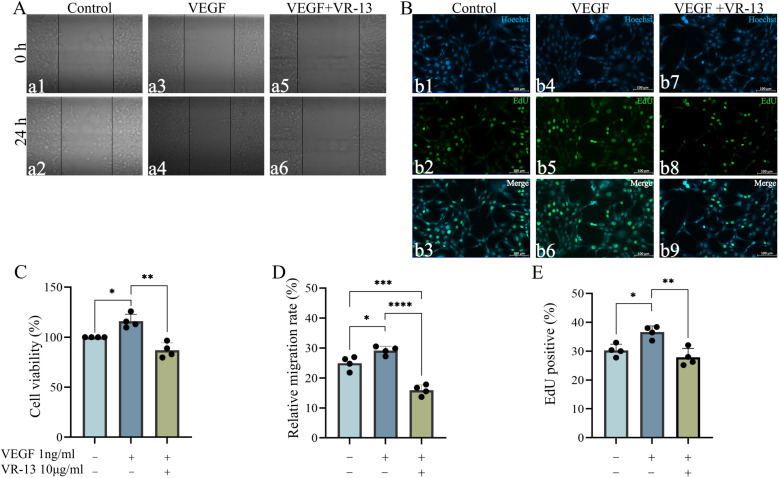
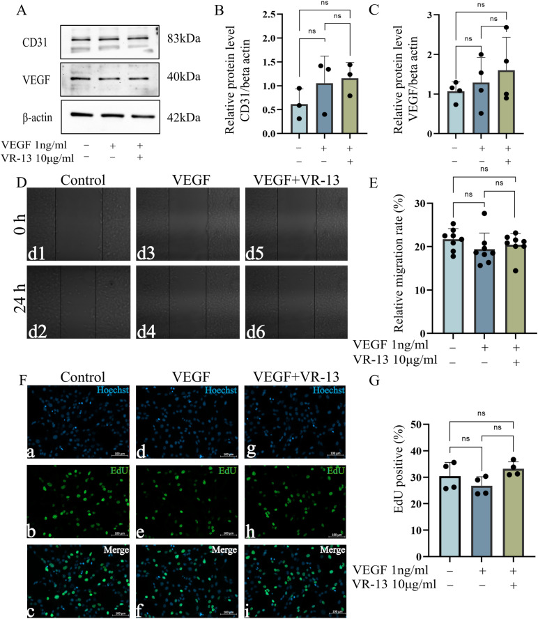
Results: According to the wound-healing and CCK-8 assays, VR-13 was the most effective in inhibiting the proliferation and migration of endothelial cells. Furthermore, VR-13 peptide effectively inhibited the pathological development of CNV without the detection of retinal toxicity in the rat CNV model.

Conclusions: Overall, it was found that VR-13 exhibit significant effects on the inducing of apoptosis and the inhibition of the progression of angiogenesis by regulating the expression of VEGF and CD31 via CD36 signaling pathway.


Conflict of interest statement

The authors have declared that no competing interests exist.

Figures

Fig 1
Fig 1. Different concentrations of thrombospondin-1 (TSP-1)-derived peptides (VR-13) inhibited the proliferation and migration of RF/6A.
A wound-healing assay was employed in the assessment of the inhibitory effects of different concentrations of VR-13 on the proliferation and migration of RF/6A cells, n = 12. VR-13 inhibited cell proliferation and migration with the concentrations of 10 μg/mL, 25 μg/mL, 50 μg/mL, 75 μg/mL, and 100 μg/mL (A, B). CCK-8 cell viability assay showed that the inhibitory effects of VR-13 peptide on cell proliferation exhibited an increasing trend with the increase of concentrations, n = 3 (C) (*, P < 0.05; **, P < 0.01; ***, P < 0.001; ****, P < 0.0001).
Fig 2
Fig 2. VR-13 peptide inhibited the cell proliferation and migration caused by exogenously supplied vascular endothelial growth factor (VEGF).
CCK-8, wound-healing, and 5-ethynyl-2′-deoxyuridine (EdU) assays(n = 4) were used to evaluate inhibition of proliferation and migration of RF/6A cells, in which the RF/6A cells were incubated with 1 ng/mL of exogenously supplied VEGF and 10 μg/mL of VR-13 peptide for 24 h. Exogenous VEGF could significantly stimulate the proliferation and migration of RF/6A, while VR-13 exhibited the ability to inhibit this function. (*, P < 0.05; **, P < 0.01; ***, P < 0.001; ****, P < 0.0001).
Fig 3
Fig 3. In RF/6A cells, the VR-13 peptide inhibited the neovascularization by regulating angiogenesis-related protein levels, such as those of VEGF (n = 6) and CD31 (n = 5), alongside the inducing of cell apoptosis.
(A–C) The expression of angiogenesis-related proteins in each group was examined by western blotting. (D, E) The total apoptosis rate in each group was calculated by flow cytometry, n = 6 (*, P < 0.05; **, P < 0.01; ***, P < 0.001; ****, P < 0.0001).
Fig 4
Fig 4. In a rat Matrigel CNV model, the VR-13 peptide exhibited inhibitory effects on the CNV pathological progression by regulating the VEGF and CD31 levels.
(A–C) Western blot for the detection of the VEGF (n = 7) and CD31 (n = 3) levels in each group (D) Fluorescein isothiocyanate-dextran angiography for the observation of the pattern of the retinal vasculature n = 3. (*, P < 0.05; **, P < 0.01; ***, P < 0.001; ****, P < 0.0001).
Fig 5
Fig 5. VR-13 peptide was bound to CD36 to regulate endothelial cell proliferation and migration, alongside the inhibition of angiogenesis by controlling the levels of the angiogenesis-related proteins VEGF and CD31.
(A–C) Western blots for the detection of the VEGF (n = 4) and CD31(n = 3) levels; (D–G) wound-healing (n = 8) and EdU assays(n = 4) for the evaluation of the inhibitory ability of VR-13 on cell proliferation and migration in CD36 knockdown cells. (*, P < 0.05; **, P < 0.01; ***, P < 0.001; ****, P < 0.0001).

Similar articles

References

    1. Sacconi R, Fragiotta S, Sarraf D, Sadda SR, Freund KB, Parravano M, et al. Towards a better understanding of non-exudative choroidal and macular neovascularization. Prog Retin Eye Res. 2023;92:101113. doi: 10.1016/j.preteyeres.2022.101113 - DOI - PubMed
    1. Sunaga T, Maeda M, Saulle R, Ng SM, Sato MT, Hasegawa T, et al. Anti-vascular endothelial growth factor biosimilars for neovascular age-related macular degeneration. Cochrane Database Syst Rev. 2024;6(6):CD015804. doi: 10.1002/14651858.CD015804.pub2 - DOI - PMC - PubMed
    1. Torres-Costa S, Ramos D, Brandão E, Carneiro Â, Rosas V, Rocha-Sousa A, et al. Incidence of endophthalmitis after intravitreal injection with and without topical antibiotic prophylaxis. Eur J Ophthalmol. 2021;31(2):600–6. doi: 10.1177/1120672120902028 - DOI - PubMed
    1. Ricci F, Bandello F, Navarra P, Staurenghi G, Stumpp M, Zarbin M. Neovascular age-related macular degeneration: therapeutic management and new-upcoming approaches. Int J Mol Sci. 2020;21(21):8242. doi: 10.3390/ijms21218242 - DOI - PMC - PubMed
    1. Sharma D, Zachary I, Jia H. Mechanisms of acquired resistance to anti-VEGF therapy for neovascular eye diseases. Invest Ophthalmol Vis Sci. 2023;64(5):28. doi: 10.1167/iovs.64.5.28 - DOI - PMC - PubMed

MeSH terms

LinkOut - more resources