. 2025 Sep;100(9):1502-1512.
doi: 10.1002/ajh.27743. Epub 2025 Jun 20.

International Consensus Histopathological Criteria for Subtyping Idiopathic Multicentric Castleman Disease Based on Machine Learning Analysis

Affiliations

International Consensus Histopathological Criteria for Subtyping Idiopathic Multicentric Castleman Disease Based on Machine Learning Analysis

Midori Filiz Nishimura et al. Am J Hematol. 2025 Sep.

Abstract

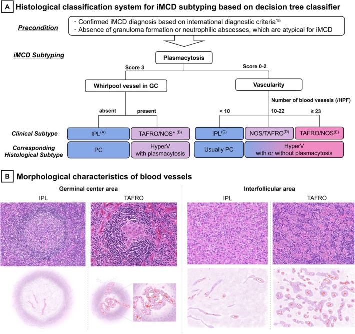
Idiopathic multicentric Castleman disease (iMCD) is a rare lymphoproliferative disorder classified into three recognized clinical subtypes-idiopathic plasmacytic lymphadenopathy (IPL), TAFRO, and NOS. Although clinical criteria are available for subtyping, diagnostically challenging cases with overlapping histopathological features highlight the need for an improved classification system integrating clinical and histopathological findings. We aimed to develop an objective histopathological subtyping system for iMCD that closely correlates with the clinical subtypes. Excisional lymph node specimens from 94 Japanese iMCD patients (54 IPL, 28 TAFRO, 12 NOS) were analyzed for five key histopathological parameters: germinal center (GC) status, plasmacytosis, vascularity, hemosiderin deposition, and "whirlpool" vessel formation in GC. Using hierarchical clustering, we visualized subgroups and developed a machine learning-based decision tree to differentiate the clinical subtypes and validated it in an external cohort of 12 patients with iMCD. Hierarchical cluster analysis separated the IPL and TAFRO cases into mutually exclusive clusters, whereas the NOS cases were interspersed between them. Decision tree modeling identified plasmacytosis, vascularity, and whirlpool vessel formation as key features distinguishing IPL from TAFRO, achieving 91% and 92% accuracy in the training and test sets, respectively. External validation correctly classified all IPL and TAFRO cases, confirming the reproducibility of the system. Our histopathological classification system closely aligns with the clinical subtypes, offering a more precise approach to iMCD subtyping. It may enhance diagnostic accuracy, guide clinical decision-making for predicting treatment response in challenging cases, and improve patient selection for future research. Further validation of its versatility and clinical utility is required.

Keywords: clinical subtype; histopathological criteria; idiopathic multicentric castleman disease; lymphoproliferative disease; machine‐learning.


Conflict of interest statement

E.O. has served as a consultant for Recordati Rare Diseases and as an expert on the advisory board for CSL Behring. D.C.F. has received research funding and consulting fees from Recordati Rare Diseases. The remaining authors declare no competing financial interests. F.v.R. has served on advisory boards for Adicet Bio, Bristol Myers Squibb, Castleman Disease Collaborative Network, EUSA Pharma, GlaxoSmithKline, Janssen Pharmaceuticals Inc., Kite Pharma, Secura Bio, Myeloma360, and Recordati. Additionally, F.v.R. has received donated study drugs for clinical trials from Pfizer.

Figures

FIGURE 1
FIGURE 1
Histological Findings in each Clinical Subtype of iMCD. (A) Histological Findings in a patient with IPL subtype. (a) The germinal center is hyperplastic with a sheet‐like proliferation of mature plasma cells observed in the expanded interfollicular area (HE, 100x). (b) Magnified view of a sheet‐like proliferation of mature plasma cells. Hemosiderin deposition is observed (HE, 400x). (c) CD138‐positive plasma cells proliferate densely in a sheet‐like pattern within the interfollicular area (CD138 staining, 200x). (d) IL‐6 staining shows strong positivity in the plasma cells within the interfollicular area (IL‐6 staining, 200x). (B) Histological Findings in a patient with TAFRO subtype. (a) The germinal center is atrophic, with prominent angiogenesis both inside and outside the follicles (H&E, 100x). (b) Blood vessels enter the germinal center, forming a characteristic “whirlpool” vessel. The endothelial cells are plump. Plasma cell infiltration is not observed (HE, 200x). (C) Histological Findings in patients with NOS subtype. (a), (b), and (c) show the histological findings of different patients with NOS. (a) In the interfollicular area, a sheet‐like proliferation of mature plasma cells is observed, presenting a pattern corresponding to plasma cell‐type histology (HE, 200x). (b) Both angiogenesis and plasma cell proliferation (upper part of the field) were observed in the interfollicular area. Angiogenesis and whirlpool vessel formation were also observed within the germinal center. This pattern has been referred to as the “mixed” type, but we define it as “HyperV with plasmacytosis” (HE, 200x). (c) Prominent angiogenesis with plump endothelial cells is observed, along with blood vessels entering the germinal center and forming a “whirlpool” vessel, consistent with HyperV type histology. Plasma cell proliferation is not observed, and we define this as “HyperV without plasmacytosis” (HE, 200x). [Color figure can be viewed at wileyonlinelibrary.com]
FIGURE 2
FIGURE 2
Evaluated Histological Parameters and their Scoring Criteria. Tissue evaluation was performed based on five criteria: (A) germinal center (GC) status (0–3); (B) plasmacytosis (0–3); (C) vascularity (absolute number of blood vessels per HPF in the interfollicular area); (D) hemosiderin deposition (0–2); and (E) whirlpool vessel formation in the GC (0 or 1). “Whirlpool” vessels vary and include those that form small spiral structures within the germinal center without penetrating vessels, those where vessels enter the germinal center and create a whirlpool‐like appearance at the center, and those where vessels extend radially and develop a whirlpool‐like pattern at the center of the GC. [Color figure can be viewed at wileyonlinelibrary.com]
FIGURE 3
FIGURE 3
Heatmap and Hierarchical Cluster Analysis based on Histological Findings in 94 iMCD Patients. Through hierarchical cluster analysis, the cases were classified into three groups. All IPL cases grouped into the same cluster, demonstrating a highly homogeneous population. While TAFRO and IPL were never mixed within the same cluster, NOS cases were mixed within the TAFRO cluster. Additionally, the second annotation represents responsiveness to tocilizumab, and the third annotation indicates the intensity of IL‐6 immunostaining. The IPL group showed low vascularity, marked plasmacytosis, strong and wide IL‐6 expression on immunostaining, and high responsiveness to tocilizumab, which is clearly visible in the heatmap. [Color figure can be viewed at wileyonlinelibrary.com]
FIGURE 4
FIGURE 4
Histological Classification System for iMCD Subtyping and Key Morphological Characteristics of Blood Vessels. (A) Histological classification system for iMCD subtyping based on decision tree classifier. The application of this classification system requires patients to meet the international diagnostic criteria for iMCD [15]. A top‐to‐bottom flowchart was created. For example, there is no need to apply the whirlpool vessel criterion to individuals with a plasmacytosis score of 0–2 (although it is not problematic to apply it, the whirlpool vessel is more effectively used as an exclusion criterion for the IPL subtype). When emphasizing the correlation with clinical subtypes, PC‐type histology was defined as either the presence of score 3 plasmacytosis [Endpoint (A)] or score 2 plasmacytosis with < 10 vessels/HPF (C), both lacking whirlpool vessels. HyperV type histology is defined as lacking score 3 plasmacytosis and exhibiting ≥ 10 vessels/HPF (D, E). Rarely, HyperV may include a score of 3 plasmacytosis if vascular proliferation is prominent in whirlpool vessels (B). (B) Morphological characteristics of blood vessels (HE and schematic illustration). In the IPL subtype, vascular proliferation is minimal and the endothelium appeared flat. In contrast, the TAFRO subtype is characterized by prominent vascular proliferation, with plump endothelial cells observed in both the regressed germinal centers and interfollicular areas. Vessels penetrate the germinal centers and often exhibit a whirlpool appearance. [Color figure can be viewed at wileyonlinelibrary.com]

Similar articles

References

    1. Tumour‐like lesions with B‐cell predominance , WHO Classification of Haematolymphoid Tumours, 5th ed. (International Agency for Research on Cancer (IARC), 2024).
    1. Lang E. and van Rhee F., “Idiopathic Multicentric Castleman Disease: An Update in Diagnosis and Treatment Advances,” Blood Reviews 64 (2024): 101161. - PubMed
    1. Otsuka M., Koga T., Sumiyoshi R., et al., “Exploring the Clinical Diversity of Castleman Disease and TAFRO Syndrome: A Japanese Multicenter Study on Lymph Node Distribution Patterns,” American Journal of Hematology 100 (2025): 592–605. - PMC - PubMed
    1. Mohri S. Mori, N., Uchida T., and Shimamine T., “Idiopathic Plasmacytic Lymphadenopathy With Polyclonal Hyperimmunoglobulinemia: A Syndrome Related to Giant Lymph Node Hyperplasia of Plasma Cell Type [in Japanese],” Japanese Journal of Sociological Research 20, no. suppl (1980): 55–65.
    1. Kojima M., Nakamura N., Tsukamoto N., et al., “Clinical Implications of Idiopathic Multicentric Castleman Disease Among Japanese: A Report of 28 Cases,” International Journal of Surgical Pathology 16 (2008): 391–398. - PubMed

Supplementary concepts

LinkOut - more resources