. 2025 Jun 20;11(25):eads5272.
doi: 10.1126/sciadv.ads5272. Epub 2025 Jun 20.

Senataxin and DNA-PKcs redundantly promote non-homologous end joining repair of DNA double strand breaks during V(D)J recombination

Affiliations

Senataxin and DNA-PKcs redundantly promote non-homologous end joining repair of DNA double strand breaks during V(D)J recombination

Bo-Ruei Chen et al. Sci Adv. .

Abstract

Nonhomologous end joining (NHEJ) is required for repairing DNA double strand breaks (DSBs) generated by the RAG endonuclease during lymphocyte antigen receptor gene assembly by V(D)J recombination. The ataxia telangiectasia-mutated (ATM) and DNA-dependent protein kinase catalytic subunit (DNA-PKcs) kinases regulate functionally redundant pathways required for NHEJ. Here, we report that loss of the senataxin helicase leads to a strong defect in RAG DSB repair upon inactivation of DNA-PKcs. The NHEJ function of senataxin is redundant with the RECQL5 helicase and the HLTF translocase and is epistatic with ATM. Co-inactivation of ATM, RECQL5, and HLTF results in an NHEJ defect similar to that from the combined deficiency of DNA-PKcs and senataxin or losing senataxin, RECQL5, and HLTF. These data suggest that ATM and DNA-PKcs regulate the functions of senataxin and RECQL5/HLTF, respectively, to provide redundant support for NHEJ.


Figures

Fig. 1.
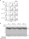
Fig. 1.. A Genome-wide CRISPR/Cas9 screen for identifying genes required for V(D)J recombination in DNA-PKcs inhibited abl pre-B cells.
(A) Schematic of the retroviral V(D)J recombination substrate pMG-INV. The open and filled triangles represent the RSSs. The retroviral LTRs, CEs, and SEs upon RAG cleavage and CJs and SJs after NHEJ-mediated repair are indicated. The anti-sense GFP cDNA is inverted to the sense orientation upon completion of the recombination and its expression driven by the retroviral LTR (green). (B) Schematic diagram of a genome-wide CRISPR-Cas9 screen in WT abl pre-B cells for identifying genes important for V(D)J recombination upon DNA-PKcs inhibition with NU7441. (C) Fold enrichments of selected gRNAs from screens conducted in DMSO and NU7441-treated WT abl pre-B cells (both also treated with imatinib). The fold enrichment is calculated as the ratio of the normalized read counts of a gRNA from GFP cells over that from GFP+ cells. Asterisk denotes (*) five gRNAs to each gene. (D) Flow cytometric analysis for GFP expression in WT and three independently isolated Setx−/− abl pre-B cell lines with pMG-INV and treated with imatinib (imat.) in the presence or absence of the DNA-PKcs kinase inhibitor NU7441 for the indicated times. The percentages of GFP+ cells are indicated in the top left corners of the histograms.
Fig. 2.
Fig. 2.. Loss of senataxin severely impairs NHEJ-mediated RAG DSB repair in DNA-PKcs-inhibited abl pre-B cells during V(D)J recombination.
(A and B) Schematic of the retroviral V(D)J recombination substrates pMX-DELCJ (A) and pMX-DELSJ (B). The open and filled triangles represent the RSSs. The red bar indicates the probe (hCD4 cDNA) for Southern blot. The Es denote the Eco RV restriction sequences. The retroviral LTRs, CEs, and SEs upon RAG cleavage and CJs and SJs after NHEJ-mediated repair are indicated. (C and D) Southern blot analysis of genomic DNA from WT and three clonal Setx−/− abl pre-B cell lines with pMX-DELCJ (C) or pMX-DELSJ (D) treated with imatinib in the presence or absence of the DNA-PKcs kinase inhibitor NU7441 for the indicated times. The genomic DNA samples were digested with Eco RV and hybridized with the hCD4 probe. The restriction fragments corresponding to unrearranged reporters (UR), repaired CJs or SJs, and unrepaired CEs or SEs are indicated. For all Southern blot analysis of pMX-DELSJ or pMX-DELCJ, the percentages of unrepaired (full length and resected) SEs in total RAG cleaved recombination substrates (SE + SJ) are shown below the Southern blot images. β T cell receptor (TCRβ): loading control.
Fig. 3.
Fig. 3.. Combined loss of senataxin and DNA-PKcs proteins impairs NHEJ-mediated RAG DSB repair.
(A) Western blot analysis of cell lysates from WT, Prkdc−/−, Setx−/−, and Setx−/−: Prkdc−/− abl pre-B cells using DNA-PKcs and KAP1 antibodies. (B and C) Southern blot analysis of Eco RV–digested genomic DNA isolated from WT, Prkdc−/−, Setx−/−, and Setx−/−: Prkdc−/− abl pre-B cells with pMX-DECJ (B) or pMX-DESJ (C) and treated with imatinib in the presence or absence of the DNA-PKcs inhibitor NU7441 for the indicated times.
Fig. 4.
Fig. 4.. ATM inhibition in Setx−/− abl pre-B cells does not affect V(D)J recombination.
(A) Flow cytometric analysis for GFP expression in WT and Setx−/− abl pre-B cell lines with pMG-INV and treated with imatinib in the presence or absence of the ATM kinase inhibitor KU55933 for the indicated times. The percentages of GFP+ cells are indicated in the top left corners of the histograms. (B) Southern blot analysis of Eco RV–digested genomic DNA isolated from WT and Setx−/− abl pre-B cell lines with pMX-DESJ and treated with imatinib in the presence or absence of KU55933 for the indicated times.
Fig. 5.
Fig. 5.. Senataxin helicase activity is required for RAG DSB repair.
(A) Western blot analysis of cell lysates from proliferating or imatinib-treated Setx−/−, Setx3HA/3HA, and SetxK1945R/K1945R and SetxL2131W/L2131W abl pre-B cells using HA and KAP1 antibodies. (B) Southern blot analysis of Eco RV–digested genomic DNA from cells described in (A) with pMX-DELSJ and treated with imatinib in the presence or absence of the DNA-PKcs kinase inhibitor NU7441 for the indicated times.
Fig. 6.
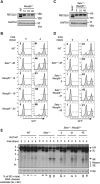
Fig. 6.. Loss of RECQL5 modestly impairs RAG-DSB repair in Setx−/− abl pre-B cells.
(A) Western blot analysis of cell lysates from WT and three independently isolated Recql5−/− abl pre-B cells using RECQL5 and glyceraldehyde-3-phosphate dehydrogenase (GAPDH) antibodies. (B) Flow cytometric analysis for GFP expression in cells described in (A) with pMG-INV and treated with imatinib for the indicated times. The percentages of GFP+ cells are indicated in the top left corners of the histograms. (C) Western blot analysis of cell lysates from Setx−/− and three independently isolated Setx−/−: Recql5−/− abl pre-B cells using RECQL5 and GAPDH antibodies. (D) Flow cytometric analysis for GFP expression in cells described in (C) treated with imatinib for the indicated times. (E) Southern blot analysis of Eco RV–digested genomic DNA from cells described in (D) with pMX-DELSJ and treated with imatinib in the presence or absence of the DNA-PKcs kinase inhibitor NU7441 for the indicated times.
Fig. 7.
Fig. 7.. Combined loss of senataxin, RECQL5, and HLTF results in defected RAG DSB repair.
(A) Western blot analysis of cell lysates from Setx−/− and three independently isolated Setx−/−: Recql5−/−: Hltf−/− abl pre-B cells using RECQL5, HLT, and GAPDH antibodies. (B) Flow cytometric analysis for GFP expression in WT, Setx−/− and Setx−/−: Recql5−/−: Hltf−/− abl pre-B cells with pMG-INV and treated with imatinib for the indicated times. The percentages of GFP+ cells are indicated in the top left corners of the histograms. (C) Southern blot analysis of EcoRV-digested genomic DNA from abl pre-B cells described in (B) with pMX-DESJ and treated with imatinib in the presence or absence of the DNA-PKcs kinase inhibitor NU7441 for the indicated times. (D) Schematic for Southern blot analysis of Cas9 DSB repair in the Eb region of the Tcrb gene. The gEb binding and restriction enzyme Hind III cutting locations are shown. The Eb probe is indicated as the gray bar (top). Southern blot analysis of Hind III–digested genomic DNA from imatinib-treated WT, Setx−/−: Recql5−/−: Hltf−/−, and Lig4−/− abl pre-B cells expressing Cas9 and gEb using the Eb probe (bottom). (E) Western blot analysis of HLTF co-immunoprecipitation with ectopically expressed HA-RECQL5 in imatinib-treated abl pre-B cell lysate with HA and HLTF antibodies. The asterisk indicates the nonspecific recognizing bands by the HLTF antibody. IP, immunoprecipitation.
Fig. 8.
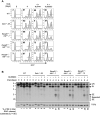
Fig. 8.. ATM activity is required for RAG DSB repair in Recql5−/−: Hltf/− abl pre-B cells.
(A) Flow cytometric analysis for GFP expression from pMG-INV in WT, Setx/−, and three independently isolated Recql5−/−: Hltf/− abl pre-B cell lines treated with imatinib in the presence or absence of the DNA-PKcs kinase inhibitor NU7441 or the ATM kinase inhibitor KU55933 for the indicated times. The percentages of GFP+ cells are indicated in the top left corners of the histograms. (B) Southern blot analysis of EcoRV-digested genomic DNA from cells described in (A) with pMX-DESJ and treated with imatinib in the presence or absence of KU55933 for the indicated times.
Fig. 9.
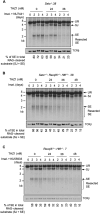
Fig. 9.. Partial inhibition of RNA polymerase II improves NHEJ-mediated RAG DSB repair in helicase-deficient abl pre-B cells.
Southern blot analysis of Eco RV–digested genomic DNA from pMX-DELSJ containing, imatinib treated Setx−/− (with the DNA-PKcs kinase inhibitor NU7441) (A), Setx−/−: Recql5−/−: Hltf−/− (B), and Recql5−/−: Hltf−/− (with the ATM kinase inhibitor KU55933) (C) abl pre-B cells with increasing concentrations (24, 36, and 48 nM) of the CDK7 inhibitor THZ1 for the indicated times.
Fig. 10.
Fig. 10.. Aberrant DNA end joining in abl pre-B cells deficient in senataxin, RECQL5, and HLTF.
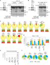
(A and B) Left: Western blot analysis of cell lysate from Setx−/− (A) or Setx−/−: Recql5−/−: Hltf−/− (B) abl pre-B cells expressing Cas9 and gRNAs to Rosa26 (gRosa26) or Mre11 (gMre11) using MRE11 and GAPDH antibodies. Right: Southern blot analysis of Eco RV–digested genomic DNA from the aforementioned cells with pMX-DELSJ and treated with imatinib in the presence or absence of the DNA-PKcs kinase inhibitor NU7441 for the indicated times. (C) Percentages of precise joints (P, yellow), joints with deletions (D, green), and joints with insertions (I, red) in SJs amplified from pMG-INV in the indicated abl pre-B cells after imatinib treatment for 4 days in the presence or absence of DNA-PKcs kinase inhibitor NU7441 or ATM kinase inhibitor KU55933. (D) Percentages of long (11 to >50 bp) nucleotide loss in deletion-containing SJs amplified from pMG-INV in the indicated abl pre-B cell lines. The numbers above each bar indicate the average percentages of SJ sequences with the specific length distributions of deletion. (E) Percentages of SJs with microhomology at joining junctions in the indicated abl pre-B cell lines with the average percentages of SJs with microhomology shown on top of each bar. (F) Microhomology length distribution (% of all microhomology) of the SJs shown in (F). Number of samples sequenced per genotype: WT: 10, WT + NU7441: 3, WT + KU55933: 3, Setx−/−: 6, Setx−/− + NU7441: 3, Recql5−/−: 3, Hltf−/−: 3, Setx−/−: Recql5−/−: 4, Setx−/−: Hltf−/−: 3, Recql5−/−: Hltf−/−: 3, Recql5−/−: Hltf/− + KU55933: 3, and Setx−/−: Recql5−/−: Hltf−/−: 4.

Update of

Similar articles

References

    1. Bassing C. H., Swat W., Alt F. W., The mechanism and regulation of chromosomal V(D)J recombination. Cell 109, S45–S55 (2002). - PubMed
    1. Christie S. M., Fijen C., Rothenberg E., V(D)J recombination: Recent insights in formation of the recombinase complex and recruitment of DNA repair machinery. Front. Cell Dev. Biol. 10, 886718 (2022). - PMC - PubMed
    1. Lieber M. R., Ma Y., Pannicke U., Schwarz K., The mechanism of vertebrate nonhomologous DNA end joining and its role in V(D)J recombination. DNA Repair 3, 817–826 (2004). - PubMed
    1. Rooney S., Chaudhuri J., Alt F. W., The role of the non-homologous end-joining pathway in lymphocyte development. Immunol. Rev. 200, 115–131 (2004). - PubMed
    1. Chang H. H. Y., Pannunzio N. R., Adachi N., Lieber M. R., Non-homologous DNA end joining and alternative pathways to double-strand break repair. Nat. Rev. Mol. Cell Biol. 18, 495–506 (2017). - PMC - PubMed

MeSH terms

Substances