. 2025 Jun 20;11(25):eadr6399.
doi: 10.1126/sciadv.adr6399. Epub 2025 Jun 20.

Metabolically driven flows enable exponential growth in macroscopic multicellular yeast

Affiliations

Metabolically driven flows enable exponential growth in macroscopic multicellular yeast

Nishant Narayanasamy et al. Sci Adv. .

Abstract

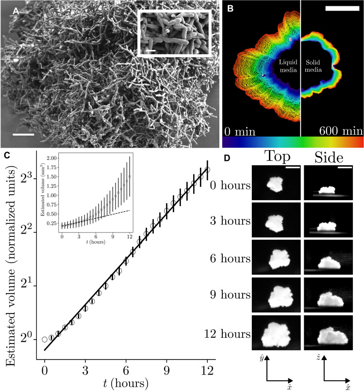
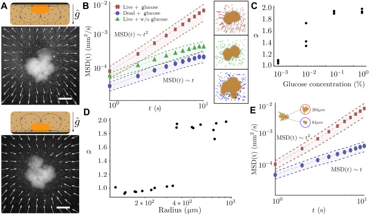
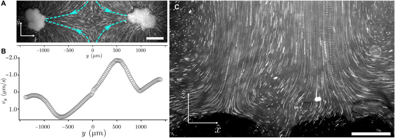
The ecological and evolutionary success of multicellular lineages stems substantially from their increased size relative to unicellular ancestors. However, large size poses biophysical challenges, especially regarding nutrient transport: These constraints are typically overcome through multicellular innovations. Here, we show that an emergent biophysical mechanism-spontaneous fluid flows arising from metabolically generated density gradients-can alleviate constraints on nutrient transport, enabling exponential growth in nascent multicellular clusters of yeast lacking any multicellular adaptations for nutrient transport or fluid flow. Beyond a threshold size, the metabolic activity of experimentally evolved snowflake yeast clusters drives large-scale fluid flows that transport nutrients throughout the cluster at speeds comparable to those generated by ciliary actuation in extant multicellular organisms. These flows support exponential growth at macroscopic sizes that theory predicts should be diffusion limited. This demonstrates how simple physical mechanisms can act as a "biophysical scaffold" to support the evolution of multicellularity by opening up phenotypic possibilities before genetically encoded innovations.


Figures

Fig. 1.
Fig. 1.. Fluid environments allow for exponential growth of the snowflake yeast clusters.
(A) Scanning electron microscope image of a snowflake yeast cluster. Scale bar, 20 μm. Inset: Higher-resolution image of single cells within the cluster. Scale bar, 5 μm. (B) Cluster outline visualized as a function of time in nondeformable [yeast extract peptone dextrose (YEPD) agar] medium and fluid (YEPD liquid) environment for a single measurement over 60 min with a 10-min interval. (C) Quantification of different growth phenotypes in deformable (N = 8) versus nondeformable (N = 10) media. Scale bar, 1 mm (details in the Supplementary Materials). Estimated volume (area of top view times average height of side view) of snowflake yeast clusters over time, normalized to that at the first time point. Error bars represent SE of 3 replicates. Inset: Estimated volume, without normalization. The dashed line shows a linear fit to the first 3.5 hours. (D) Microscopy images of the top and side view over time of one of the clusters measured for (C). Scale bars, 1 mm.
Fig. 2.
Fig. 2.. Macroscopic snowflake yeast advectively mixes its ambient fluid environment.
Macroscopic snowflake yeast generate three-dimensional flows in the ambient fluid as visualized by tracer particle streaks and particle tracking data from a (A) side view and a (B) top view. Scale bar, 500 μm. (C) Flow speeds are comparable to those generated by ciliated and flagellated multicellular organisms: S. coeruleus (1 to 2 mm), Volvox carteri (0.5 to 2 mm) Chlamydomonas reinhardtii (10 to 20 μm), and Synaptula rosetta colony (10 to 30 μm). Graphics are obtained from the Database Center for Life Sciences (CC BY-SA 4.0). (D) Flow remains constant over a period of nearly 500 min during which the growth measurements were made (single observation).
Fig. 3.
Fig. 3.. Snowflake yeast beyond a threshold size drive buoyant flows due to their metabolic activity.
(A) Flows around snowflake yeast immobilized in agar (black region). The dotted line shows the imaging plane in which the flows are measured. The top and bottom panels show the flows in the same imaging plane when the experimental setup is flipped with respect to the direction of gravity. Scale bar, 500 μm. Reversal of flow in the same imaging plane hints at a gravity sensitive flow mechanism such as buoyant flows. (B) Metabolic activity is necessary for the flows, which are quantified by the MSD of tracer beads around the snowflake yeast. Tracer beads around live, metabolically active snowflake yeast exhibit ballistic motion i.e., α ≈ 2 (square data points), while tracer beads diffuse, i.e., α ≈ 1, around metabolically inactive and dead clusters (triangles, circles respectively). (C) Metabolically active flows emerge at high enough glucose concentration in the ambient medium. (D) Flows emerge around clusters beyond a certain threshold size along the evolutionary lineage of the MuLTEE. The exponent α of the tracer particle MSDs exhibits a clear and sharp transition from a diffusive behavior (i.e., α ≈ 1) to a ballistic behavior (i.e., α ≈ 2). (E) When clusters large enough to produce flows are broken to sizes below the threshold size identified in (D), the clusters no longer generate flows (α ~ 1, circles). On the other hand, clusters that are individually below the threshold size do create a flow when aggregated together to form a larger group (α ~ 2, squares).
Fig. 4.
Fig. 4.. Snowflake yeast clusters act as individual metabolically powered density pumps.
(A) Flows generated by the snowflake yeast are localized around individual clusters. The flows around two neighboring clusters exhibit a stagnation point between the clusters. Scale bar, 500 μm. (B) Velocity along the line connecting the two clusters shows a singular stagnation point of the flow. (C) Presence of such a stagnation point is also seen from the traces of the vertical plumes of two clusters that are within a cluster-radius distance from each other. Scale bar, 500 μm.

Update of

Similar articles

References

    1. Grosberg R. K., Strathmann R. R., The evolution of multicellularity: A minor major transition? Annu. Rev. Ecol. Evol. Syst. 38, 621–654 (2007).
    1. M. D. Herron, P. L. Conlin, W. C. Ratcliff, Eds., The Evolution of Multicellularity (CRC Press, 2022).
    1. West S. A., Fisher R. M., Gardner A., Toby Kiers E., Major evolutionary transitions in individuality. Proc. Natl. Acad. Sci. U.S.A. 112, 10112–10119 (2015). - PMC - PubMed
    1. Kai Tong G., Bozdag O., Ratcliff W. C., Selective drivers of simple multicellularity. Curr. Opin. Microbiol. 67, 102141 (2022). - PubMed
    1. Fisher S. A., Burggren W. W., Role of hypoxia in the evolution and development of the cardiovascular system. Antioxid. Redox Signal. 9, 1339–1352 (2007). - PubMed

MeSH terms

LinkOut - more resources